. 2020 May 6;6(19):eaay8828.
doi: 10.1126/sciadv.aay8828. eCollection 2020 May.

A 3D human brain-like tissue model of herpes-induced Alzheimer's disease

Affiliations

A 3D human brain-like tissue model of herpes-induced Alzheimer's disease

Dana M Cairns et al. Sci Adv. .

Abstract

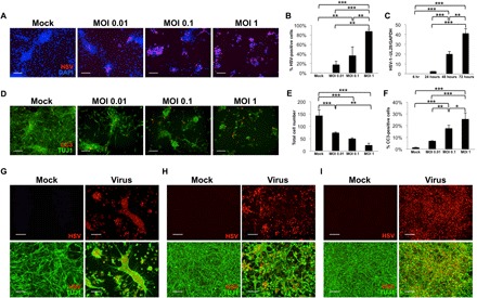
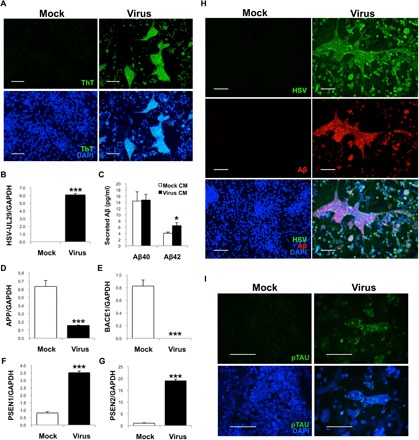
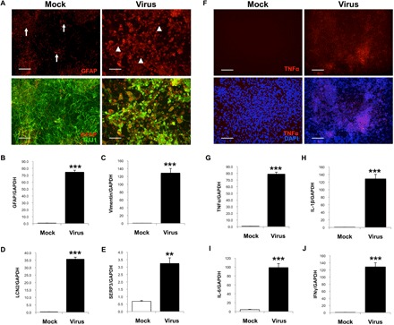
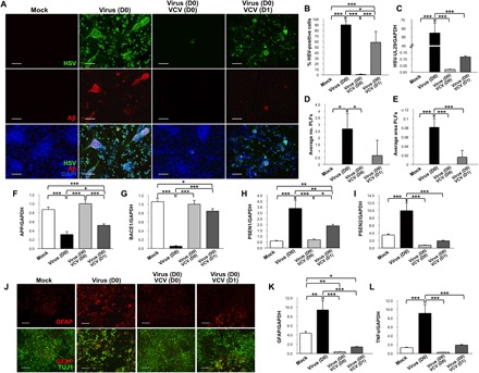
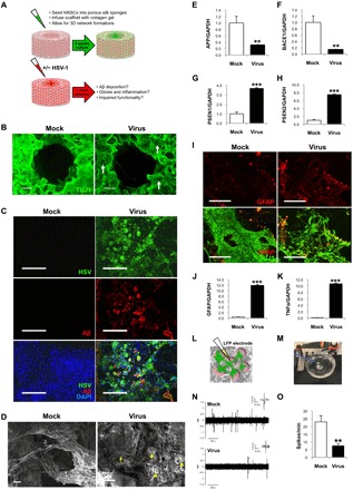
Alzheimer's disease (AD) is a neurodegenerative disorder that causes cognitive decline, memory loss, and inability to perform everyday functions. Hallmark features of AD-including generation of amyloid plaques, neurofibrillary tangles, gliosis, and inflammation in the brain-are well defined; however, the cause of the disease remains elusive. Growing evidence implicates pathogens in AD development, with herpes simplex virus type I (HSV-1) gaining increasing attention as a potential causative agent. Here, we describe a multidisciplinary approach to produce physiologically relevant human tissues to study AD using human-induced neural stem cells (hiNSCs) and HSV-1 infection in a 3D bioengineered brain model. We report a herpes-induced tissue model of AD that mimics human disease with multicellular amyloid plaque-like formations, gliosis, neuroinflammation, and decreased functionality, completely in the absence of any exogenous mediators of AD. This model will allow for future studies to identify potential downstream drug targets for treating this devastating disease.


Figures

Fig. 1
Fig. 1. hiNSCs are highly infectable and responsive to HSV-1 exposure.
hiNSCs were cultured with varying MOIs for 24 hours and assayed for virus expression by immunostaining (A and B) and quantitative polymerase chain reaction (qPCR) showing viral HSV-1–UL29 expression over time (C). Results from β-III tubulin (TUJ1) and cleaved Caspase3 (CC3) immunostaining (D) indicate that relatively high MOI (~1 or greater) results in rapid and robust cell death in 24 hours, with corresponding quantification of cell number (E) and percentage of CC3-positive cells in response to increasing MOI in hiNSCs (F). Low-level HSV-1 infection causes morphological changes. hiNSCs were cultured 1 day (G), 4 days (H), or 7 days (I) before HSV-1 exposure (MOI of 0.0001) for 2 to 3 days. Immature hiNSCs form HSV-1–positive cell conglomerates in response to infection. Scale bars, 100 μm. Asterisks indicate statistically significant differences with error bars showing means ± SD (*P ≤ 0.05, **P ≤ 0.01, and ***P ≤ 0.001). DAPI, 4′,6-diamidino-2-phenylindole; GAPDH, glyceraldehyde-3-phosphate dehydrogenase.
Fig. 2
Fig. 2. HSV infection causes Aβ+ PLFs and specific regulation of AD mediators in hiNSCs.
(A) hiNSCs were cultured for 4 days then subjected to HSV-1 infection for 3 days. HSV-1–infected hiNSCs demonstrated large areas of thioflavin T (ThT)–positive regions. (B) qPCR confirms HSV-1 infection in hiNSCs. (C) Results from an ELISA against Aβ isoforms Aβ1–40 and Aβ1–42, using CM from mock- or HSV-1–infected hiNSCs, reveals a statistically significant increase in Aβ1–42, but not Aβ1–40, in HSV-1–infected CM. qPCR analysis reveals significant down-regulation of APP and BACE1 (D and E) and robust up-regulation of γ-secretase subunits PSEN1/2 (F and G). (H) Further immunostaining reveals large multicellular conglomerates of Aβ+ PLFs that overlaps with HSV staining. (I) Those PLFs also stain positive for hyperphosphorylated Tau. Scale bars, 100 μm. Asterisks indicate statistically significant differences with error bars showing means ± SD (*P ≤ 0.05 and ***P ≤ 0.001).
Fig. 3
Fig. 3. HSV-1 causes reactive gliosis and inflammation reminiscent of neurodegeneration in hiNSCs.
(A and B) HSV-1–infected hiNSCs highly express glia marker GFAP. Immunostaining results reveal increased number of GFAP+ cells and an altered morphology suggestive of gliosis. Other known markers of reactive astrocytes were also up-regulated in HSV-1–infected hiNSCs: (C) vimentin, (D) LCN2, and (E) SERP3. (F and G) Pro-inflammatory marker TNFα was also up-regulated. In HSV-1–infected hiNSCs, qPCR analysis revealed similarly high expression of other inflammatory markers known to be involved in AD: (H) IL-1β, (I) IL-6, and (J) IFNγ. Scale bars, 100 μm. Asterisks indicate statistically significant differences with error bars showing means ± SD (**P ≤ 0.01 and ***P ≤ 0.001).
Fig. 4
Fig. 4. VCV treatment results in reduced AD-like phenotype induced by HSV-1 infection in hiNSCs.
(A) Treatment with VCV reduces HSV-1 infection as well as generation of Aβ+ PLFs. (B) Quantification of infection and (C) qPCR for HSV-UL29. VCV causes a decrease in (D) total number and (E) average area of PLFs. VCV helps to normalize expression of AD mediators back to control levels in HSV-1–infected hiNSCs: (F) APP, (G) BACE1, (H) PSEN1, and (I) PSEN2. (J) VCV treatment rescues gliosis phenotype in HSV-1–infected hiNSCs. qPCR analysis reveals restoration of (K) GFAP and (L) TNFα expression. Scale bars, 100 μm. Asterisks indicate statistically significant differences with error bars showing means ± SD (*P ≤ 0.05, **P ≤ 0.01, and ***P ≤ 0.001).
Fig. 5
Fig. 5. hiNSCs cultured in a 3D brain model develop an AD-like phenotype in response to low-level HSV-1 infection.
(A) Model of 3D human brain–like model. hiNSCs were cultured in the donut model for 4 weeks before HSV-1 infection for 1 week. (B) Low-magnification images of brain model showing β-III tubulin (TUJI1) and beta amyloid (Aβ) immunostaining. Arrows point to regions of neuronal loss in HSV-1–infected tissues. (C) Confocal images showing HSV and Aβ immunostaining in both mock- and virus-infected constructs. (D) Scanning electron micrographs reveal morphological changes in HSV-1–infected tissue constructs. Arrowheads and arrow indicate the presence of both small and relatively larger PLFs, respectively. Scale bars, 10 μm. (E) APP and (F) BACE1 were down-regulated, and (G and H) PSEN1/2 were up-regulated, similar to results in monolayer cultures. HSV-1 induces gliosis and neuroinflammation in 3D cultures. Glial marker GFAP is up-regulated as evidenced by (I) immunostaining and (J) qPCR, as well as (K) pro-inflammatory marker TNFα. LFP recordings were performed to assess functionality in 3D cultures. Schematic representation of microelectrode placement for LFP assessments (L) and an image of constructs placed on electrophysiology rigs for recordings (M) (photo credit: Dana M Cairns, Tufts University). (N and O) Representative traces of mock versus infected brain models shows that HSV-1–treated donuts show significantly less activity reminiscent of impaired functionality in patients with AD. Asterisks indicate statistically significant differences with error bars showing means ± SD (**P ≤ 0.01 and ***P ≤ 0.001).

References

    1. Alzheimer's Association , 2016 Alzheimer’s disease facts and figures. Alzheimers Dement. 12, 459–509 (2016). - PubMed
    1. Naj A. C., Schellenberg G. D.; Alzheimer’s Disease Genetics Consortium (ADGC) , Genomic variants, genes, and pathways of Alzheimer’s disease: An overview. Am. J. Med. Genet. B Neuropsychiatr. Genet. 174, 5–26 (2017). - PMC - PubMed
    1. Weyer S. W., Klevanski M., Delekate A., Voikar V., Aydin D., Hick M., Filippov M., Drost N., Schaller K. L., Saar M., Vogt M. A., Gass P., Samanta A., Jäschke A., Korte M., Wolfer D. P., Caldwell J. H., Müller U. C., APP and APLP2 are essential at PNS and CNS synapses for transmission, spatial learning and LTP. EMBO J. 30, 2266–2280 (2011). - PMC - PubMed
    1. Kelleher R. J. III, Shen J., Presenilin-1 mutations and Alzheimer’s disease. Proc. Natl. Acad. Sci. U.S.A. 114, 629–631 (2017). - PMC - PubMed
    1. De Strooper B., Vassar R., Golde T., The secretases: Enzymes with therapeutic potential in Alzheimer disease. Nat. Rev. Neurol. 6, 99–107 (2010). - PMC - PubMed

Publication types

Substances