. 2020 May 22;6(21):eaba6357.
doi: 10.1126/sciadv.aba6357. eCollection 2020 May.

In situ repair abilities of human umbilical cord-derived mesenchymal stem cells and autocrosslinked hyaluronic acid gel complex in rhesus monkeys with intrauterine adhesion

Affiliations

In situ repair abilities of human umbilical cord-derived mesenchymal stem cells and autocrosslinked hyaluronic acid gel complex in rhesus monkeys with intrauterine adhesion

Lingjuan Wang et al. Sci Adv. .

Abstract

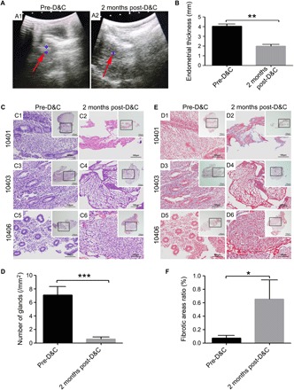
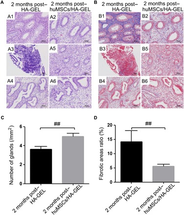
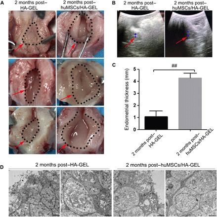
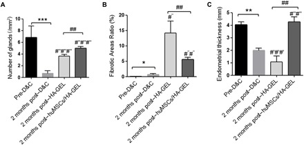
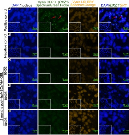
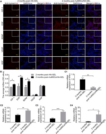
Increasing occurrence of moderate to severe intrauterine adhesion (IUA) is seriously affecting the quality of human life. The aim of the study was to establish IUA models in nonhuman primates and to explore the dual repair effects of human umbilical cord-derived mesenchymal stem cells (huMSCs) loaded on autocrosslinked hyaluronic acid gel (HA-GEL) on endometrial damage and adhesion. Here, we recorded the menstrual cycle data in detail with uterine cavities observed and endometrial tissues detected after intervention, and the thicker endometria, decreased amount of fibrotic formation, increased number of endometrium glands, etc., suggested that both HA-GEL and huMSC/HA-GEL complexes could partially repair IUA caused by mechanical injury, but huMSC/HA-GEL complex transplantation had notable dual repair effects: a reliable antiadhesion property and the promotion of endometrial regeneration.


Figures

Fig. 1<b>The evaluation of IUA model establishment.</b>
Fig. 1The evaluation of IUA model establishment.
(A) Detection of Doppler ultrasound. A1: Representative image of endometrial thickness for pre-D&C; A2: Representative image of endometrial thickness at 2 months post-D&C (red arrow, the endometrium echo; blue area, the largest cross section of endometrium). (B) Comparisons of endometrial thickness for pre- or post-D&C. (C) H&E staining of endometria for pre-D&C (C1, C3, and C5) and post-D&C (C2, C4, and C6); 10401, 10403, and 10406, respectively; see table S3 for details. Inserted overview pictures are of lower magnification; black squares are highly magnified regions. (D) Masson staining of endometria for pre-D&C (D1, D3, and D5) and post-D&C (D2, D4, and D6); 10401, 10403, and 10406, respectively; see table S3 for details. Inserted overview pictures are of lower magnification; black squares are highly magnified regions. (E) Comparisons of endometrial gland numbers per unit area for pre- or post-D&C. (F) Comparisons of fibrotic area ratios for pre- or post-D&C. *P < 0.05, **P < 0.01, and ***P < 0.001 versus the pre-D&C group, and the results shown are the mean ± SEM of three technical replicates from each animal.
Fig. 2<b>Histological inspection of different interventions.</b>
Fig. 2Histological inspection of different interventions.
(A) Endometrial H&E staining at 2 months after HA-GEL transplantation (A1, A3, and A4 correspond to 10401, 10403, and 10404, respectively) and huMSCs/HA-GEL transplantation (A2, A5, and A6 correspond to 10402, 10405, and 10406, respectively); 10401 to 10406, see table S3 for details. (B) Endometrial Masson staining at 2 months after HA-GEL transplantation (B1, B3, and B4 correspond to 10401, 10403, and 10404, respectively) and huMSCs/HA-GEL transplantation (B2, B5, and B6 correspond to 10402, 10405, and 10406, respectively); 10401 to 10406, see table S3 for details. (C) Comparisons of endometrial gland numbers per unit area between the HA-GEL transplantation group and the huMSC/HA-GEL transplantation group. (D) Comparisons of fibrotic area ratios between the HA-GEL transplantation group and the huMSCs/HA-GEL transplantation group. ##P < 0.01 versus HA-GEL transplantation group, and the results shown are the mean ± SEM of three technical replicates from each animal.
Fig. 3 <b>The endometrial effects of different interventions on morphology and ultrastructure.</b>
Fig. 3 The endometrial effects of different interventions on morphology and ultrastructure.
(A) Representative images of uterine cavities in the HA-GEL transplantation and huMSCs/HA-GEL transplantation groups (the dotted area and the red arrow mark the endometrial area). (B) Representative images of endometrial thickness for ultrasound detection in the HA-GEL transplantation and huMSCs/HA-GEL transplantation groups (the red arrow marks the endometrial echo; the blue area marks the largest cross section of the endometrium). (C) Comparisons of endometrial thickness between the HA-GEL transplantation group and the huMSCs/HA-GEL transplantation group. (D) Representative images of ultrastructural changes in the HA-GEL transplantation and huMSCs/HA-GEL transplantation groups (the left panel shows the surface of epithelial cells; the right panel shows the intercellular changes). ##P < 0.01 versus 2 months post–HA-GEL, and the results shown are the mean ± SEM of three technical replicates from each animal. Photos provided by Lingjuan Wang and Chengliang Xiong (Institute of Reproductive Health, Center of Reproductive Medicine, Tongji Medical College, Huazhong University of Science and Technology).
Fig. 4<b>Comprehensive assessment for the effects of different interventions on endometrial regeneration and remodeling.</b>
Fig. 4Comprehensive assessment for the effects of different interventions on endometrial regeneration and remodeling.
(A) Comparisons of endometrial gland numbers per unit area. (B) Comparisons of ratios of fibrotic area (%). (C) Comparisons of endometrial thickness (mm). *P < 0.05, **P < 0.01, and ***P < 0.001, all versus pre-D&C; #′#′#′P < 0.001 versus pre-D&C; #″P < 0.05, #″#″P < 0.01, and #″#″#″P < 0.001, all versus 2 months post-D&C; ##P < 0.01 versus 2 months post–HA-GEL, and the results shown are the mean ± SEM of three technical replicates from each animal.
Fig. 5 <b>FISH analysis for tracking transplanted huMSCs in the endometrium.</b>
Fig. 5 FISH analysis for tracking transplanted huMSCs in the endometrium.
(A) Positive control, human endometrium (containing XX chromosomes); the red arrow indicates the green signal for Vysis CEP X (DXZ1). (B) Endometrial localization of huMSCs in the HA-GEL transplantation group (negative control). (C to E) Distribution of huMSCs in endometria 2 months after huMSCs/HA-GEL co-transplantation. Double/single-labeled staining (orange/green signal or just green signal) cells were defined as huMSCs. For details on 10402, 10405, and 10406, see table S3. Inserted overview pictures show a lower magnification.
Fig. 6 <b>The expression of cytokines in the endometria.</b>
Fig. 6 The expression of cytokines in the endometria.
(A) The expression and localization of the potential cytokines secreted by huMSCs in the HA-GEL transplantation group and the huMSCs/HA-GEL transplantation group; inserted overview pictures show a lower magnification. (B) The optical density (OD) value of IGF-1, EGF, BDNF, VEGF, and HGF 2 months after HA-GEL transplantation or huMSCs/HA-GEL co-transplantation. (C1) Comparison of IFN-γ mRNA expression between the HA-GEL transplantation group and the huMSC/HA-GEL transplantation group. (C2) Comparison of IL-4 mRNA expression between the HA-GEL transplantation group and the huMSC/HA-GEL transplantation group. (C3) Comparison of IGF-1 mRNA expression between the HA-GEL transplantation group and the huMSC/HA-GEL transplantation group. (C4) Comparison of EGF mRNA expression between the HA-GEL transplantation group and the huMSC/HA-GEL transplantation group. *P < 0.05, **P < 0.01, and ***P < 0.001 versus 2 months post–HA-GEL; the results shown are the mean ± SEM of three technical replicates from each animal.

References

    1. Evans-Hoeker E. A., Young S. L., Endometrial receptivity and intrauterine adhesive disease. Semin. Reprod. Med. 32, 392–401 (2014). - PubMed
    1. Hooker A. B., Lemmers M., Thurkow A. L., Heymans M. W., Opmeer B. C., Brölmann H. A. M., Mol B. W., Huirne J. A. F., Systematic review and meta-analysis of intrauterine adhesions after miscarriage: Prevalence, risk factors and long-term reproductive outcome. Hum. Reprod. Update 20, 262–278 (2014). - PubMed
    1. Guo E. J., Chung J. P. W., Poon L. C. Y., Li T. C., Reproductive outcomes after surgical treatment of Asherman syndrome: A systematic review. Best Pract. Res. Clin. Obstet. Gynaecol. 59, 98–114 (2019). - PubMed
    1. Yu D., Wong Y.-M., Cheong Y., Xia E., Li T.-C., Asherman syndrome—One century later. Fertil. Steril. 89, 759–779 (2008). - PubMed
    1. Zhu R., Duan H., Gan L., Wang S., Comparison of intrauterine suitable balloon and Foley balloon in the prevention of adhesion after hysteroscopic adhesiolysis. Biomed. Res. Int. 2018, 1–6 (2018). - PMC - PubMed

Publication types

Substances