. 2020 Sep 3;30(10):5484-5501.
doi: 10.1093/cercor/bhaa130.

Bipartite Functional Fractionation within the Default Network Supports Disparate Forms of Internally Oriented Cognition

Affiliations

Bipartite Functional Fractionation within the Default Network Supports Disparate Forms of Internally Oriented Cognition

Rocco Chiou et al. Cereb Cortex. .

Abstract

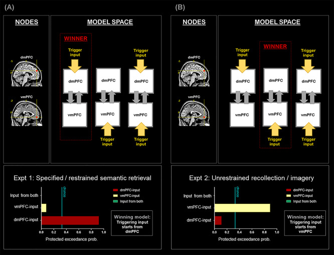
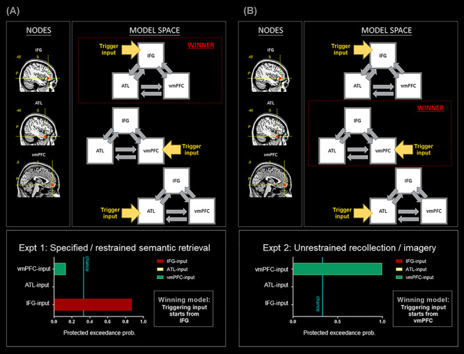
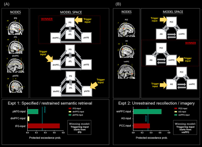
Our understanding about the functionality of the brain's default network (DN) has significantly evolved over the past decade. Whereas traditional views define this network based on its suspension/disengagement during task-oriented behavior, contemporary accounts have characterized various situations wherein the DN actively contributes to task performance. However, it is unclear how different task-contexts drive componential regions of the DN to coalesce into a unitary network and fractionate into different subnetworks. Here we report a compendium of evidence that provides answers to these questions. Across multiple analyses, we found a striking dyadic structure within the DN in terms of the profiles of task-triggered fMRI response and effective connectivity, significantly extending beyond previous inferences based on meta-analysis and resting-state activities. In this dichotomy, one subset of DN regions prefers mental activities "interfacing with" perceptible events, while the other subset prefers activities "detached from" perceptible events. While both show a common "aversion" to sensory-motoric activities, their differential preferences manifest a subdivision that sheds light upon the taxonomy of the brain's memory systems. This dichotomy is consistent with proposals of a macroscale gradational structure spanning across the cerebrum. This gradient increases its representational complexity, from primitive sensory-motoric processing, through lexical-semantic representations, to elaborated self-generated thoughts.

Keywords: connectivity; default-mode network; memory; semantic cognition; topography.


Figures

Figure 1
Figure 1
(A) Brain regions showing significantly greater activity for mind-wandering (rest), compared to the visuospatial control task, thresholded at P < 0.05 (FWE-corrected for whole-brain voxel intensity). Significant clusters in Experiment 1 (green), Experiment 2 (blue), and their conjunctions (cyan). (B) ROI analysis of the dmPFC versus vmPFC. Note that negative β-weight means more active for mind-wandering (rest), compared to the visuospatial control tasks. (C) ROI analysis of the three core DN areas (the vmPFC, PCC, and AG), as well as the three core SN areas (the ATL, IFG, and pMTG). See Supplementary Material for the locations of ROIs that are rendered on a template brain. ***P < 0.001.
Figure 2
Figure 2
(A) Brain regions showing significantly greater activity for the two types of self-related tasks: The self-concept evaluation task (red), for the autobiographical memory task (yellow), and their conjunctions (orange). All contrasts were against the baseline of mind-wandering (rest), thresholded at P < 0.05 (FWE-corrected for whole-brain voxel intensity). (B) ROI analysis reveals an interaction between brain regions (dmPFC vs. vmPFC) and the types of self-related tasks (self-concept vs. autobiographical memory). (C) ROI analysis of the three core DN areas (the vmPFC, PCC, and AG), as well as the three core SN areas (the ATL, IFG, and pMTG). (D) The significant interaction between the two networks and the two types of self-processing. (E) The six ROIs ranked by their preference for autobiographical memory over self-concept, with all of the three DN regions showing a noticeably greater preference than all of the three SN regions. ***P < 0.001, *P < 0.05.
Figure 3
Figure 3
All contrasts were thresholded at P < 0.05 (FWE-corrected for whole-brain voxels). The upper row illustrates the contrast of (left to right) “self-knowledge > visuospatial task,” “self-knowledge > other-referential knowledge,” and “self-knowledge > resting-state baseline.” Note that the choice of baseline affects the size of vmPFC cluster, with the rest-baseline engaging the vmPFC most, the visuospatial-baseline engaging the vmPFC least, and the other-baseline being intermediate. A highly similar pattern is found in the contrast of (lower row, left to right) “Rest > Visuospatial,” “Rest > Other,” and “Rest > Self,” indicating that mind-wandering (rest) equally engages the vmPFC as self-knowledge. The image follows the neurological convention—the left/right side on the image represents the left/right hemisphere.
Figure 4
Figure 4
(A) A direct comparison between the two introspective tasks reveals the regions showing significantly greater activity for the autobiographical task (yellow clusters: Autobiographical Memory > Theory of Mind) and for the mentalizing task (red clusters: Theory of Mind > Autobiographical Memory). Statistics are thresholded at thresholded at P < 0.05 (FWE-corrected for whole-brain voxel intensity). In addition to the key finding that we elaborate in the main text (i.e., greater DN activity during autobiographical memory vs. greater SN activity during theory of mind), it is noteworthy that, in the mentalizing task, participants simulate the thoughts/feelings of the protagonist in the photograph while viewing/interpreting the semantic meaning of the visual scene. This is a multifaceted operation that entails visual attention and scene recognition, semantic interpretation, and theory of mind. Our results—namely, the red clusters—reflect this multifaceted nature: Relative to the autobiographical task, the mentalizing task imposes greater demand on visual processing, hence greater activation of the posterior vision- and attention-related cortices. (B) ROI analysis reveals an interaction between brain regions (dmPFC vs. vmPFC) and introspective tasks (autobiographical memory vs. theory of mind). (C) ROI analysis of the three core DN areas (the vmPFC, PCC, and AG), as well as the three core SN areas (the ATL, IFG, and pMTG), revealing a significant interaction between networks and tasks. (D) The significant interaction between the two inferior parietal subregions (the AG vs. TPJ) and the two types of introspective processing. (E) The significant interaction between the regions preferring other-referential tasks (the dmPFC, left TPJ, and right TPJ) and the two types of other-referential processes. Note that all of the ROI analyses here are based on β-weights compared against resting baseline. ***P < 0.001, **P < 0.01, *P < 0.05.
Figure 5
Figure 5
(A) DCM-1: All analyses are based on the data of Experiment 1. (B) DCM-2: All analyses are based on the data of Experiment 2. Note that the nodes of the two DCM have the same coordinates, suggesting a drastic change of network dynamics as a result of experimental contexts.
Figure 6
Figure 6
(A) DCM-3: All analyses are based on the data of Experiment 1. (B) DCM-4: All analyses are based on the data of Experiment 2. Note that the nodes of the two DCM have the same coordinates, suggesting a drastic change of network dynamics as a result of experimental contexts.
Figure 7
Figure 7
(A) DCM-5: All analyses are based on the data of Experiment 1. All of the nodes belong to the SN. (B) DCM-6: All analyses are based on the data of Experiment 2. All of the nodes belong to the DN.

Similar articles

Cited by

References

    1. Andreasen NC, O'leary DS, Cizadlo T, Arndt S, Rezai K, Watkins GL, Boles Ponto LL, Hichwa RD. 1995. Remembering the past: two facets of episodic memory explored with positron emission tomography. Am J Psychiatry. 152:1576–1585. - PubMed
    1. Andrews-Hanna JR, Reidler JS, Sepulcre J, Poulin R, Buckner RL. 2010. Functional-anatomic fractionation of the brain's default network. Neuron. 65:550–562. - PMC - PubMed
    1. Anticevic A, Repovs G, Shulman GL, Barch DM. 2010. When less is more: TPJ and default network deactivation during encoding predicts working memory performance. NeuroImage. 49:2638–2648. - PMC - PubMed
    1. Bajada CJ, Trujillo-Barreto NJ, Parker GJ, Cloutman LL, Lambon Ralph MA. 2019. A structural connectivity convergence zone in the ventral and anterior temporal lobes: data-driven evidence from structural imaging. Cortex. 120:298–307. - PMC - PubMed
    1. Baldassano C, Chen J, Zadbood A, Pillow JW, Hasson U, Norman KA. 2017. Discovering event structure in continuous narrative perception and memory. Neuron. 95:709–721.e705. - PMC - PubMed

Publication types