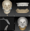
Fusion of intra-oral scans in cone-beam computed tomography scans
- PMID: 32495223
- PMCID: PMC7785548
- DOI: 10.1007/s00784-020-03336-y
Fusion of intra-oral scans in cone-beam computed tomography scans
Abstract
Purpose: The purpose of this study was to evaluate the clinical accuracy of the fusion of intra-oral scans in cone-beam computed tomography (CBCT) scans using two commercially available software packages.
Materials and methods: Ten dry human skulls were subjected to structured light scanning, CBCT scanning, and intra-oral scanning. Two commercially available software packages were used to perform fusion of the intra-oral scans in the CBCT scan to create an accurate virtual head model: IPS CaseDesigner® and OrthoAnalyzer™. The structured light scanner was used as a gold standard and was superimposed on the virtual head models, created by IPS CaseDesigner® and OrthoAnalyzer™, using an Iterative Closest Point algorithm. Differences between the positions of the intra-oral scans obtained with the software packages were recorded and expressed in six degrees of freedom as well as the inter- and intra-observer intra-class correlation coefficient.
Results: The tested software packages, IPS CaseDesigner® and OrthoAnalyzer™, showed a high level of accuracy compared to the gold standard. The accuracy was calculated for all six degrees of freedom. It was noticeable that the accuracy in the cranial/caudal direction was the lowest for IPS CaseDesigner® and OrthoAnalyzer™ in both the maxilla and mandible. The inter- and intra-observer intra-class correlation coefficient showed a high level of agreement between the observers.
Clinical relevance: IPS CaseDesigner® and OrthoAnalyzer™ are reliable software packages providing an accurate fusion of the intra-oral scan in the CBCT. Both software packages can be used as an accurate fusion tool of the intra-oral scan in the CBCT which provides an accurate basis for 3D virtual planning.
Keywords: CAD; Digital imaging/radiology; Oral implants/implantology; Orthodontic(s); Orthognathic surgery; Treatment planning.
Conflict of interest statement
The authors declare that they have no conflict of interest.
Figures


Similar articles
-
The impact of different cone beam computed tomography and multi-slice computed tomography scan parameters on virtual three-dimensional model accuracy using a highly precise ex vivo evaluation method.J Craniomaxillofac Surg. 2016 May;44(5):632-6. doi: 10.1016/j.jcms.2016.02.005. Epub 2016 Feb 13. J Craniomaxillofac Surg. 2016. PMID: 27017101
-
Reliability and accuracy of three imaging software packages used for 3D analysis of the upper airway on cone beam computed tomography images.Dentomaxillofac Radiol. 2017 Aug;46(6):20170043. doi: 10.1259/dmfr.20170043. Epub 2017 Jun 14. Dentomaxillofac Radiol. 2017. PMID: 28467118 Free PMC article.
-
Three-dimensional maxillary and mandibular regional superimposition using cone beam computed tomography: a validation study.Int J Oral Maxillofac Surg. 2016 May;45(5):662-9. doi: 10.1016/j.ijom.2015.12.006. Epub 2016 Jan 12. Int J Oral Maxillofac Surg. 2016. PMID: 26794399
-
A novel method for fusion of intra-oral scans and cone-beam computed tomography scans for orthognathic surgery planning.J Craniomaxillofac Surg. 2016 Feb;44(2):160-6. doi: 10.1016/j.jcms.2015.11.017. Epub 2015 Dec 8. J Craniomaxillofac Surg. 2016. PMID: 26732637
-
Registration of digital dental models and cone-beam computed tomography images using 3-dimensional planning software: Comparison of the accuracy according to scanning methods and software.Am J Orthod Dentofacial Orthop. 2020 Jun;157(6):843-851. doi: 10.1016/j.ajodo.2019.12.013. Am J Orthod Dentofacial Orthop. 2020. PMID: 32487314
Cited by
-
[Research progress of digital occlusion setup in orthognathic surgery].Zhongguo Xiu Fu Chong Jian Wai Ke Za Zhi. 2023 Feb 15;37(2):247-251. doi: 10.7507/1002-1892.202210086. Zhongguo Xiu Fu Chong Jian Wai Ke Za Zhi. 2023. PMID: 36796824 Free PMC article. Review. Chinese.
-
Registration-free workflow for electromagnetic and optical navigation in orbital and craniofacial surgery.Sci Rep. 2021 Sep 10;11(1):18080. doi: 10.1038/s41598-021-97706-5. Sci Rep. 2021. PMID: 34508161 Free PMC article.
-
Plaster Casts vs. Intraoral Scans: Do Different Methods of Determining the Final Occlusion Affect the Simulated Outcome in Orthognathic Surgery?J Pers Med. 2022 Aug 5;12(8):1288. doi: 10.3390/jpm12081288. J Pers Med. 2022. PMID: 36013237 Free PMC article.
-
Optimizing Orthognathic Surgery: Leveraging the Average Skull as a Dynamic Template for Surgical Simulation and Planning in 30 Patient Cases.J Clin Med. 2023 Dec 18;12(24):7758. doi: 10.3390/jcm12247758. J Clin Med. 2023. PMID: 38137827 Free PMC article.
-
Efficacy of Constructing Digital Hybrid Skull-Dentition Images Using an Intraoral Scanner and Cone-Beam Computed Tomography.Scanning. 2022 Mar 3;2022:8221514. doi: 10.1155/2022/8221514. eCollection 2022. Scanning. 2022. PMID: 35316954 Free PMC article.
References
-
- Jheon AH, Oberoi S, Solem RC, Kapila S. Moving towards precision orthodontics: An evolving paradigm shift in the planning and delivery of customized orthodontic therapy. Orthod. Craniofacial Res. 2017;20(March):106–113. - PubMed
-
- Verhamme LM, Meijer GJ, Bergé SJ, Soehardi RA, Xi T, de Haan AFJ, Schutyser F, Maal TJJ. An accuracy study of computer-planned implant placement in the augmented maxilla using mucosa-supported surgical templates. Clin Implant Dent Relat Res. 2015;17(6):1154–1163. - PubMed
-
- Stokbro K, Aagaard E, Torkov P, Bell RB, Thygesen T. Virtual planning in orthognathic surgery. Int J Oral Maxillofac Surg. 2014;43(8):957–965. - PubMed
-
- Liebregts J, Xi T, Timmermans M, de Koning M, Bergé S, Hoppenreijs T, Maal T. Accuracy of three-dimensional soft tissue simulation in bimaxillary osteotomies. J Craniomaxillofac Surg. 2015;43(3):329–335. - PubMed
MeSH terms
LinkOut - more resources
Full Text Sources
Other Literature Sources
Miscellaneous

