

Blockade of PAR-1 Signaling Attenuates Cardiac Hypertrophy and Fibrosis in Renin-Overexpressing Hypertensive Mice
- PMID: 32495720
- PMCID: PMC7429042
- DOI: 10.1161/JAHA.119.015616
Blockade of PAR-1 Signaling Attenuates Cardiac Hypertrophy and Fibrosis in Renin-Overexpressing Hypertensive Mice
Abstract
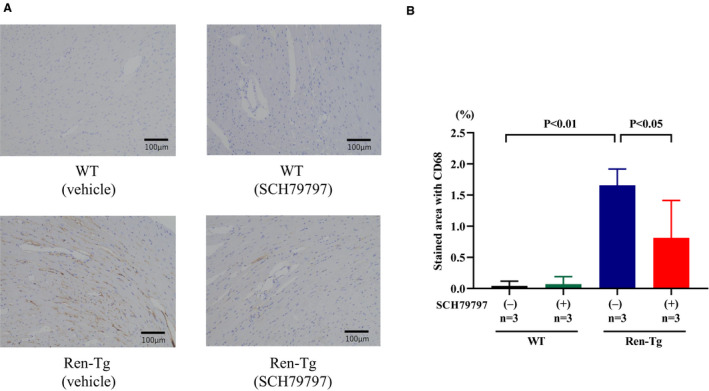
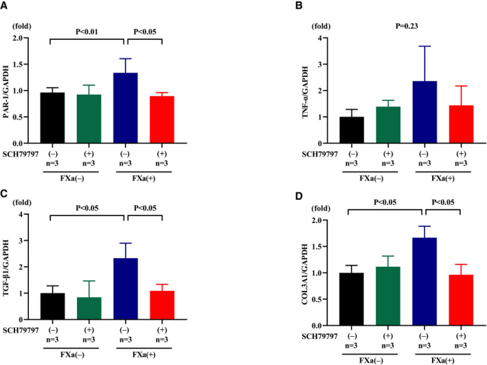
Background Although PAR-1 (protease-activated receptor-1) exerts important functions in the pathophysiology of the cardiovascular system, the role of PAR-1 signaling in heart failure development remains largely unknown. We tested the hypothesis that PAR-1 signaling inhibition has protective effects on the progression of cardiac remodeling induced by chronic renin-angiotensin system activation using renin-overexpressing hypertensive (Ren-Tg) mice. Methods and Results We treated 12- to 16-week-old male wild-type (WT) mice and Ren-Tg mice with continuous subcutaneous infusion of the PAR-1 antagonist SCH79797 or vehicle for 4 weeks. The thicknesses of interventricular septum and the left ventricular posterior wall were greater in Ren-Tg mice than in WT mice, and SCH79797 treatment significantly decreased these thicknesses in Ren-Tg mice. The cardiac fibrosis area and monocyte/macrophage deposition were greater in Ren-Tg mice than in WT mice, and both conditions were attenuated by SCH79797 treatment. Cardiac mRNA expression levels of PAR-1, TNF-α (tumor necrosis factor-α), TGF-β1 (transforming growth factor-β1), and COL3A1 (collagen type 3 α1 chain) and the ratio of β-myosin heavy chain (β-MHC) to α-MHC were all greater in Ren-Tg mice than in WT mice; SCH79797 treatment attenuated these increases in Ren-Tg mice. Prothrombin fragment 1+2 concentration and factor Xa in plasma were greater in Ren-Tg mice than in WT mice, and both conditions were unaffected by SCH79797 treatment. In isolated cardiac fibroblasts, both thrombin and factor Xa enhanced ERK1/2 (extracellular signal-regulated kinase 1/2) phosphorylation, and SCH79797 pretreatment abolished this enhancement. Furthermore, gene expression of PAR-1, TGF-β1, and COL3A1 were enhanced by factor Xa, and all were inhibited by SCH79797. Conclusions The results indicate that PAR-1 signaling is involved in cardiac remodeling induced by renin-angiotensin system activation, which may provide a novel therapeutic target for heart failure.
Keywords: cardiac fibrosis; cardiac hypertrophy; factor Xa; protease‐activated receptor; renin–angiotensin system.
Figures


References
-
- Yancy CW, Jessup M, Bozkurt B, Butler J, Casey DE Jr, Colvin MM, Drazner MH, Filippatos GS, Fonarow GC, Givertz MM, et al. 2017 ACC/AHA/HFSA focused update of the 2013 ACCF/AHA guideline for the management of heart failure: a report of the American College of Cardiology/American Heart Association Task Force on clinical practice guidelines and the Heart Failure Society of America. Circulation. 2017;136:e137–e161. - PubMed
-
- Mosterd A, Cost B, Hoes AW, de Bruijne MC, Deckers JW, Hofman A, Grobbee DE. The prognosis of heart failure in the general population: the Rotterdam Study. Eur Heart J. 2001;22:1318–1327. - PubMed
-
- Vasan RS, Larson MG, Benjamin EJ, Evans JC, Reiss CK, Levy D. Congestive heart failure in subjects with normal versus reduced left ventricular ejection fraction: prevalence and mortality in a population‐based cohort. J Am Coll Cardiol. 1999;33:1948–1955. - PubMed
-
- Steffel J, Luscher TF, Tanner FC. Tissue factor in cardiovascular diseases: molecular mechanisms and clinical implications. Circulation. 2006;113:722–731. - PubMed
-
- Takada M, Tanaka H, Yamada T, Ito O, Kogushi M, Yanagimachi M, Kawamura T, Musha T, Yoshida F, Ito M, et al. Antibody to thrombin receptor inhibits neointimal smooth muscle cell accumulation without causing inhibition of platelet aggregation or altering hemostatic parameters after angioplasty in rat. Circ Res. 1998;82:980–987. - PubMed
MeSH terms
Substances
LinkOut - more resources
Full Text Sources
Medical
Research Materials
Miscellaneous
