. 2020 Jul 9;48(12):6458-6470.
doi: 10.1093/nar/gkaa479.

Dynamic topology of double-stranded telomeric DNA studied by single-molecule manipulation in vitro

Affiliations

Dynamic topology of double-stranded telomeric DNA studied by single-molecule manipulation in vitro

Xiaonong Zhang et al. Nucleic Acids Res. .

Abstract

The dynamic topological structure of telomeric DNA is closely related to its biological function; however, no such structural information on full-length telomeric DNA has been reported due to difficulties synthesizing long double-stranded telomeric DNA. Herein, we developed an EM-PCR and TA cloning-based approach to synthesize long-chain double-stranded tandem repeats of telomeric DNA. Using mechanical manipulation assays based on single-molecule atomic force microscopy, we found that mechanical force can trigger the melting of double-stranded telomeric DNA and the formation of higher-order structures (G-quadruplexes or i-motifs). Our results show that only when both the G-strand and C-strand of double-stranded telomeric DNA form higher-order structures (G-quadruplexes or i-motifs) at the same time (e.g. in the presence of 100 mM KCl under pH 4.7), that the higher-order structure(s) can remain after the external force is removed. The presence of monovalent K+, single-wall carbon nanotubes (SWCNTs), acidic conditions, or short G-rich fragments (∼30 nt) can shift the transition from dsDNA to higher-order structures. Our results provide a new way to regulate the topology of telomeric DNA.


Figures

Figure 1.
Figure 1.
(A) Schematics of the construction of the recombinant plasmid. Short (TTAGGG)5 (blue fragment) and (CCCTAA)5 (red fragment) DNAs were extended by EM-PCR and then inserted into the T-Vector. The recombinant plasmid that contains double-stranded telomeric DNA can be obtained after overnight transfection and incubation. (B) Agarose gel analysis of the extension products produced from (TTAGGG)5 and (CCCTAA)5 during EM-PCR. Lane 1: 4 cycles; lane 2: 7 cycles; lane 3: 10 cycles; lane 4: 14 cycles; lane 5: 17 cycles; and lane 6: 20 cycles. (C) The T-Vector and the recombinant plasmid were double-digested by BspQI and BstAPI restriction endonucleases. The products were analyzed by agarose gel as shown in lane 1 (T-Vector) and lane 2 (recombinant, pUC18-T2AG3), respectively. (D) AFM imaging of telomeric-sequence-containing DNA (∼1200 bp) on mica.
Figure 2.
Figure 2.
(A) Experimental setup for investigating double-stranded telomeric DNA. The DNA molecule is chemically bound to a gold surface (via thiol-gold chemistry) and picked up by an AFM tip through the streptavidin-biotin interactions. (B) Typical force-extension curve of telomeric dsDNA in 100 mM KCl, at pH 4.7. The red trace is a WLC fit to the experimental curve with a persistence length of P = 50 nm. (C) Histogram of transition force (n = 97).
Figure 3.
Figure 3.
Typical force-extension curves of telomeric dsDNA obtained by repeatedly manipulating the same DNA molecule in: (A and B) 100 mM KCl, 10 mM Tris–HCl, pH 7.7; (C and D) 100 mM KCl, 10 mM MES, pH 4.7. Stretching curves obtained at pH 7.7 can be superimposed, and those obtained at pH 4.7 cannot be superimposed. At pH 4.7, the force plateau of the stretching curve is decreased with an increasing number of stretching–relaxation cycles. The curves obtained in different stretching-relaxation cycles are marked in different colors, and the order of the stretching–relaxation cycle is marked numerically.
Figure 4.
Figure 4.
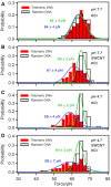
The plateau-force histograms obtained during repeated stretching-relaxation cycles on random DNA (black) and telomeric DNA (red) in different experimental buffer: (A) 100 mM KCl, 10 mM Tris, pH 7.7; (B) 100 mM KCl, 10 mM Tris, pH 7.7 with 10 μg/ml SWCNT-COOH; (C) 100 mM KCl, 10 mM MES, pH 4.7; (D) 100 mM KCl, 10 mM MES, pH 4.7 with 10 μg/ml SWCNT-COOH. The Gaussian fittings on the force distributions of telomeric DNA and random DNA are colored in blue and green, respectively. At least 90 single-molecule stretching curves were used for the construction of each of the force histogram.
Figure 5.
Figure 5.
(A) Typical stretching (black) and relaxation (gray) curves of telomeric DNA in 10 μg/ml SWCNT-COOH and 100 mM KCl (pH 4.7) at room temperature. (B) The probabilities of low-force value plateau (red in circles, left) and double-plateau (blue in triangles, right) in stretching curves in different experimental buffers. The solid and open markers represent telomeric DNA and random DNA, respectively. (C) The plateau-force histogram of stretching curves of the double-plateau (n = 128). Multiple Gaussian peaks are shown in red with the smoothed curve shown in black. Each Gaussian peak is labeled with a letter (peaks a–c) and the mean force value with standard deviation. (D) The proportion of three force regions (L, H, D) in the double-plateau under different experimental conditions.
Figure 6.
Figure 6.
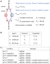
(A) Schematic of the dsDNA stretching and corresponding force analysis. (B) Possible unfolding force combinations corresponding to the L/H/D regions. (C) Major structures under different conditions. GQ and iM represent G-quadruplex and i-motif, respectively.
Figure 7.
Figure 7.
A probable model of telomeric DNA topology after force-induced melting. Different conditions in the renaturation process eventually induce telomeric dsDNA to form different topologies.

References

    1. Shay J.W., Wright W.E.. Senescence and immortalization: role of telomeres and telomerase. Carcinogenesis. 2005; 26:867–874. - PubMed
    1. Greider C.W. Telomerase activity, cell proliferation, and cancer. Proc. Natl. Acad. Sci. U.S.A. 1998; 95:90–92. - PMC - PubMed
    1. Moyzis R.K., Buckingham J.M., Cram L.S., Dani M., Deaven L.L., Jones M.D., Meyne J., Ratliff R.L., Wu J.R.. A highly conserved repetitive DNA sequence, (TTAGGG)n, present at the telomeres of human chromosomes. Proc. Natl. Acad. Sci. U.S.A. 1988; 85:6622–6626. - PMC - PubMed
    1. Morin G.B. The human telomere teriminal transferase enzyme is a ribonucleoprotein that synthesizes TTAGGG repeats. Cell. 1989; 59:521–529. - PubMed
    1. McElligott R., Wellinger R.J.. The terminal DNA structure of mammalian chromosomes. EMBO J. 1997; 16:3705–3714. - PMC - PubMed

Publication types