. 2020 Aug 20;48(14):e79.
doi: 10.1093/nar/gkaa472.

A universal method for the rapid isolation of all known classes of functional silencing small RNAs

Affiliations

A universal method for the rapid isolation of all known classes of functional silencing small RNAs

Thomas Grentzinger et al. Nucleic Acids Res. .

Abstract

Diverse classes of silencing small (s)RNAs operate via ARGONAUTE-family proteins within RNA-induced-silencing-complexes (RISCs). Here, we have streamlined various embodiments of a Q-sepharose-based RISC-purification method that relies on conserved biochemical properties of all ARGONAUTEs. We show, in multiple benchmarking assays, that the resulting 15-min benchtop extraction procedure allows simultaneous purification of all known classes of RISC-associated sRNAs without prior knowledge of the samples-intrinsic ARGONAUTE repertoires. Optimized under a user-friendly format, the method - coined 'TraPR' for Trans-kingdom, rapid, affordable Purification of RISCs - operates irrespectively of the organism, tissue, cell type or bio-fluid of interest, and scales to minute amounts of input material. The method is highly suited for direct profiling of silencing sRNAs, with TraPR-generated sequencing libraries outperforming those obtained via gold-standard procedures that require immunoprecipitations and/or lengthy polyacrylamide gel-selection. TraPR considerably improves the quality and consistency of silencing sRNA sample preparation including from notoriously difficult-to-handle tissues/bio-fluids such as starchy storage roots or mammalian plasma, and regardless of RNA contaminants or RNA degradation status of samples.


Figures

Figure 1.
Figure 1.
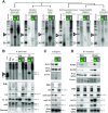
(A) Schematics of the three-steps, 15 min Microspin™ anion-exchange chromatography procedure. (B) Left: immunoblot analysis of native AGOs extracted from Arabidopsis inflorescences using the procedure in (A). Right: anti-Flag immunoblot analysis of genomic FLAG-AGO3 expressed under the AGO3 promoter extracted from 1–5 days after pollination (DAP) siliques (top) or genomic FLAG-AGO7 expressed under the AGO7 promoter extracted from 2 weeks-old seedlings (bottom) using the procedure in (A). (C) RNA extracted from fractions purified in (B) was radiolabeled with T4 Polynucleotide Kinase (PNK) prior to migration on 17% denaturing polyacrylamide and transfer onto a nylon membrane. (D) Low molecular weight (LMW) RNA blot analysis of RNA extracted from fractions purified in (B). Radiolabeled oligonucleotides were used as probes to detect known Arabidopsis silencing sRNAs. Nomenclature for panels B–D. I: clarified lysate, E: AGO-eluted fraction, HS: High-salt resin wash after AGO elution. Clarified lysate used in lane I represents 20% of the material used for the TraPR purification; Ambion® DECADE™: RNA ruler, in nucleotides. EtBr: total RNA ethidium bromide staining. RNA spike: synthetic 22-nt RNA added in fractions post-TraPR purification as a control for RNA extraction. U6 RNA hybridization provides an internal control for successful RISCs isolation in the E-fraction. Prot. spike: FLAG-tagged protein added post-purification as a control for protein extraction, detected with an anti-Flag antibody. Coom.: total protein Coomassie blue staining.
Figure 2.
Figure 2.
(A) PNK radiolabeling (as in Figure 1C) of total RNAs from the I-, E- and HS-fractions extracted via the procedure schematized in Figure 1A as applied to various organisms. (B) Top: as in (A) but with exponentially growing Paramecium cells (Veg.) and Paramecium cells at the onset of sexual events (T 0 arbitrarily defined as the time when 50% of the cells begin maternal macronucleus fragmentation, as evaluated cytologically). Two synthetic radiolabeled RNAs were used as rulers. Bottom: LMW RNA blot analysis of sRNAs co-extracted in (B, top). Radiolabeled oligonucleotides were used as probes to detect known Paramecium silencing sRNAs (Cl22, mtA, Sardine) accumulating under the indicated condition/stage. (C) Top: immunoblot analysis of native ALG1 extracted from C. elegans mixed (eggs+larvae+adults) populations using the procedure schematized in Figure 1A. Bottom: LMW RNA blot analysis of sRNAs co-extracted in (C, top). Radiolabeled oligonucleotides were used as probes to detect known C. elegans silencing sRNAs. (D) Top: immunoblot analysis of native AGO1 and AGO2 from mouse neuroblastoma cells (N2a; left) or dissected brain (right), extracted via the procedure schematized in Figure 1A. Bottom: LMW RNA blot analysis of sRNAs co-extracted in (D, top). Radiolabeled oligonucleotides were used as probes to detect known mouse miRNAs (let-7a, miR-16). Nomenclature for panels A–D. I: clarified lysate, E: AGO-eluted fraction, HS: High-salt resin wash after AGO elution. Clarified lysate used in lane I represent 20% of the material used for the TraPR purification; Ambion® DECADE™: RNA ruler, in nucleotides. EtBr: total RNA ethidium bromide staining. RNA spike: synthetic 22-nt RNA added in fractions post-TraPR purification as a control for RNA extraction. U6 RNA hybridization provides an internal control for successful RISCs isolation in E-fraction and equal RNA loading. Prot. spike: FLAG-tagged protein added post-purification as a control for protein extraction, detected with an anti-Flag antibody. Coom: total protein Coomassie blue staining.
Figure 3.
Figure 3.
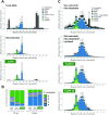
(A) Size distribution and annotation of Arabidopsis inflorescence-derived sRNAs following TRUseq-based deep-sequencing of TRI®-Reagent-extracted total RNA (Total-RNA, top), polyacrylamide gel-selected sRNA (Gel-selected, middle) or sRNAs directly cloned after TraPR purification (bottom). n = 3 technical replicates for each condition, bars indicate the standard deviation. (B) 5′-terminal nucleotide composition of deep-sequenced sRNAs prepared by the methods indicated in (A). Nucleotide identity is displayed for 21-nt-long (left) and 24-nt-long (right) sRNAs. Bars indicate the standard deviation. (C) Size distribution and annotation of Drosophila ovary sRNAs following deep-sequencing, via an in-house protocol, of Gel-selected+ribo-depleted sRNAs (top), Gel-selected+ribo-depleted+oxidized sRNAs (middle), or sRNAs directly cloned after TraPR purification (bottom). 10 μg of total RNA were used for the first two methods whereas TraPR-based purification involved either 5 (TraPR 5) or 50 (TraPR 50) ovary pairs. n = 2 technical replicates for each preparation method, bars indicate the two independent datapoints.
Figure 4.
Figure 4.
(A) Northern LMW RNA blot analysis of sRNAs found in the I-, E-, and HS-fractions following TraPR-based extraction conducted in mouse liver lysates treated for 30 min at room temperature with 100 U RNase T1 or not (Intact). TRI®-Reagent-extracted total RNA (Total-RNA) from identically treated lysates was analyzed in parallel. Radiolabeled oligonucleotides were used as probes to detect known mouse liver miRNAs (let-7a; miR-122). I: clarified lysate, E: AGO-eluted fraction, HS: High-salt resin wash after AGO elution. Clarified lysate used in lane I represent 20% of the material used for the TraPR purification; EtBr: total RNA ethidium bromide staining. U6 RNA hybridization provides an internal control for successful RISCs isolation in E- fraction and equal RNA loading. (B) Size distribution and genomic origin of mouse liver sRNAs following deep-sequencing of TRI®-Reagent-extracted total RNA (Total-RNA, left) or sRNAs directly purified by TraPR (right) from intact (top) or RNAse T1-treated (bottom) mouse liver lysates. n = 3 biological replicates for each condition, bars indicate the standard deviation. (C) Extended Pearson correlation matrix of rRNAs (black), miRNAs (green) and other sRNAs (gray) based on the sRNA reads count over each considered annotated loci in the libraries prepared in (B).
Figure 5.
Figure 5.
(A) Size distribution and genomic origin of mouse plasma sRNA following Lexogen-based sequencing of TRI®-Reagent-extracted total RNA (Total-RNA, left) or sRNAs directly purified by TraPR (right). n = 4 biological replicates for each condition, bars indicate the standard deviation. (B) miRNA-dispersion analysis, based on the standard deviation of miRNA counts, in the libraries prepared in (A) as depicted per quartile reflecting miRNA abundance (C) Proportion of plasmatic tRNA-derived reads according to their sizes (<20-nt; 20-to-24-nt; > 24-nt) in the libraries prepared in (A). (D) 5′-terminal nucleotide composition of plasmatic tRNA-derived RNA in the 20-to-24-nt size range in the libraries prepared in (A). For panels B, C and D Wilcoxon rank sum test, n.s.: non-significant, *P < 10−3. (E) Proportion of plasmatic tRNA Val-AAC-derived sRNA reads according to their sizes (<20-nt; 20-to-24-nt; >24-nt) in the libraries prepared in (A). (F) 5′-terminal nucleotide composition of plasmatic tRNA Val-AAC-derived sRNAs in the 20-to-24-nt size range in the libraries prepared in (A). Bars indicate the standard deviation. (G) Single nucleotide sRNA profiles over tRNA Val-AAC using the libraries prepared in (A), displayed by size range (grey <20-nt; green 20-to-24-nt; black >24-nt). RPM: reads per million. For panels B–D, boxes indicate the first to third quartiles and whiskers show the highest and lowest values within 1.5 times the interquartile range (IQR).

References

    1. Wilson R.C., Doudna J.A.. Molecular mechanisms of RNA interference. Annu. Rev. Biophys. 2013; 42:217–239. - PMC - PubMed
    1. Chalker D.L., Yao M.C.. DNA elimination in ciliates: transposon domestication and genome surveillance. Annu. Rev. Genet. 2011; 45:227–246. - PubMed
    1. Matzke M.A., Mosher R.A.. RNA-directed DNA methylation: an epigenetic pathway of increasing complexity. Nat. Rev. Genet. 2014; 15:394–408. - PubMed
    1. Ow M.C., Lau N.C., Hall S.E.. Small RNA library cloning procedure for deep sequencing of specific endogenous siRNA classes in Caenorhabditis elegans. Methods Mol. Biol. 2014; 1173:59–70. - PMC - PubMed
    1. McGinn J., Czech B.. Small RNA library construction for high-throughput sequencing. Methods Mol. Biol. 2014; 1093:195–208. - PubMed

Publication types

MeSH terms