. 2020 Jun 4;15(6):e0232200.
doi: 10.1371/journal.pone.0232200. eCollection 2020.

Developmental alterations in the transcriptome of three distinct rodent models of schizophrenia

Affiliations

Developmental alterations in the transcriptome of three distinct rodent models of schizophrenia

Jennifer J Donegan et al. PLoS One. .

Abstract

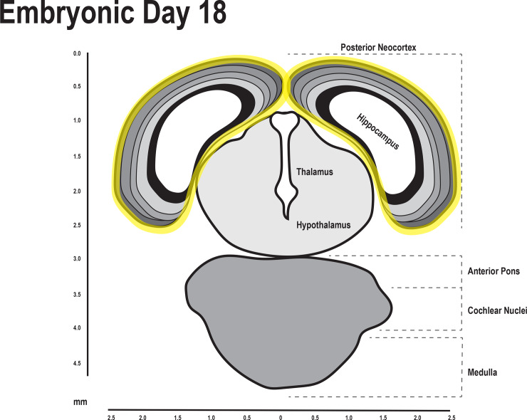
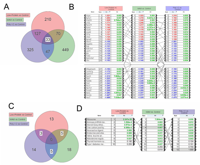
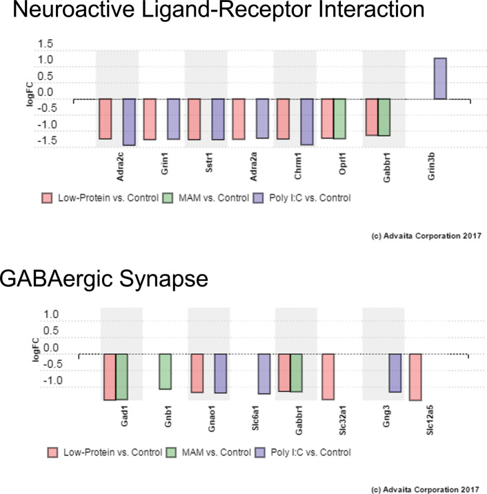
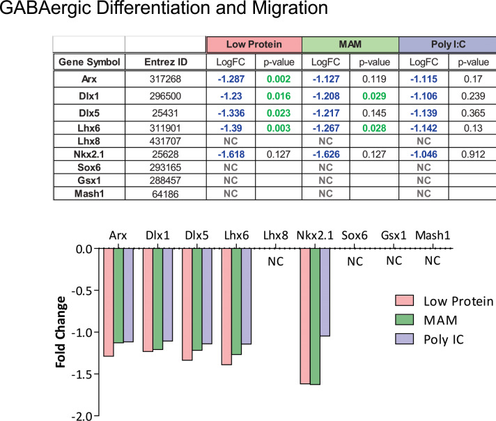
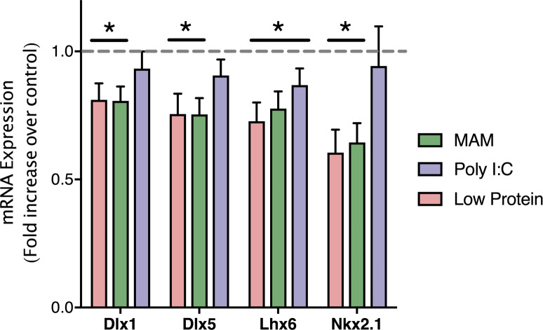
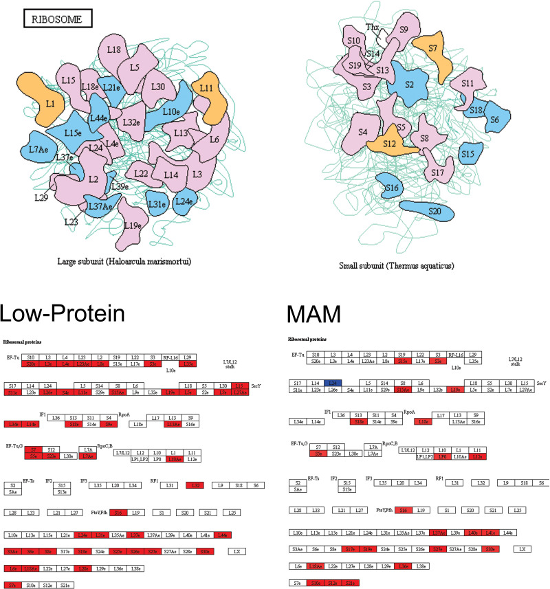
Schizophrenia is a debilitating disorder affecting just under 1% of the population. While the symptoms of this disorder do not appear until late adolescence, pathological alterations likely occur earlier, during development in utero. While there is an increasing literature examining transcriptome alterations in patients, it is not possible to examine the changes in gene expression that occur during development in humans that will develop schizophrenia. Here we utilize three distinct rodent developmental disruption models of schizophrenia to examine potential overlapping alterations in the transcriptome, with a specific focus on markers of interneuron development. Specifically, we administered either methylazoxymethanol acetate (MAM), Polyinosinic:polycytidylic acid (Poly I:C), or chronic protein malnutrition, on GD 17 and examined mRNA expression in the developing hippocampus of the offspring 18 hours later. Here, we report alterations in gene expression that may contribute to the pathophysiology of schizophrenia, including significant alterations in interneuron development and ribosome function.


Conflict of interest statement

The authors have declared that no competing interests exist.

Figures

Fig 1
Fig 1. The hippocampus and neocortex was dissected for RNA sequencing.
Diagram of the fetal brain on embryonic day 18. The hippocampal and neocortical regions that were dissected are outlined in yellow[35].
Fig 2
Fig 2. Transcriptional analysis identified consistent and overlapping changes in gene expression across three developmental models of schizophrenia.
(A) Venn Diagram showing the number of genes that were differentially expressed in each schizophrenia model compared to control animals. Of these, 33 genes were differentially expressed in all three models. (B) The differentially expressed genes that were affected by all three prenatal manipulations, were ranked. (C) Venn Diagram depicting the pathways differentially altered by all three conditions. (D) Those pathways that showed overlap between at least 2 treatment groups are ranked.
Fig 3
Fig 3. Pathway analysis identified expression changes in genes associated with Neuroactive Ligand-Receptor Interactions and GABAergic Synapses.
The unbiased pathway analyses identified pathways with relevance to schizophrenia, including GO terms ‘Neuroactive Ligand-Receptor Interactions’ (ranked 5th) and ‘GABAergic Synapses’ (Ranked 15th) (A) Graph showing the differentially expressed genes in the Neuroactive Ligand-Receptor Interaction pathway. (B) Graph showing the differentially expressed genes in the GABAergic Synapse pathway.
Fig 4
Fig 4. Alterations in genes associated with GABAergic development.
In addition to the independent pathway analyses, we had an a priori hypothesis that genes involved in interneuron development and migration would be altered in these groups. Specifically, those genes associated with MGE-derived interneurons were downregulated (Arx, Dlx1, Dlx5, Lhx6, & Nkx2.1) whereas markers of CGE-derived interneurons (Sox6, Gsx1, & Mash1) were not significantly affected.
Fig 5
Fig 5. qPCR confirmation of genes associated with GABAergic development.
qPCR confirmed that the expression of genes associated with the development and migration of GABAergic interneurons (Dlx1, Dlx5, Lhx6, and Nkx2.1) were decreased by developmental disruption models of schizophrenia. * is p<0.05 compared to saline-treated controls. n = 5–6 per group.
Fig 6
Fig 6. Pathway analysis identified expression changes in ribosomal proteins.
The most significant pathway identified was that of Ribosomal function. (Top) Cartoon depicting proteins in the large (left) and small (right) subunits of the ribosome. (Bottom) Both Low-Protein and MAM caused changes in the expression of genes associated with ribosomal proteins. For each treatment group, an increased expression of genes encoding ribosomal proteins are highlighted in red while decreased expression is shown in blue.

References

    1. Moreno-Kustner B, Martin C, Pastor L. Prevalence of psychotic disorders and its association with methodological issues. A systematic review and meta-analyses. PloS one. 2018;13(4):e0195687 10.1371/journal.pone.0195687 - DOI - PMC - PubMed
    1. Feinberg I. Schizophrenia: caused by a fault in programmed synaptic elimination during adolescence? Journal of psychiatric research. 1982;17(4):319–34. 10.1016/0022-3956(82)90038-3 - DOI - PubMed
    1. Weinberger DR. Implications of normal brain development for the pathogenesis of schizophrenia. Archives of general psychiatry. 1987;44(7):660–9. 10.1001/archpsyc.1987.01800190080012 - DOI - PubMed
    1. Brown AS. Prenatal infection as a risk factor for schizophrenia. Schizophrenia Bulletin. 2006;32(2):200–2. 10.1093/schbul/sbj052 - DOI - PMC - PubMed
    1. Brown AS, Begg MD, Gravenstein S, Schaefer CA, Wyatt RJ, Bresnahan M, et al. Serologic evidence of prenatal influenza in the etiology of schizophrenia. Archives of General Psychiatry. 2004;61(8):774–80. 10.1001/archpsyc.61.8.774 - DOI - PubMed

Publication types