. 2020 Jun 4;15(6):e0229549.
doi: 10.1371/journal.pone.0229549. eCollection 2020.

Expression of dlx genes in the normal and regenerating brain of adult zebrafish

Affiliations

Expression of dlx genes in the normal and regenerating brain of adult zebrafish

Hellen Weinschutz Mendes et al. PLoS One. .

Abstract

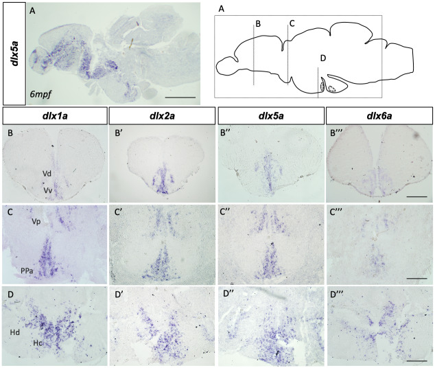
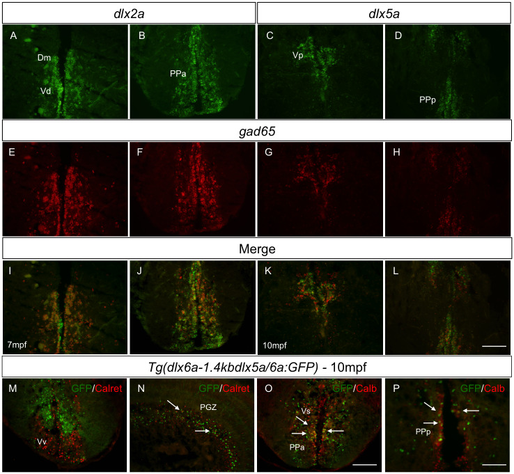
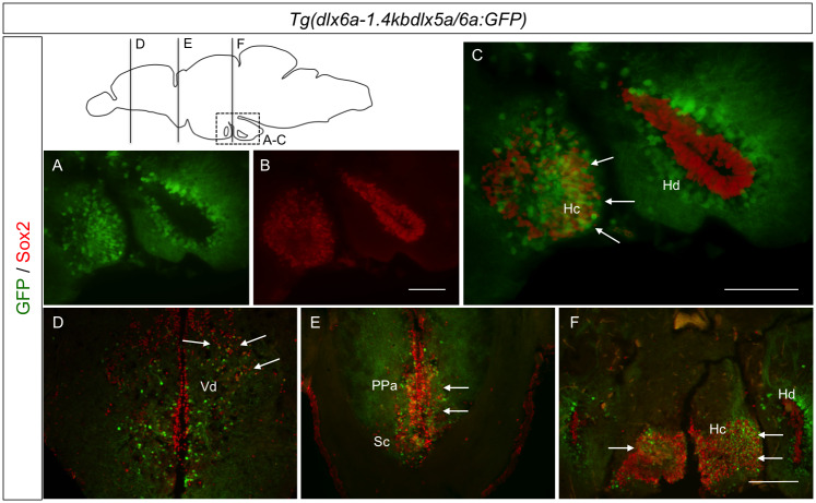
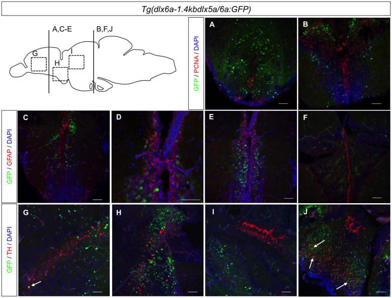
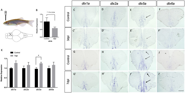
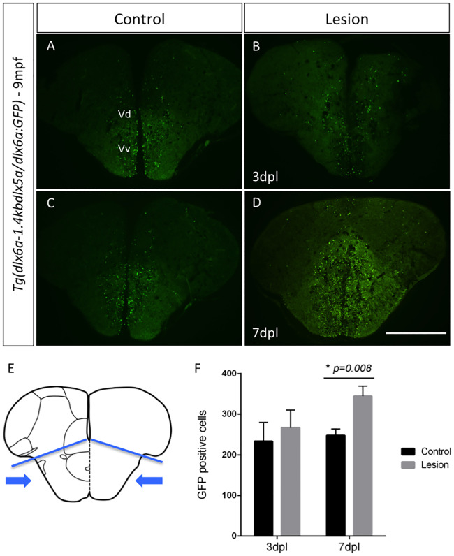
Dysfunctions in the GABAergic system lead to various pathological conditions and impaired inhibitory function is one of the causes behind neuropathies characterized by neuronal hyper excitability. The Dlx homeobox genes are involved in the development of nervous system, neural crest, branchial arches and developing appendages. Dlx genes also take part in neuronal migration and differentiation during development, more precisely, in the migration and differentiation of GABAergic neurons. Functional analysis of dlx genes has mainly been carried out in developing zebrafish embryos and larvae, however information regarding the expression and roles of these genes in the adult zebrafish brain is still lacking. The extensive neurogenesis that takes place in the adult zebrafish brain, makes them a good model for the visualization of mechanisms involving dlx genes during adulthood in physiological conditions and during regeneration of the nervous system. We have identified the adult brain regions where transcripts of dlx1a, dlx2a, dlx5a and dlx6a genes are normally found and have confirmed that within telencephalic domains, there is high overlapping expression of the four dlx paralogs with a marker for GABAergic neurons. Co-localization analyses carried with the Tg(dlx6a-1.4kbdlx5a/dlx6a:GFP) reporter line have also shown that in some areas of the diencephalon, cells expressing the dlx5a/6a bigene may have a neural stem cell identity. Furthermore, investigations in a response to stab wound lesions, have demonstrated a possible participation of the dlx5a/6a bigene, most likely of dlx5a, during regeneration of the adult zebrafish brain. These observations suggest a possible participation of dlx-expressing cells during brain regeneration in adult zebrafish and also provide information on the role of dlx genes under normal physiological conditions in adults.


Conflict of interest statement

The authors have declared that no competing interests exist.

Figures

Fig 1
Fig 1. dlx1a, dlx2a, dlx5a and dlx6a are expressed in the adult zebrafish brain.
Schematic representation of sections depicted in the top right panel. In situ hybridization shows dlx expression in cryosections of the adult zebrafish brain. Sagittal section showing dlx5a expression in a 6 mpf fish (A). Transverse sections showing expression throughout areas of the forebrain, midbrain and hindbrain of dlx1a (B-D), dlx2a (B’-D’), dlx5a (B”-D”) and dlx6a (B”’-D”’) in 1 ypf zebrafish (N = 6 for each dlx gene). Hc.: caudal zone of the periventricular hypothalamus; Hd.: dorsal zone of the periventricular hypothalamus; PPa.: anterior part of parvocellular preoptic nucleus; V.: ventral telencephalic area; Vd.: dorsal nucleus of V.; Vp.: parvocellular nucleus of V.; Vv.: ventral nucleus of V. Scale bar (A): 1mm; (B-D”’): 400μm.
Fig 2
Fig 2. Co-expression of dlx paralogs with markers of GABAergic neurons in adult zebrafish.
Double fluorescence ISH of transverse sections of the forebrain showing expression of dlx2a (A-B) and dlx5a (C-D) in green and expression of gad65 in red (E-H). Anatomical parts indicated. Merged images showing co-localization of dlx and gad65 in yellow [I-L] (N = 4 for dlx2a and dlx5a). Double IHC with Tg(dlx6a-1.4kbdlx5a/6a:GFP) and Calretinin or Calbindin shows co-localization, indicated by white arrows [N-P] (N = 6 for Calbindin and Calretinin). Merged images were created with ImageJ(32) software. Calret.: Calretinin and Calb.: Calbindin. Dm.: medial zone of dorsal telencephalic area; PGZ.: periventricular gray zone; PPa.: anterior part of parvocellular preoptic nucleus; PPp.: posterior part of parvocellular preoptic nucleus; V.: ventral telencephalic area; Vd.: dorsal nucleus of V.; Vp.: parvocellular nucleus of V.;Vs.: supracommissural nucleus of V.; Vv.: ventral nucleus of V. Scale bar: 400μm.
Fig 3
Fig 3. Co-localization of GFP and Sox2 in the Tg(dlx6a-1.4kbdlx5a/6a:GFP) adult zebrafish brain.
Double IHC with Tg(dlx6a-1.4kbdlx5a/6a:GFP) and Sox2 shows co-localization, indicated by white arrows, in merged images of GFP and Sox2 (C-F) (N = 8). Merged images were created with ImageJ(32) software. Hc.: caudal hypothalamus; Hd.: dorsal zone of periventricular hypothalamus; PPa.: anterior part of parvocellular preoptic nucleus; Sc.: suprachiasmatic nucleus; Vd.: dorsal nucleus of V. Scale bar: 400μm.
Fig 4
Fig 4. Immunohistochemical labeling of PCNA, GFAP and TH in Tg(dlx6a-1.4kbdlx5a/6a:GFP) adult zebrafish.
Double IHC with Tg(dlx6a-1.4kbdlx5a/6a:GFP) in combination with either PCNA, GFAP or TH. Labeling of GFP with PCNA (A-D) and GFAP (E-H), shows no co-localization of the two markers with GFP (N = 6). Labeling of GFP and TH (I-L) shows a few co-localizations of the two markers, indicated by white arrows. Merged images created with NIS-Elements Advanced Research Software. Scale bar: 200μm.
Fig 5
Fig 5. Expression of dlx1a, dlx2a, dlx5a and dlx6a post-lesion in adult zebrafish.
Top left panel (A) shows the location of the mechanical lesion. Expression of the four dlx paralogs in controls (C-F and G-J) and lesioned brains at 3 dpl (C’-F’) and 7 dpl (G’-J’). Down-regulation of dlx5a is suggested at 3dpl (E’ indicated by gray arrow) and confirmed by RT-qPCR (B) (Student’s t-test, n = 5, p = 0.004). A slight up-regulation of dlx2a and dlx5a is seen at 7dpl compared to controls. For each gene, and for each time point, assays were carried out with at least 2 biological replicates in 3 different experimental repetitions (N = 6). RT-qPCR analyses at 7dpl (K) reveals no significant changes in expression levels of dlx1a, dlx2a and dlx6a (N = 7 for each gene each). A significant increase in dlx5a expression was observed at 7dpl (Student’s t-test, n = 7, p = 0.008). Scale bar: 400 μM.
Fig 6
Fig 6. GFP labeling in Tg(dlx6a-1.4kbdlx5a/6a:GFP) adult zebrafish at 7 days post-lesion and cell quantification.
Expression of GFP in Tg(dlx6a-1.4kbdlx5a/6a:GFP) determined with a GFP antibody in controls (A and C) and regenerating brains at 3 dpl (B) and 7dpl (D). Schematic representation of the telencephalon where lesion is inflicted and areas used for cell counting of all GFP-positive cells (E) (area indicated by blue arrows bellow blue lines). Quantification of GFP+ cells in the regenerating brain at 3dpl and 7dpl (P = 0.008, N = 6) (F). Scale bar: 400 μm.

References

    1. Panganiban G, Rubenstein JLR. Developmental functions of the Distal-less/Dlx homeobox genes. Development. 2002;129: 4371–86. Available: http://www.ncbi.nlm.nih.gov/pubmed/12223397 - PubMed
    1. Pleasure SJ, Anderson S, Hevner R, Bagri A, Marin O, Lowenstein DH, et al. Cell migration from the ganglionic eminences is required for the development of hippocampal GABAergic interneurons. Neuron. 2000;28: 727–740. 10.1016/s0896-6273(00)00149-5 - DOI - PubMed
    1. Anderson SA, Eisenstat DD, Shi L, Rubenstein JLR. Interneuron migration from basal forebrain to neocortex: Dependence on Dlx genes. Science. 1997;278: 474–476. 10.1126/science.278.5337.474 - DOI - PubMed
    1. Anderson S a., Qiu M, Bulfone A, Eisenstat DD, Meneses J, Pedersen R, et al. Mutations of the homeobox genes Dlx-1 and Dlx-2 disrupt the striatal subventricular zone and differentiation of late born striatal neurons. Neuron. 1997;19: 27–37. 10.1016/s0896-6273(00)80345-1 - DOI - PubMed
    1. Akimenko M a, Ekker M, Wegner J, Lin W, Westerfield M. Combinatorial expression of three zebrafish genes related to distal-less: part of a homeobox gene code for the head. J Neurosci. 1994;14: 3475–3486. 10.1523/JNEUROSCI.14-06-03475.1994 - DOI - PMC - PubMed

Publication types

Substances

Grants and funding

LinkOut - more resources