. 2020 Nov;92(11):2693-2701.
doi: 10.1002/jmv.26139. Epub 2020 Jul 2.

Increasing host cellular receptor-angiotensin-converting enzyme 2 expression by coronavirus may facilitate 2019-nCoV (or SARS-CoV-2) infection

Affiliations

Increasing host cellular receptor-angiotensin-converting enzyme 2 expression by coronavirus may facilitate 2019-nCoV (or SARS-CoV-2) infection

Meng-Wei Zhuang et al. J Med Virol. 2020 Nov.

Abstract

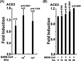
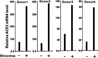
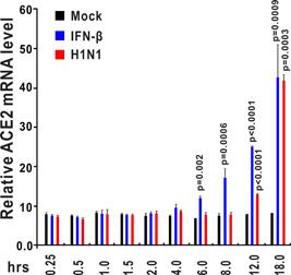
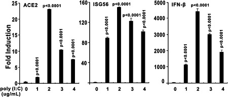
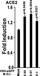
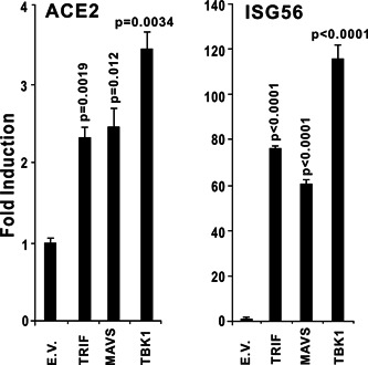
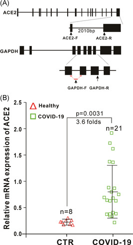
The ongoing outbreak of a new coronavirus (2019-nCoV, or severe acute respiratory syndrome coronavirus 2 [SARS-CoV-2]) has caused an epidemic of the acute respiratory syndrome known as coronavirus disease (COVID-19) in humans. SARS-CoV-2 rapidly spread to multiple regions of China and multiple other countries, posing a serious threat to public health. The spike (S) proteins of SARS-CoV-1 and SARS-CoV-2 may use the same host cellular receptor, angiotensin-converting enzyme 2 (ACE2), for entering host cells. The affinity between ACE2 and the SARS-CoV-2 S protein is much higher than that of ACE2 binding to the SARS-CoV S protein, explaining why SARS-CoV-2 seems to be more readily transmitted from human to human. Here, we report that ACE2 can be significantly upregulated after infection of various viruses, including SARS-CoV-1 and SARS-CoV-2, or by the stimulation with inflammatory cytokines such as interferons. We propose that SARS-CoV-2 may positively induce its cellular entry receptor, ACE2, to accelerate its replication and spread; high inflammatory cytokine levels increase ACE2 expression and act as high-risk factors for developing COVID-19, and the infection of other viruses may increase the risk of SARS-CoV-2 infection. Therefore, drugs targeting ACE2 may be developed for the future emerging infectious diseases caused by this cluster of coronaviruses.

Keywords: 2019-nCoV; ACE2; COVID-19; IFN; ISG; SARS-CoV-2.


Conflict of interest statement

The authors declare that there are no conflict of interests.

Figures

Figure 1
Figure 1
The expression of angiotensin‐converting enzyme 2 (ACE2) is upregulated by coronaviruses. A, ACE2 expression is upregulated in the lungs of severe acute respiratory syndrome coronavirus (SARS‐CoV) MA15‐infected C57Bl/6 mice. At 24 hours after the intranasal instillation of 102 or 103 PFU of SARS‐CoV MA15 in 50 µL of phosphate buffered saline (PBS) or mock infection with PBS alone, lungs were harvested, and total RNA was isolated and subjected to microarray analysis. ACE2 expression data were retrieved from the Gene Expression Omnibus (GEO) microarray database (National Center for Biotechnology Information [NCBI] accession: PRJNA149057; GEO: GSE33266). B, ACE2 expression is upregulated in primary human airway epithelial cells in response to wild‐type Middle East respiratory syndrome coronavirus (MERS‐CoV; icMERS‐CoV EMC2012). Human airway epithelial cells were infected with a multiplicity of infection of 5 PFU per cell. Infected samples were collected at the time points as indicated above. ACE2 expression data were retrieved from the GEO microarray database (NCBI accession: PRJNA391962; GEO: GSE100504)
Figure 2
Figure 2
ACE2 expression is upregulated in human airway epithelial cells in response to rhinovirus infection. Primary epithelial cells from four donors were exposed to rhinovirus or medium alone (used as a control). Twenty‐four hours after infection, gene expression was assessed using Affymetrix U133 plus 2.0 human GeneChips. ACE2 expression data were retrieved from the GEO microarray database (NCBI accession: PRJNA137767; GEO: GSE27973). ACE2, angiotensin‐converting enzyme 2; GEO, Gene Expression Omnibus; NCBI, National Center for Biotechnology Information
Figure 3
Figure 3
ACE2 expression is upregulated in human bronchial epithelial cells (HBECs) after interferon‐β (IFN‐β) stimulation and H1N1 influenza infection. HBECs were treated with IFN‐β (1000 U/mL) or wild‐type H1N1 influenza (A/PR/8/34). Control samples were incubated with fresh medium under the same conditions. HBECs were harvested at the indicated time points (0.25, 0.5, 1, 1.5, 2, 4, 6, 8, 12, and 18 hours after treatment). Total RNA was extracted and analyzed using an Affymetrix High Throughput Array. ACE2 expression data were retrieved from GEO microarray database (NCBI accession: PRJNA121751; GEO: GSE19392). ACE2, angiotensin‐converting enzyme 2; GEO, Gene Expression Omnibus; NCBI, National Center for Biotechnology Information
Figure 4
Figure 4
ACE2 expression is upregulated in human primary keratinocytes exposed to IFN‐α and IFN‐γ. Primary keratinocytes from three donors were either untreated (control) or exposed to the IFN‐α or IFN‐γ cytokines. Twenty‐four hours after treatment, total RNA was extracted for subsequent microarray analysis. ACE2 expression data were retrieved from the GEO microarray database (NCBI accession: PRJNA153023; GEO: GSE36287). ACE2, angiotensin‐converting enzyme 2; GEO, Gene Expression Omnibus; IFN, interferon; NCBI, National Center for Biotechnology Information
Figure 5
Figure 5
Transfection of poly (I:C) induces ACE2 expression. HEK293 cells were cultured in six‐well plates and transfected with poly (I:C) as indicated above using Lipofectamine 2000. HEK293 cells were collected for RNA isolation and reverse‐transcription quantitative polymerase chain reaction (RT‐qPCR) analysis at 9 hours after transfection. The results in each panel are representative of three independent experiments. The graphs show the means ± SD (n = 3). The results in each panel are representative of three independent experiments. ACE2, angiotensin‐converting enzyme 2
Figure 6
Figure 6
Vesicular stomatitis virus (VSV)‐enhanced green fluorescent protein (eGFP) infection induces ACE2 expression. HEK293 cells were cultured in six‐well plates and infected with VSV‐eGFP as indicated above. After 6 hours, HEK293 cells were collected for RNA isolation and RT‐qPCR analysis. The graphs show the means ± SD (n = 3). The results in each panel are representative of three independent experiments. ACE2, angiotensin‐converting enzyme 2; RT‐qPCR, reverse‐transcription quantitative polymerase chain reaction
Figure 7
Figure 7
Overexpression of TRIF, MAVS, or TBK1 induces ACE2 expression. TRIF and MAVS are the essential adaptors of Toll‐like receptor 3 (TLR3) and RIG‐I/MDA‐5, respectively. TBK1 is an crucial activator of TLR3 and RIG‐I/MDA‐5 pathways downstream of TRIF and MAVS. HEK293 cells were cultured in 24‐well plates. After 24 hours, the plasmids indicated above were transfected into HEK293T cells. Thirty‐six hours after transfection, total RNA was isolated for subsequent RT‐qPCR analysis. The graphs show the means ± SD (n = 3). The results in each panel are representative of three independent experiments. ACE2, angiotensin‐converting enzyme 2; RT‐qPCR, reverse‐transcription quantitative polymerase chain reaction
Figure 8
Figure 8
The promoter of ACE2 is activated by RNA‐sensing pathways and inflammatory cytokine stimulation. A, Schematic diagram of the human ACE2 luciferase reporter. Nucleotides −1281 to +300 (with transcript start site set as 1) of the human ACE2 promoter region were cloned into the pGL3‐Basic luciferase reporter to construct ACE‐luc. B, Induction patterns of the human ACE2 promoter by RIG‐IN (a truncated active form of RIG‐I), MAVS, TRIF, and TBK1. HEK293T cells were transfected with plasmids containing the empty vector (EV), RIG‐IN, MAVS, TRIF, or TBK1 together with ACE‐luc. C, The promoter activities of ACE2 was induced by recombinant protein of IFN‐α (10 ng/mL), IFN‐β (10 ng/mL), IFN‐γ (10 ng/mL), TNF‐α (10 ng/mL), IL‐6 (10 ng/mL), IL‐1α (10 ng/mL), or IL‐1β (10 ng/mL). The graphs show the means ± SD (n = 3). The results in each panel are representative of three independent experiments. ACE2, angiotensin‐converting enzyme 2; IFN, interferon
Figure 9
Figure 9
ACE2 is upregulated in severe acute respiratory syndrome coronavirus 2 (SARS‐CoV‐2)‐infected patients. A, Schematic diagrams of the RT‐qPCR primer positions of human ACE2 and glyceraldehyde 3‐phosphate dehydrogenase (GAPDH). The RT‐qPCR primers of ACE2 located in two different exons with a large intron between them to make sure that the primers can only amplify a fragment of ACE2 from complementary DNA (cDNA) only but not genomic DNA. The forward primer of GAPDH (GAPDH‐F) span an exon‐exon boundary as indicated, which only amplify a fragment of GAPDH from cDNA only but not genomic DNA. B, Expression of ACE2 in nasopharyngeal swab specimens from healthy (control, CTR) and SARS‐CoV‐2‐infected individuals. Nasopharyngeal swab specimens collected from patients with COVID‐19 were confirmed to be SARS‐CoV‐2 RNA‐positive. Expression of ACE2 in theses samples was detected by RT‐qPCR. ACE2, angiotensin‐converting enzyme 2; RT‐qPCR, reverse‐transcription quantitative polymerase chain reaction
Figure 10
Figure 10
Proposed working model of ACE2 during SARS‐CoV‐2 infection. In the first wave of infection, SARS‐CoV‐2 enters host cells through ACE2 and releases its viral RNAs, which are subsequently sensed by TLR3 and RIG‐I/MDA‐5. The activation of TLR3 and RIG‐I/MDA‐5 leads to the activation of innate immune signaling pathways, resulting in the production of IFNs and other inflammatory cytokines. Then, ACE2 was upregulated by these cytokines through the Janus kinase/signal transducers and activators of transcription (JAK/STAT) pathway or other pathways, which subsequently mediated the enhanced entry of SARS‐CoV‐2 into host cells. ACE2, angiotensin‐converting enzyme 2; IFN, interferon; SARS‐CoV‐2, severe acute respiratory syndrome coronavirus 2; TLR3, Toll‐like receptor 3

Similar articles

Cited by

References

    1. Zhu N, Zhang D, Wang W, et al. A novel coronavirus from patients with pneumonia in China, 2019. N Engl J Med. 2020;382(8):727‐733. - PMC - PubMed
    1. Zhou P, Yang XL, Wang XG, et al. A pneumonia outbreak associated with a new coronavirus of probable bat origin. Nature. 2020;579(7798):270‐273. - PMC - PubMed
    1. Wu F, Zhao S, Yu B, et al. A new coronavirus associated with human respiratory disease in China. Nature. 2020;579(7798):265‐269. - PMC - PubMed
    1. Chan JF, Yuan S, Kok KH, et al. A familial cluster of pneumonia associated with the 2019 novel coronavirus indicating person‐to‐person transmission: a study of a family cluster. Lancet. 2020;395(10223):514‐523. - PMC - PubMed
    1. Huang C, Wang Y, Li X, et al. Clinical features of patients infected with 2019 novel coronavirus in Wuhan, China. Lancet. 2020;395(10223):497‐506. - PMC - PubMed

Publication types

MeSH terms