

Urethral luminal epithelia are castration-insensitive cells of the proximal prostate
- PMID: 32497356
- PMCID: PMC7339731
- DOI: 10.1002/pros.24020
Urethral luminal epithelia are castration-insensitive cells of the proximal prostate
Abstract
Background: Castration-insensitive epithelial progenitors capable of regenerating the prostate have been proposed to be concentrated in the proximal region based on facultative assays. Functional characterization of prostate epithelial populations isolated with individual cell surface markers has failed to provide a consensus on the anatomical and transcriptional identity of proximal prostate progenitors.
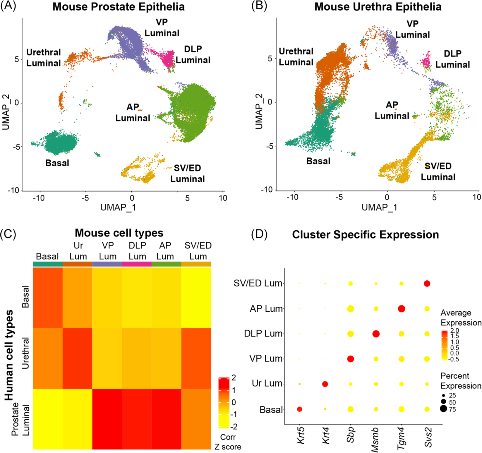
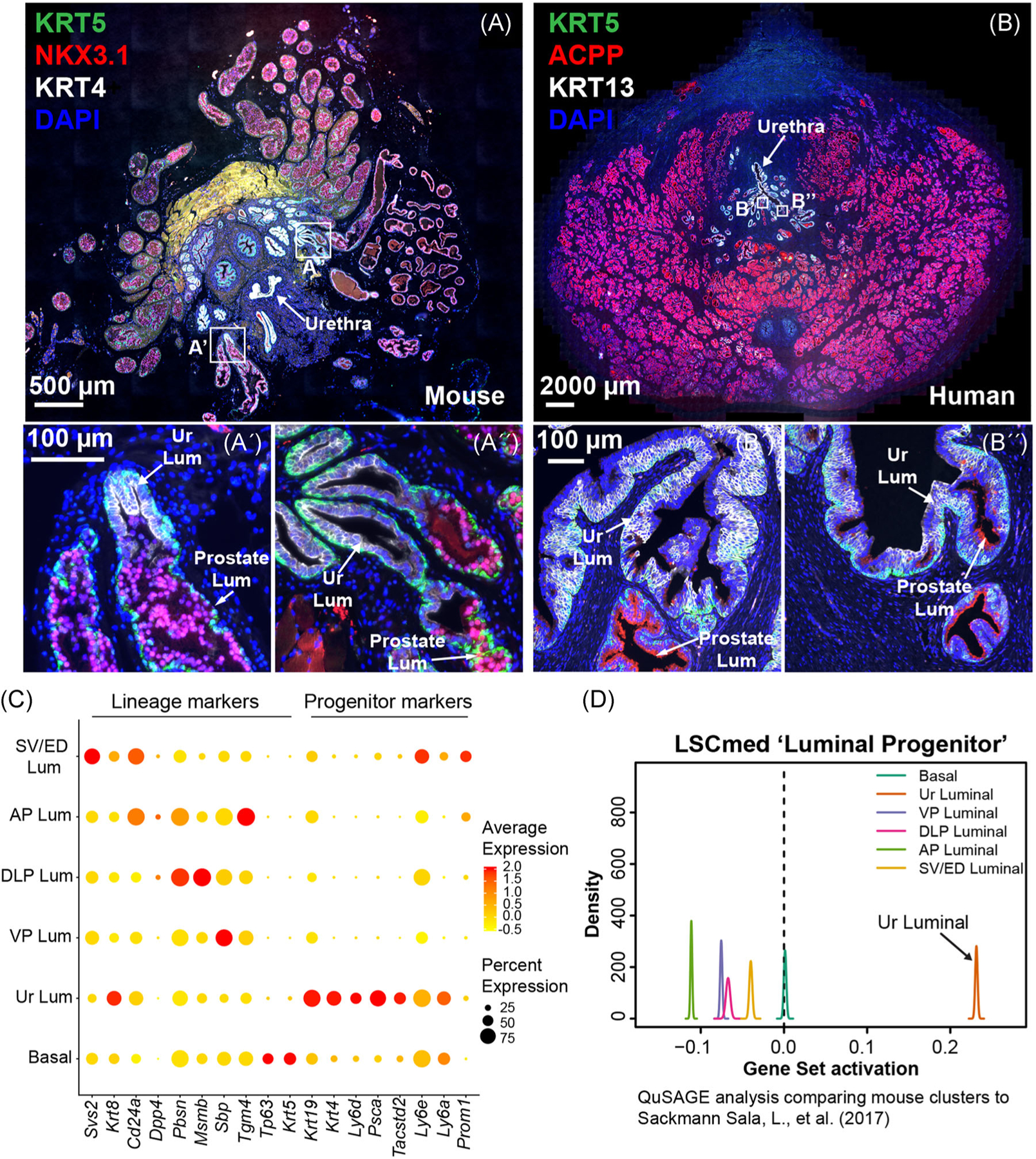
Methods: Here, we use single-cell RNA sequencing to obtain a complete transcriptomic profile of all epithelial cells in the mouse prostate and urethra to objectively identify cellular subtypes. Pan-transcriptomic comparison to human prostate cell types identified a mouse equivalent of human urethral luminal cells, which highly expressed putative prostate progenitor markers. Validation of the urethral luminal cell cluster was performed using immunostaining and flow cytometry.
Results: Our data reveal that previously identified facultative progenitors marked by Trop2, Sca-1, KRT4, and PSCA are actually luminal epithelial cells of the urethra that extend into the proximal region of the prostate, and are resistant to castration-induced androgen deprivation. Mouse urethral luminal cells were identified to be the equivalent of previously identified human club and hillock cells that similarly extend into proximal prostate ducts. Benign prostatic hyperplasia (BPH) has long been considered an "embryonic reawakening," but the cellular origin of the hyperplastic growth concentrated in the periurethral region is unclear. We demonstrate an increase in urethral luminal cells within glandular nodules from BPH patients. Urethral luminal cells are further increased in patients treated with a 5-α reductase inhibitor.
Conclusions: Our data demonstrate that cells of the proximal prostate that express putative progenitor markers, and are enriched by castration in the proximal prostate, are urethral luminal cells and that these cells may play an important role in the etiology of human BPH.
Keywords: benign prostatic hyperplasia; castration; prostate stem cell; prostatic urethra; single-cell RNA sequencing.
© 2020 Wiley Periodicals LLC.
Conflict of interest statement
CONFLICT OF INTERESTS
The authors declare that there are no conflict of interests.
Figures


Similar articles
-
The influence of prostatic anatomy and neurotrophins on basal prostate epithelial progenitor cells.Prostate. 2016 Jan;76(1):114-21. doi: 10.1002/pros.23109. Epub 2015 Oct 7. Prostate. 2016. PMID: 26444457
-
Trop2 identifies a subpopulation of murine and human prostate basal cells with stem cell characteristics.Proc Natl Acad Sci U S A. 2008 Dec 30;105(52):20882-7. doi: 10.1073/pnas.0811411106. Epub 2008 Dec 16. Proc Natl Acad Sci U S A. 2008. PMID: 19088204 Free PMC article.
-
Stem Cell Antigen-1 Identifies a Distinct Androgen-Independent Murine Prostatic Luminal Cell Lineage with Bipotent Potential.Stem Cells. 2016 Jan;34(1):191-202. doi: 10.1002/stem.2217. Epub 2015 Oct 27. Stem Cells. 2016. PMID: 26418304 Free PMC article.
-
Progenitors in prostate development and disease.Dev Biol. 2021 May;473:50-58. doi: 10.1016/j.ydbio.2020.11.012. Epub 2021 Jan 30. Dev Biol. 2021. PMID: 33529704 Free PMC article. Review.
-
Prostate luminal progenitor cells: from mouse to human, from health to disease.Nat Rev Urol. 2022 Apr;19(4):201-218. doi: 10.1038/s41585-021-00561-2. Epub 2022 Jan 25. Nat Rev Urol. 2022. PMID: 35079142 Review.
Cited by
-
Male Lower Urinary Tract Dysfunction: An Underrepresented Endpoint in Toxicology Research.Toxics. 2022 Feb 16;10(2):89. doi: 10.3390/toxics10020089. Toxics. 2022. PMID: 35202275 Free PMC article. Review.
-
Understanding and targeting prostate cancer cell heterogeneity and plasticity.Semin Cancer Biol. 2022 Jul;82:68-93. doi: 10.1016/j.semcancer.2021.11.001. Epub 2021 Nov 26. Semin Cancer Biol. 2022. PMID: 34844845 Free PMC article. Review.
-
Mouse-Geneformer: A deep learning model for mouse single-cell transcriptome and its cross-species utility.PLoS Genet. 2025 Mar 19;21(3):e1011420. doi: 10.1371/journal.pgen.1011420. eCollection 2025 Mar. PLoS Genet. 2025. PMID: 40106407 Free PMC article.
-
RUNX1 marks a luminal castration-resistant lineage established at the onset of prostate development.Elife. 2020 Oct 7;9:e60225. doi: 10.7554/eLife.60225. Elife. 2020. PMID: 33025905 Free PMC article.
-
Transcriptomic Signature and Growth Factor Regulation of Castration-Tolerant Prostate Luminal Progenitor Cells.Cancers (Basel). 2022 Aug 3;14(15):3775. doi: 10.3390/cancers14153775. Cancers (Basel). 2022. PMID: 35954439 Free PMC article.
References
-
- Sugimura Y, Cunha GR, Donjacour AA. Morphogenesis of ductal networks in the mouse prostate. Biol Reprod. 1986;34(5):961–971. - PubMed
-
- Thomson AA, Marker PC. Branching morphogenesis in the prostate gland and seminal vesicles. Differentiation. 2006;74(7):382–392. - PubMed
-
- Timms BG, Mohs TJ, Didio LJ. Ductal budding and branching patterns in the developing prostate. J Urol. 1994;151(5):1427–1432. - PubMed
Publication types
MeSH terms
Substances
Grants and funding
- R01DK099328/DK/NIDDK NIH HHS/United States
- F30DK122686/DK/NIDDK NIH HHS/United States
- R01DK115477/DK/NIDDK NIH HHS/United States
- F31 ES030968/ES/NIEHS NIH HHS/United States
- U54DK104310/DK/NIDDK NIH HHS/United States
- F30 DK122686/DK/NIDDK NIH HHS/United States
- P30 CA142543/CA/NCI NIH HHS/United States
- R01 DK127589/DK/NIDDK NIH HHS/United States
- R01 DK115477/DK/NIDDK NIH HHS/United States
- R01 DK059164/DK/NIDDK NIH HHS/United States
- TL1 TR002375/TR/NCATS NIH HHS/United States
- R01 DK099328/DK/NIDDK NIH HHS/United States
- U01DK110807/DK/NIDDK NIH HHS/United States
- U01 DK110807/DK/NIDDK NIH HHS/United States
- R01 ES001332/ES/NIEHS NIH HHS/United States
- TL1TR002375/NH/NIH HHS/United States
- U54 DK104310/DK/NIDDK NIH HHS/United States
- 5P30CA142543/CA/NCI NIH HHS/United States
LinkOut - more resources
Full Text Sources
Medical
Molecular Biology Databases
Research Materials
Miscellaneous
