. 2020 Aug 15:345:577279.
doi: 10.1016/j.jneuroim.2020.577279. Epub 2020 May 27.

Imbalance in T follicular helper cells producing IL-17 promotes pro-inflammatory responses in MuSK antibody positive myasthenia gravis

Affiliations

Imbalance in T follicular helper cells producing IL-17 promotes pro-inflammatory responses in MuSK antibody positive myasthenia gravis

Yingkai Li et al. J Neuroimmunol. .

Abstract

A detailed understanding of the role of Tfh cells in MuSK-antibody positive myasthenia gravis (MuSK-MG) is lacking. We characterized phenotype and function of Tfh cells in MuSK-MG patients and controls. We found similar overall Tfh and follicular regulatory (Tfr) T cell frequencies in MuSK-MG and healthy controls, but MuSK-MG patients exhibited higher frequencies of Tfh17 cells and a higher ratio of Tfh:Tfr cells. These results suggest imbalanced Tfh cell regulation, further supported by increased frequencies of CD4 T cells co-producing IL-21/IL-17 and IL-17/IFN-γ, and increased Tfh-supported IgG production. These results support a role for Tfh cell dysregulation in MuSK-MG immunopathology.

Keywords: Autoimmunity; MuSK antibody; Myasthenia gravis; T follicular helper cells; Tfh17 cells.


Figures

Figure 1
Figure 1
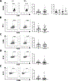
Comparable frequencies of circulating Tfh cells in healthy controls and MuSK-MG. (1A) Tfh cells were identified by gating CXCR5 on CD4 T cells. Percentages of Tfh cells in CD4 T cells in lymphocytes in MG patients were not different than healthy controls. The overall frequencies of Tfh cells in MuSK-MG patients according to MGFA classification were similar. Frequencies of PD-1+/ICOS+ Tfh cells (1B), CD38+/ICOS+ Tfh cells (1C), Ki-67+/ICOS+ Tfh cells (1D) and Ki-67+/CD38+ Tfh cells (1E) were not different in MuSK-MG patients compared with controls.
Figure 2
Figure 2
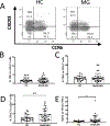
Tfh subsets in MuSK-MG patients. (2A) Gating strategy of Tfh subsets in CD4 T cells using CXCR3 and CCR6 in MuSK-MG and healthy controls. Tfh1 was defined as CXCR3+CCR6-. Tfh2 was defined as CXCR3-CCR6-. Tfh17 was defined as CXCR3-CCR6+. (2B, 2C) Tfh1 and Tfh2 frequencies were not different between MuSK-MG and healthy controls. (2D) Tfh17 frequencies were significantly increased in MuSK-MG patients. (2E) The ratio of Tfh17/Tfh1 subsets is increased in MuSK-MG patients compared with healthy controls. *, significant difference, p<0.05; **, significant difference, p<0.01.
Figure 3
Figure 3
Comparison of Tfr subsets between MuSK-MG and healthy control groups. (3A) Gating strategy Tfr cells in controls and MuSK-MG patients. (3B) Overall Tfr frequencies and (3C) Tfr frequencies among MuSK-MG patients with different MGFA classifications were not significantly different compared to controls. (3D) The overall Tfh:Tfr ratio is increased in MuSK-MG patients compared to controls. (3E) The Tfh17/Tfr ratio is also increased in MuSK-MG patients. **, significant difference, p<0.01; ***, significant difference, p<0.001.
Figure 4
Figure 4
CD4 T cell function assessed by intracellular cytokine production in MuSK-MG and healthy controls. (4A) IL-21, (4B) IFN-γ and (4C) IL-17 cytokine production is increased in CD4 T cells in MuSK-MG compared with healthy controls. (4D) Demonstration of IL-17+/IFN-γ+ gating strategy among CD4 T cells (left). IL-17+/IFN-γ+ CD4+ pathogenic Th17 cells are increased in MuSK-MG compared with controls (right). (4E) IL-17+/IL-21+ gating strategy in CD4 T cells between controls and patients with MuSK-MG (left). IL-17+/IL-21+ CD4 T cells are significantly increased in MuSK-MG patients (right). *, significant difference, p<0.05.
Figure 5
Figure 5
Tfh cells promote B cell IgG production in vitro. Sorted peripheral CD19+CD127-IgD- naïve B cells (2*105/well) were co-cultured with Tfh cells (2*105/well) from MuSK-MG patients (N=4) or healthy controls (N=4) in the presence of CD3/CD28. Naïve B cells only served as a control condition. Production of IgG Abs in cultured supernatants was detected by ELISA. In the B cell only condition IgG production was not different between MuSK-MG patients or healthy controls. As expected, adding Tfh cells to naïve B cells resulted in increased IgG production in both controls and MuSK-MG patients. However, IgG production was significantly increased in MuSK-MG patients compared with controls. *, significant difference, p<0.05; **, significant difference, p<0.01.

References

    1. Balandina A, Lécart S, Dartevelle P, Saoudi A, Berrih-Aknin S, 2005. Functional defect of regulatory CD4+CD25+ T cells in the thymus of patients with autoimmune myasthenia gravis. Blood. 105, 735–741. 10.1182/blood-2003-11-3900. - DOI - PMC - PubMed
    1. Bentebibel S-E, Lopez S, Obermoser G, Schmitt N, Mueller C, Harrod C, Flano E, Mejias A, Albrecht RA, Blankenship D, Xu H, Pascual V, Banchereau J, Garcia-Sastre A, Palucka AK, Ramilo O, Ueno H, 2013. Induction of ICOS+CXCR3+CXCR5+ TH Cells Correlates with Antibody Responses to Influenza Vaccination. Sci. Transl. Med 5, 176ra132–176ra132. 10.1126/scitranslmed.3005191. - DOI - PMC - PubMed
    1. Berrih S, Morel E, Gaud C, Raimond F, Le Brigand H, Bach JF, 1984. Anti-AChR antibodies, thymic histology, and T cell subsets in myasthenia gravis. Neurology. 34, 66–71. 10.1212/wnl.34.1.66. - DOI - PubMed
    1. Boswell KL, Paris R, Boritz E, Ambrozak D, Yamamoto T, Darko S, Wloka K, Wheatley A, Narpala S, McDermott A, Roederer M, Haubrich R, Connors M, Ake J, Douek DC, Kim J, Petrovas C, Koup RA, 2014. Loss of circulating CD4 T cells with B cell helper function during chronic HIV infection. PLoS Pathog. 10, e1003853 10.1371/journal.ppat.1003853. - DOI - PMC - PubMed
    1. Brenna E, Davydov AN, Ladell K, McLaren JE, Bonaiuti P, Metsger M, Ramsden JD, Gilbert SC, Lambe T, Price DA, Campion SL, Chudakov DM, Borrow P, McMichael AJ, 2020. CD4(+) T Follicular Helper Cells in Human Tonsils and Blood Are Clonally Convergent but Divergent from Non-Tfh CD4(+) Cells. Cell reports. 30, 137–152.e135. 10.1016/j.celrep.2019.12.016. - DOI - PMC - PubMed

Publication types