. 2020 Jun 2;9(6):1703.
doi: 10.3390/jcm9061703.

Unleashing the Diagnostic, Prognostic and Therapeutic Potential of the Neuronostatin/GPR107 System in Prostate Cancer

Affiliations

Unleashing the Diagnostic, Prognostic and Therapeutic Potential of the Neuronostatin/GPR107 System in Prostate Cancer

Prudencio Sáez-Martínez et al. J Clin Med. .

Abstract

Certain components of the somatostatin-system play relevant roles in Prostate Cancer (PCa), whose most aggressive phenotype (Castration-Resistant-PCa (CRPC)) remains lethal nowadays. However, neuronostatin and the G protein-coupled receptor 107 (GPR107), two novel members of the somatostatin-system, have not been explored yet in PCa. Consequently, we investigated the pathophysiological role of NST/GPR107-system in PCa. GPR107 expression was analyzed in well-characterized PCa patient's cohorts, and functional/mechanistic assays were performed in response to GPR107-silencing and NST-treatment in PCa cells (androgen-dependent (AD: LNCaP) and androgen-independent (AI: 22Rv1/PC-3), which are cell models of hormone-sensitive and CRPC, respectively), and normal prostate cells (RWPE-1 cell-line). GPR107 was overexpressed in PCa and associated with key clinical parameters (e.g., advance stage of PCa, presence of vascular invasion and metastasis). Furthermore, GPR107-silencing inhibited proliferation/migration rates in AI-PCa-cells and altered key genes and oncogenic signaling-pathways involved in PCa aggressiveness (i.e., KI67/CDKN2D/MMP9/PRPF40A, SST5TMD4/AR-v7/In1-ghrelin/EZH2 splicing-variants and AKT-signaling). Interestingly, NST treatment inhibited proliferation/migration only in AI-PCa cells and evoked an identical molecular response than GPR107-silencing. Finally, NST decreased GPR107 expression exclusively in AI-PCa-cells, suggesting that part of the specific antitumor effects of NST could be mediated through a GPR107-downregulation. Altogether, NST/GPR107-system could represent a valuable diagnostic and prognostic tool and a promising novel therapeutic target for PCa and CRPC.

Keywords: G protein-coupled receptor GPR107; castration resistant prostate cancer; diagnostic/prognostic biomarker; neuronostatin; prostate cancer; somatostatin-system; splicing; therapeutic target.


Conflict of interest statement

The authors declare no conflict of interest. The funders had no role in the design of the study; in the collection, analyses, or interpretation of data; in the writing of the manuscript, or in the decision to publish the results.

Figures
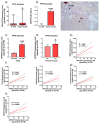
Figure 1
Figure 1
Expression levels of G protein-coupled receptor 107 (GPR107) in human prostate cancer samples. (a) Comparison of GPR107 expression levels between formalin-fixed paraffin embedded (FFPE) samples from Prostate Cancer (PCa) tissues and non-tumor adjacent regions (N-TAR) (n = 84). (b) Comparation of GPR107 protein levels by Immunohistochemistry (IHC) between a representative set of PCa samples (n = 16) and its N-TAR (n = 16). A representative image is also included. (c,d) Association between GPR107 expression levels and advance stage of PCa (c) and vascular invasion (d). (eh) Correlation of GPR107 expression levels and Matrix Metallopeptidase 3 (MMP3; (e), Cyclin Dependent Kinase 2 and 4 (CDK2 and CDK4, respectively; (f), Interleukin 6 Receptor (IL6-R; (g) and Vascular endothelial growth factor Receptor (VEGFR; (h) expression levels in the same cohort of FFPE samples. Messenger RNA (mRNA) levels were determined by quantitative Polymerase Chain Reaction (qPCR) and adjusted by Actin Beta (ACTB) expression levels. Asterisks (* p < 0.05; **** p < 0.0001) indicate statistically significant differences between groups. ND: Non-detected.
Figure 2
Figure 2
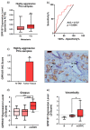
Expression levels of G protein-coupled receptor 107 (GPR107) in primary prostate cancer samples from patients with metastasis. (a) Association between GPR107 expression levels and the presence of metastasis in a cohort of fresh samples from patients with Prostate Cancer (PCa) n = 67). (b) Operating characteristic (ROC) curve analysis to determine the accuracy of GPR107 to discriminate between metastatic vs. non-metastatic patients’ tumor. (c) Comparison of GPR107 protein level by Immunohistochemistry (IHC) between primary PCa samples from patients with metastasis and its non-tumor adjacent regions (N-TAR). Messenger RNA (mRNA) levels were determined by quantitative Polymerase Chain Reaction (qPCR) and adjusted by Actin Beta (ACTB) expression levels. (d-e) Comparison of GPR107 expression levels between normal prostate (n), primary prostate tumor (P) and metastatic Castration Resistant Prostate Cancer (mCRPC) samples obtained from two in silico databases (Grasso/Varambally; (d) and (e), respectively). Asterisks (* p < 0.05; ** p < 0.01; *** p < 0.001; **** p < 0.0001) indicate statistically significant differences between groups. AUC: Area Under the Curve.
Figure 3
Figure 3
Screening of G protein-coupled receptor 107 (GPR107) expression level and functional effects of its silencing in normal and tumor prostate cell lines. (a) Comparison of GPR107 expression levels between a non-tumor prostate cell line (RWPE-1) and different Prostate Cancer (PCa) cell lines (LNCaP, 22Rv1 and PC-3). Messenger RNA (mRNA) levels were determined by quantitative Polymerase Chain Reaction (qPCR) and adjusted by a normalization factor (NF) generated by the combination of Actin Beta (ACTB) and Glyceraldehyde-3-Phosphate Dehydrogenase (GAPDH) expression levels. (b)–(c) Proliferation rate of 22Rv1 (b) and PC-3 (c) cells after 24, 48 and 72 h of GPR107-silencing. (d) Migration rate of PC-3 cells after 24 h of GPR107-silencing. In (b)–(d), data were represented as percent of scrambled cells (set at 100%). Asterisks (* p < 0.05; ** p < 0.01; *** p < 0.001) indicate statistically significant differences between groups. siGPR107: small interferent RNA against GPR107.
Figure 4
Figure 4
Molecular consequences of G protein-coupled receptor 107 (GPR107)-silencing in androgen-independent Prostate cancer (PCa) cells. (a) Fold change of markers of proliferation (Marked of Proliferation Ki-67 (MKI67)), cell cycle inhibition (Cyclin Dependent Kinase Inhibitor 2D (CDKN2D)), migration (Matrix Metallopeptidase 9 and Pre-MRNA Processing Factor 40 Homolog A (MMP9 and PRPF40A, respectively)) and aggressiveness (Androgen Receptor variant 7 (AR-v7), Somatostatin Receptor 5 Transmembrane Domain 4 variant (SST5TMD4), Intron 1-retained ghrelin variant (In1-Ghrelin) and Enhancer Of Zeste Homolog 2 (EZH2)) in androgen-independent cells (22Rv1 and PC-3) in response to GPR107-silencing compared to scrambled cells. Messenger RNA (mRNA) levels were determined by quantitative Polymerase Chain Reaction (qPCR), adjusted by Actin Beta (ACTB) expression levels and represented as log 2 of fold change of expression levels small interferent RNA(siRNA)-treated/scrambled-cells). (b) Protein levels of phospho-AKT (p-AKT) in response of GPR107-silencing in androgen-independent PCa cells (22Rv1 and PC-3). p-AKT levels were normalized by total AKT protein levels. Protein data were represented as percent of scrambled cells (set at 100%). Asterisks (* p <0.05; ** p < 0.01 and *** p < 0.001) indicate statistically significant differences between GPR107-silencing and scrambled cells. siGPR107: small interferent RNA against GPR107.
Figure 5
Figure 5
Functional effects after neuronostatin (NST) treatment and after the combination of NST and G protein-coupled receptor 107 (GPR107)-silencing treatment in prostate cell lines. (a) Proliferation rate of normal prostate (RWPE-1) and Prostate Cancer (PCa) (LNCaP, 22Rv1 and PC-3) cells in response to NST treatment (10–7 M; after 24, 48 and 72 h). (b) Migration rate of normal prostate (RWPE-1) and PCa (PC-3) cells after 24 h of NST treatment (10–7 M). Proliferation (c) and migration (d) rate of androgen-independent PCa cells in response to NST and GPR107-silencing alone or in combination. Data were represented as percent of vehicle-treated cells (set at 100%). Asterisks (* p < 0.05; ** p < 0.01 and *** p < 0.001) indicate statistically significant differences between groups. siGPR107: small interferent RNA against GPR107.
Figure 6
Figure 6
Molecular consequences of neuronostatin (NST) treatment in prostate cell lines. (a) Fold change of markers of proliferation (Marked of Proliferation Ki-67 (MKI67)), cell cycle inhibition (Cyclin Dependent Kinase Inhibitor 2D (CDKN2D)), migration (Matrix Metallopeptidase 9 and Pre-MRNA Processing Factor 40 Homolog A (MMP9 and PRPF40A, respectively)) and aggressiveness (Androgen Receptor variant 7 (AR-v7), Somatostatin Receptor 5 Transmembrane Domain 4 variant (SST5TMD4), Intron 1-retained ghrelin variant (In1-Ghrelin) and Enhancer Of Zeste Homolog 2 (EZH2)) in androgen-independent Prostate Cancer (PCa) cells (22Rv1 and PC-3) after NST treatment (10−7 M) compared to vehicle-treated cells. Messenger RNA (mRNA) levels were determined by quantitative Polymerase Chain Reaction (qPCR), adjusted by Actin Beta (ACTB) expression levels and represented as log2 of fold change of expression levels (NST-treated/vehicle-treated cells). (b) Protein levels of phospho-AKT in response of NST treatment in androgen-independent PCa cells (22Rv1 and PC-3) after 5 (t = 5), 15 (t = 15) and 30 (t = 30) minutes of exposition. Phospho-AKT (p-AKT) levels were normalized by total AKT protein levels. Protein data were represented as percent of vehicle-treated cells (set at 100%). (c) GPR107 expression levels after 24 h of NST treatment in prostate cell lines. mRNA levels of GPR107 were determined by qPCR and adjusted by ACTB expression levels. Asterisks (* p < 0.05; ** p < 0.01 and *** p < 0.001) indicate statistically significant differences between NST treatment and vehicle-treated cells.

Similar articles

Cited by

References

    1. Bray F., Bray F., Ferlay J., Soerjomataram I., Siegel R.L., Torre L.A., Jemal A. Global cancer statistics 2018: GLOBOCAN estimates of incidence and mortality worldwide for 36 cancers in 185 countries. CA Cancer J. Clin. 2018;68:394–424. doi: 10.3322/caac.21492. - DOI - PubMed
    1. Quagliariello V., Rossetti S., Cavaliere C., Di Palo R., Lamantia E., Castaldo L., Nocerino F., Ametrano G., Cappuccio F., Malzone G., et al. Metabolic syndrome, endocrine disruptors and prostate cancer associations: Biochemical and pathophysiological evidences. Oncotarget. 2017;8:30606–30616. doi: 10.18632/oncotarget.16725. - DOI - PMC - PubMed
    1. Fizazi K., Scher H.I., Molina A., Logothetis C.J., Chi K.N., Jones R.J., Staffurth J.N., North S., Vogelzang N.J., Saad F., et al. Abiraterone acetate for treatment of metastatic castration-resistant prostate cancer: Final overall survival analysis of the COU-AA-301 randomised, double-blind, placebo-controlled phase 3 study. Lancet Oncol. 2012;13:983–992. doi: 10.1016/S1470-2045(12)70379-0. - DOI - PubMed
    1. Dhingra R., Sharma T., Singh S., Sharma S., Tomar P., Malhotra M., Bhardwaj T.R. Enzalutamide: A novel anti-androgen with prolonged survival rate in CRPC patients. Mini Rev. Med. Chem. 2013;13:1475–1486. doi: 10.2174/13895575113139990003. - DOI - PubMed
    1. Maximum androgen blockade in advanced prostate cancer: An overview of 22 randomised trials with 3283 deaths in 5710 patients. Prostate Cancer Trialists’ Collaborative Group. Lancet. 1995;346:265–269. doi: 10.1016/S0140-6736(95)92163-X. - DOI - PubMed