. 2020 Jun 2;21(11):3990.
doi: 10.3390/ijms21113990.

Arginase-1+ Exosomes from Reprogrammed Macrophages Promote Glioblastoma Progression

Affiliations

Arginase-1+ Exosomes from Reprogrammed Macrophages Promote Glioblastoma Progression

Juliana H Azambuja et al. Int J Mol Sci. .

Abstract

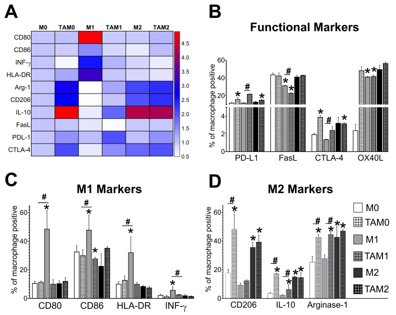
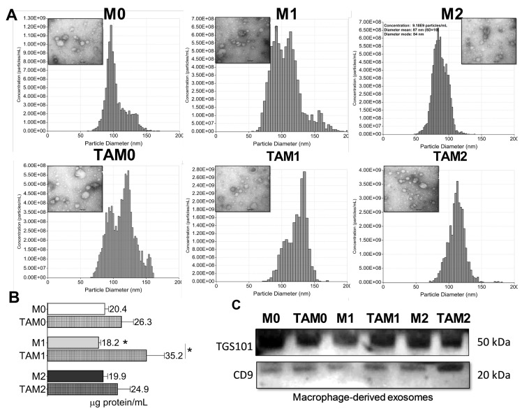
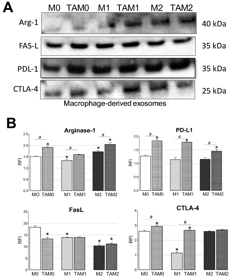
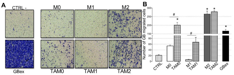
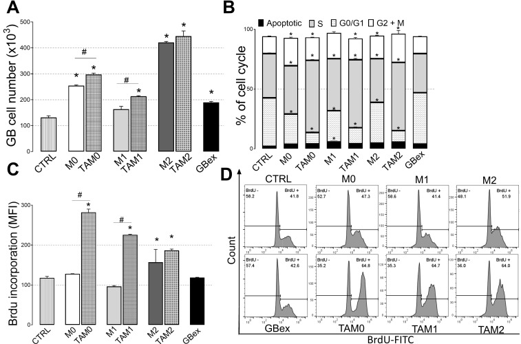
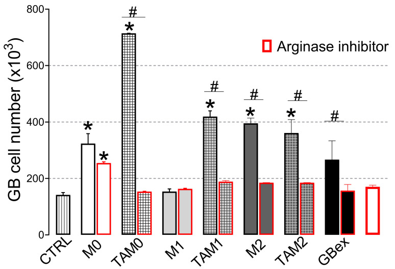
Interactions between tumor cells and tumor-associated macrophages (TAMs) are critical for glioblastoma progression. The TAMs represent up to 30% of the glioblastoma mass. The role of TAMs in tumor progression and in the mechanisms underlying tumor growth remain unclear. Using an in vitro model resembling the crosstalk between macrophages and glioblastoma cells, we show that glioblastoma-derived exosomes (GBex) reprogram M1 (mediate pro-inflammatory function) and M2 (mediate anti-inflammatory function) macrophages, converting M1 into TAMs and augmenting pro-tumor functions of M2 macrophages. In turn, these GBex-reprogrammed TAMs, produce exosomes decorated by immunosuppressive and tumor-growth promoting proteins. TAM-derived exosomes disseminate these proteins in the tumor microenvironment (TME) promoting tumor cell migration and proliferation. Mechanisms underlying the promotion of glioblastoma growth involved Arginase-1+ exosomes produced by the reprogrammed TAMs. A selective Arginase-1 inhibitor, nor-NOHA reversed growth-promoting effects of Arginase-1 carried by TAM-derived exosomes. The data suggest that GBex-reprogrammed Arginase-1+ TAMs emerge as a major source of exosomes promoting tumor growth and as a potential therapeutic target in glioblastoma.

Keywords: Arginase-1; TAM-derived exosomes; glioblastoma; glioblastoma--derived exosomes (GBex); macrophage reprogramming; tumor-associated macrophages (TAMs).


Conflict of interest statement

The authors declare no conflict of interest.

Figures

Figure 1
Figure 1
GBex induced a phenotype like TAMs in different macrophages subsets. (A) The heat map shows the fold of decrease or increase in expression of macrophage markers evaluated by flow cytometry and considering M0 as control (B) percent of macrophages positive for functional immunomodulatory markers PDL-1, FasL, CTLA-4 and OX40L); (C) M1 markers (CD86, CD80, HLA-DR INF)-γ; (D) M2 markers (CD206, Arginase-2 and IL-10). Data were analyzed by flow cytometry as described in Materials and Methods. Data represent mean ± SD of three independent experiments performed in triplicate. Data were analyzed by ANOVA followed by Tukey post hoc. *Significantly different from macrophage M0 cells (white bar); #significantly different between macrophage and TAM cells (p < 0.05).
Figure 2
Figure 2
Characteristics of exosomes produced by macrophages or GBex-reprogrammed TAMs. (A) Results of qNANO analyses and representative transmission electron microscopy (TEM) images providing concentrations and sizes of exosomes produced by macrophages or TAMs; (B) Total protein levels isolated from supernatants of macrophages or TAMs. The data are mean values ± standard error (SEM) from 3 experiments Data were analyzed by ANOVA followed by Tukey post hoc. *Significantly different from control cells at p < 0.05; (C) Western blot profiles of exosomes produced by macrophages or TAMs. Each lane was loaded with 10 μg exosome protein. Note the presence of exosome markers CD9 and TSG101.
Figure 3
Figure 3
Immunosuppressive cargos of exosomes produced by macrophages or TAMs. (A) Representative Western blots of exosomes isolated from macrophages or TAMs. Equal amounts of exosomal protein (10 μg) were loaded per lane; (B) Flow cytometry results for the detection of PDL-1, FasL, CTLA-4, and Arginase-1 carried on exosomes produced by macrophages or TAMs. Exosomes were immunocaptured with anti-CD63 mAb for on-bead flow cytometry as described in Materials and Methods. Data are relative fluorescence intensity (RFI) values ± SEM from three independent experiments Data were analyzed by ANOVA followed by Tukey post hoc. *Significantly different from the control at p < 0.05 and # Significant difference between macrophages and TAMs at p < 0.05.
Figure 4
Figure 4
Exosomes produced by TAMs enhance glioma cell migration. U251 glioma cells were suspended in Dulbecco′s Modified Eagle′s - Medium (DMEM) and were seeded in the upper chamber of transwell units. Exosomes (10 µg) produced by macrophages or were added to the lower transwell chamber. (A) Representative light microscopy images of crystal violet-stained glioblastoma cells accumulating on the lower surface of the membrane; (B) Numbers of glioblastoma cells migrating to the lower chamber. Data are mean values ± SEM from three independent experiments. Data were analyzed by ANOVA followed by post hoc comparisons (Tukey–Kramer test). *Significantly different from the control at p < 0.05 and # Significant difference between macrophages and TAMs at p < 0.05.
Figure 5
Figure 5
Exosomes produced by TAMs enhance glioma cell proliferation. Glioma cells were incubated in the presence of 10µg exosomes produced by different subsets of macrophages or TAMs. Control cells were incubated in DMEM. (A) Cell proliferation was assessed by counting cells in a flow cytometer; (B) Flow cytometry based determination of the cell cycle distribution (Apoptotic, G1/G0, S, and G2/M) in U251 cells; (C) Bromodeoxyuridine (BrdU) incorporation by glioblastoma cells. Combined data are mean values ± SEM from three independent experiments; (D). Representative flow cytometry results from one experiment showing BrdU incorporation by glioblastoma cells incubated with exosomes produced by macrophages or TAMs. Results were analyzed by one-way ANOVA, followed by Tukey’s multiple comparisons test. *Significantly different from the control at p < 0.05 and # Significant difference between macrophages and TAMs at p < 0.05.
Figure 6
Figure 6
Arginase-1+ exosomes released from TAMs mediate glioblastoma cell proliferation in vitro. Glioblastoma cells were incubated with 10 µg of exosomes produced by of macrophages or TAMs in the presence or absence of an arginase inhibitor (N-hydroxy-L-arginine) for 4 days. Control cells were exposed to DMEM. Cell proliferation was assessed by counting cells. Data are mean cell counts ± SEM from three independent experiments. Results were analyzed by one-way ANOVA, followed by Tukey’s multiple comparisons test. *Significantly different from the control at p < 0.05 and # Significant difference between macrophages and TAMs at p < 0.05.

Similar articles

Cited by

References

    1. Lapointe S., Perry A., Butowski N.A. Primary brain tumours in adults. Lancet. 2018;392:432–446. doi: 10.1016/S0140-6736(18)30990-5. - DOI - PubMed
    1. Laub C.K., Stefanik J., Doherty L. Approved Treatments for Patients with Recurrent High-grade Gliomas. Semin. Oncol. Nurs. 2018;34:486–493. doi: 10.1016/j.soncn.2018.10.005. - DOI - PubMed
    1. Chua J., Nafziger E., Leung D. Evidence-Based Practice: Temozolomide Beyond Glioblastoma. Curr. Oncol. Rep. 2019;21:30. doi: 10.1007/s11912-019-0783-5. - DOI - PubMed
    1. Tomaszewski W., Sanchez-Perez L., Gajewski T.F., Sampson J.H. Brain Tumor Microenvironment and Host State: Implications for Immunotherapy. Clin. Cancer. Res. 2019;25:4202–4210. doi: 10.1158/1078-0432.CCR-18-1627. - DOI - PMC - PubMed
    1. Guadagno E., Presta I., Maisano D., Donato A., Pirrone C.K., Cardillo G., Corrado S.D., Mignogna C., Mancuso T., Donato G., et al. Role of Macrophages in Brain Tumor Growth and Progression. Int. J. Mol. Sci. 2018;19:1005. doi: 10.3390/ijms19041005. - DOI - PMC - PubMed