Review
. 2020 Jun 2;9(6):1385.
doi: 10.3390/cells9061385.

Experimental Models of Sarcopenia: Bridging Molecular Mechanism and Therapeutic Strategy

Affiliations
Review

Experimental Models of Sarcopenia: Bridging Molecular Mechanism and Therapeutic Strategy

Sakulrat Mankhong et al. Cells. .

Abstract

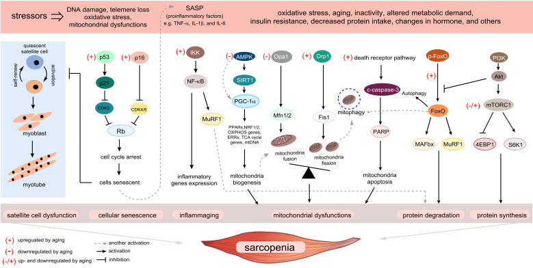
Sarcopenia has been defined as a progressive decline of skeletal muscle mass, strength, and functions in elderly people. It is accompanied by physical frailty, functional disability, falls, hospitalization, and mortality, and is becoming a major geriatric disorder owing to the increasing life expectancy and growing older population worldwide. Experimental models are critical to understand the pathophysiology of sarcopenia and develop therapeutic strategies. Although its etiologies remain to be further elucidated, several mechanisms of sarcopenia have been identified, including cellular senescence, proteostasis imbalance, oxidative stress, and "inflammaging." In this article, we address three main aspects. First, we describe the fundamental aging mechanisms. Next, we discuss both in vitro and in vivo experimental models based on molecular mechanisms that have the potential to elucidate the biochemical processes integral to sarcopenia. The use of appropriate models to reflect sarcopenia and/or its underlying pathways will enable researchers to understand sarcopenia and develop novel therapeutic strategies for sarcopenia. Lastly, we discuss the possible molecular targets and the current status of drug candidates for sarcopenia treatment. In conclusion, the development of experimental models for sarcopenia is essential to discover molecular targets that are valuable as biochemical biomarkers and/or therapeutic targets for sarcopenia.

Keywords: aging; cellular senescence; experimental model; sarcopenia; skeletal muscle.


Conflict of interest statement

The authors declare no conflict of interest.

Figures

Figure 1
Figure 1
Overview of the fundamental aging mechanisms that contribute to sarcopenia. During the aging process, a variety of stressors engage various cellular signaling cascades, ultimately facilitating the progression of sarcopenia. Several senescence-inducing stimuli cause senescence of satellite cells leading to the loss of capacity to repair upon muscle injury. Many of these stimuli cause the upregulation of p53–p21Cip1 and p16Ink4a pathways, which induce a temporal cell-cycle arrest by inhibiting cyclin-dependent kinase (CDK) 2 and CDK4/6, respectively. When cells enter a senescent state, the senescence-associated secretory phenotype (SASP) is expressed in those cells as a paracrine signaling pathway. “Inflammaging” and SASP production in senescent skeletal muscle cells converge on activation of nuclear factor kappa B (NF-κB) signaling, which induces upregulation of muscle ring finger 1 (MuRF1). Activation of the aging process causes mitochondrial dysfunction by deactivation of the sirtuin 1–peroxisome proliferator-activated receptor γ coactivator 1-α (PGC-1α) axis, which is primarily responsible for the maintenance of mitochondrial quality control. The accumulation of damaged mitochondrial DNA due to an imbalance in mitochondrial dynamics will be eliminated via mitophagy. However, under aging conditions, inefficient mitophagy eventually induces apoptosis. Aging interrupts the coordinated balance between protein synthesis and degradation, which activates and interconnects with multiple signaling pathways. Muscle atrophy F-box (MAFbx) and MuRF1, the major muscle-specific E3 ubiquitin ligases, are increased by activation of the transcription factor forkhead box O (FoxO) by aging stimuli, while the roles of Akt/mammalian target of the rapamycin (mTOR) signaling pathway in sarcopenia are controversial. Several possible mechanisms shown here could contribute to sarcopenia and be targets of intervention.
Figure 2
Figure 2
Possible molecular mechanisms of cellular senescence in skeletal muscle cells. The cell-cycle exit is mainly regulated by induction of cyclin-dependent kinase (CDK) inhibitors including p53–p21Cip, p16Ink4a/p15Ink4b, and p27Kip1. CCN1 and miRNA29 directly activate both p53 and p21Cip, while Hsp90β represses p53–p21Cip signaling. Transforming growth factor (TGF)-β signaling activates CDK inhibitors via a pSmad3-dependent pathway. Notch and pSmad3 antagonize each other, and thus, Notch signaling inhibits these CDK inhibitors. Fibroblast growth factor receptor-p38 MAPK signaling has been identified as a critical pathway of deregulated cell-cycle progression and possibly influences cellular senescence; however, its downstream target(s) should be investigated. Skeletal muscle cells undergoing senescence show senescence-associated secretory phenotype (SASP), which can impinge on nearby cells.

References

    1. Rosenberg I.H., Roubenoff R. Stalking Sacropenia. Ann. Intern. Med. 1995;123:727–728. doi: 10.7326/0003-4819-123-9-199511010-00014. - DOI - PubMed
    1. Cruz-Jentoft A.J., Bahat G., Bauer J., Boirie Y., Bruyère O., Cederholm T., Cooper C., Landi F., Rolland Y., Sayer A.A., et al. Sarcopenia: Revised European consensus on definition and diagnosis. Age Ageing. 2019;48:16–31. doi: 10.1093/ageing/afy169. - DOI - PMC - PubMed
    1. Hardee J.P., Lynch G.S. Current pharmacotherapies for sarcopenia. Expert Opin. Pharmacother. 2019;20:1645–1657. doi: 10.1080/14656566.2019.1622093. - DOI - PubMed
    1. Cruz-Jentoft A.J., Sayer A.A. Sarcopenia. Lancet. 2019;393:2636–2646. doi: 10.1016/S0140-6736(19)31138-9. - DOI - PubMed
    1. Hayflick L., Moorhead P.S. The serial cultivation of human diploid cell strains. Exp. Cell Res. 1961;25:585–621. doi: 10.1016/0014-4827(61)90192-6. - DOI - PubMed

Publication types