. 2020 Jun 2;9(6):1386.
doi: 10.3390/cells9061386.

CaSR Antagonist (Calcilytic) NPS 2143 Hinders the Release of Neuroinflammatory IL-6, Soluble ICAM-1, RANTES, and MCP-2 from Aβ-Exposed Human Cortical Astrocytes

Affiliations

CaSR Antagonist (Calcilytic) NPS 2143 Hinders the Release of Neuroinflammatory IL-6, Soluble ICAM-1, RANTES, and MCP-2 from Aβ-Exposed Human Cortical Astrocytes

Anna Chiarini et al. Cells. .

Abstract

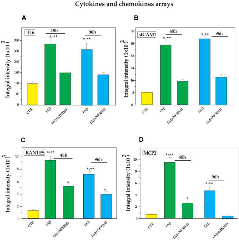
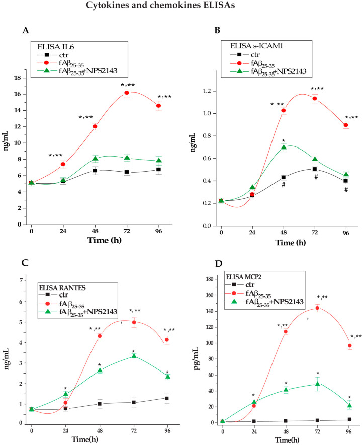
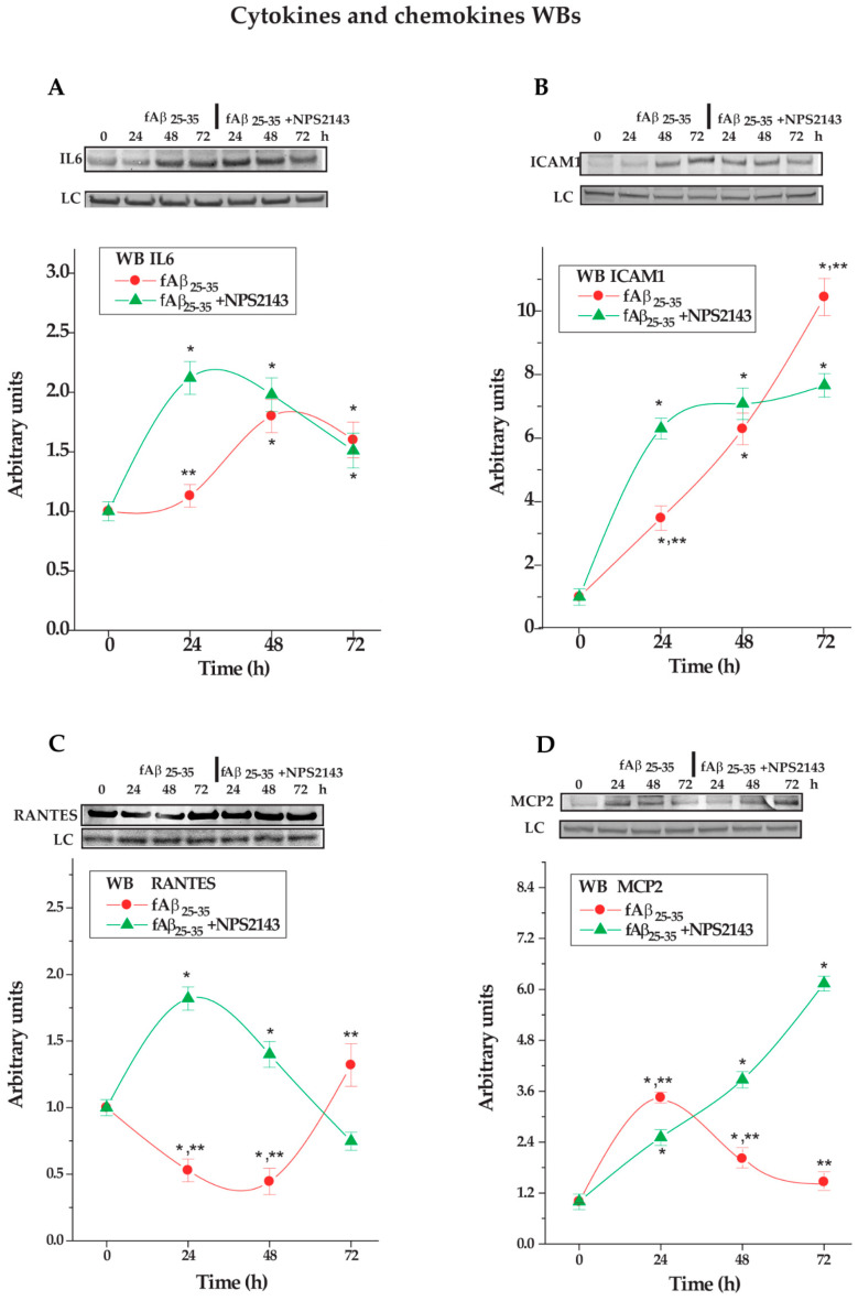
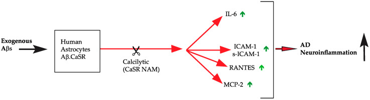
Available evidence shows that human cortical neurons' and astrocytes' calcium-sensing receptors (CaSRs) bind Amyloid-beta (Aβ) oligomers triggering the overproduction/oversecretion of several Alzheimer's disease (AD) neurotoxinseffects calcilytics suppress. We asked whether AβCaSR signaling might also play a direct pro-neuroinflammatory role in AD. Cortical nontumorigenic adult human astrocytes (NAHAs) in vitro were untreated (controls) or treated with Aβ25-35±NPS 2143 (a calcilytic) and any proinflammatory agent in their protein lysates and growth media assayed via antibody arrays, enzyme-linked immunosorbent assays (ELISAs), and immunoblots. Results show Aβ•CaSR signaling upregulated the synthesis and release/shedding of proinflammatory interleukin (IL)-6, intercellular adhesion molecule-1 (ICAM-1) (holoprotein and soluble [s] fragment), Regulated upon Activation, normal T cell Expressed and presumably Secreted (RANTES), and monocyte chemotactic protein (MCP)-2. Adding NPS 2143 (i) totally suppressed IL-6's oversecretion while remarkably reducing the other agents' over-release; and (ii) more effectively than Aβ alone increased over controls the four agents' distinctive intracellular accumulation. Conversely, NPS 2143 did not alter Aβ-induced surges in IL-1β, IL-3, IL-8, and IL-16 secretion, consequently revealing their Aβ•CaSR signaling-independence. Finally, Aβ25-35±NPS 2143 treatments left unchanged MCP-1's and TIMP-2's basal expression. Thus, NAHAs Aβ•CaSR signaling drove four proinflammatory agents' over-release that NPS 2143 curtailed. Therefore, calcilytics would also abate NAHAs' Aβ•CaSR signaling direct impact on AD's neuroinflammation.

Keywords: ICAM-1; IL-6; MCP-2; RANTES; amyloid-β; astrocytes; calcium-sensing receptor; human; neurodegeneration; neuroinflammation.


Conflict of interest statement

The authors declare no conflict of interest.

Figures

Figure 1
Figure 1
Pictures of pure (100%) in vitro cultures of cortical nontumorigenic adult human astrocytes (NAHAs) that express their cell type-specific markers glial fibrillary acid protein (GFAP) (top panels) and glutamine synthase (GS) (bottom panels) as detected in immunoblots (left panels) and via immunocytochemistry (right panels). NAHAs cultures were set up as detailed in the Materials and Methods). After two weeks of staying in vitro, they were sampled and processed for western immunoblotting and immunocytochemistry as described in the Materials and Methods. Original magnification of the microscope pictures: GFAP, X 100; GS, X 200.
Figure 2
Figure 2
Time-dependent differential expression of (A) Interleukin (IL)-6, (B) Soluble intercellular adhesion molecule-1 (s-ICAM-1), (C) Regulated upon Activation, normal T cell Expressed and presumably Secreted (RANTES), and (D) Monocyte chemoattractant protein (MCP)-2, the basal secretion of which increased after treating NAHAs with fAβ25–35, yet significantly decreased after fAβ25–35 + NPS 2143 treatment. Each neuroinflammatory agent was detected in the NAHA-conditioned media via membrane-based antibody arrays as described in the Materials and Methods. The developed antibody arrays were analyzed in an OdisseyTM (LI-COR) scanner and the positive staining intensities of IL-6, s-ICAM-1, RANTES, and MCP-2 were quantified using the Image StudioTM (version 5.2) software. The integral intensities of the positive signals from each array were normalized via comparisons to corresponding controls. Bars are means ± SEMs of three independent experiments, each carried out in duplicate. One-way ANOVA followed by post hoc Tukey’s test allowed to calculate p values. CTR, untreated controls. * p < 0.05 vs. CTR; ** p < 0.05 of fAβ25–35 alone vs. fAβ25–35 + NPS 2143 treatment.
Figure 3
Figure 3
Changes in released levels of (A) IL-6, (B) s-ICAM-1, (C) RANTES, and (D) MCP-2 in NAHAs treated with fAβ25–35 ± NPS 2143 vs. untreated controls (CTR). Each agent was assessed in the NAHA-conditioned media via a specific ELISA kit as detailed in the Materials and Methods. Points on the curves are means ± SEMs of 3–5 independent experiments, each carried out in duplicate. One-way ANOVA followed by post hoc Tukey’s test allowed us to calculate p values. * p < 0.05 vs. time-corresponding CTR values; # p < 0.05 vs. 0-h CTR value; ** p < 0.05 vs. fAβ25–35 + NPS 2143 values.
Figure 4
Figure 4
Changes in protein lysates’ levels of (A) IL-6, (B) s-ICAM-1, (C) RANTES, and (D) MCP-2 in NAHAs treated with fAβ25–35 ± NPS 2143 vs. untreated controls (CTR). At the top of each panel are typical immunoblots showing the densitometric changes of each agent according to treatments vs. untreated controls (CTR). LC, loading control. The graphs underneath show the densitometric evaluations of the specific bands at each time point of every treatment. Points on the curves are mean values of three independent experiments, each carried out in duplicate with control (0-h) values normalized as 1.0. One-way ANOVA followed by post hoc Tukey’s test allowed to calculate p values. * p < 0.05 vs. CTR; ** p < 0.05 vs. fAβ25–35 + NPS 2143.
Figure 5
Figure 5
Synopsis of the direct pro-neuroinflammatory effects pathological Aβ•CaSR signaling evokes in NAHAs. The administration of a CaSR negative allosteric modulators (NAM) nearly totally switches off (in the case of IL-6) or remarkably mitigates (in the case of MCP-2, s-ICAM-1, and RANTES) the Aβ•CaSR signaling-evoked noxious pro-neuroinflammatory upshots. “” denotes upregulation. Abbreviations as in the text.

References

    1. Prince M.J., Wimo A., Guerchet M.M., Ali G.C., Wu Y.-T., Prina M. World Alzheimer Report 2015: The Global Impact of Dementia: An Analysis of Prevalence, Incidence, Cost and Trends. Alzheimer’s Disease International; London, UK: 2015.
    1. Labzin L.I., Heneka M.T., Latz E. Innate immunity and neurodegeneration. Annu. Rev. Med. 2018;69:437–449. doi: 10.1146/annurev-med-050715-104343. - DOI - PubMed
    1. Becher B., Spath S., Goverman J. Cytokine networks in neuroinflammation. Nat. Rev. Immunol. 2017;17:49–59. doi: 10.1038/nri.2016.123. - DOI - PubMed
    1. Hardy J., Selkoe D.J. The amyloid hypothesis of Alzheimer’s disease: Progress and problems on the road to therapeutics. Science. 2002;297:353–356. doi: 10.1126/science.1072994. - DOI - PubMed
    1. Braak H., Del Tredici K. The preclinical phase of the pathological process underlying sporadic Alzheimer’s disease. Pt 10Brain. 2015;138:2814–2833. doi: 10.1093/brain/awv236. - DOI - PubMed

Publication types

MeSH terms