. 2020 Aug;19(8):1598-1612.
doi: 10.1158/1535-7163.MCT-20-0106. Epub 2020 Jun 4.

First-in-Class Inhibitors of Oncogenic CHD1L with Preclinical Activity against Colorectal Cancer

Affiliations

First-in-Class Inhibitors of Oncogenic CHD1L with Preclinical Activity against Colorectal Cancer

Joshua M Abbott et al. Mol Cancer Ther. 2020 Aug.

Abstract

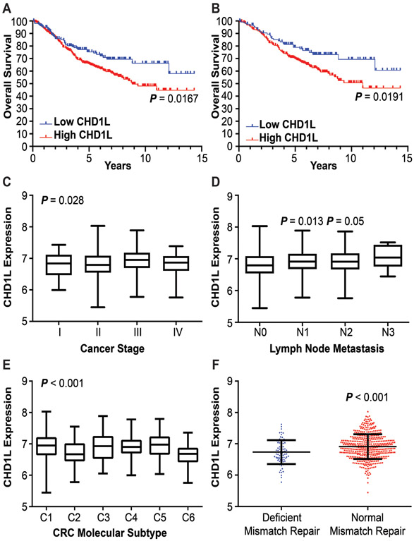
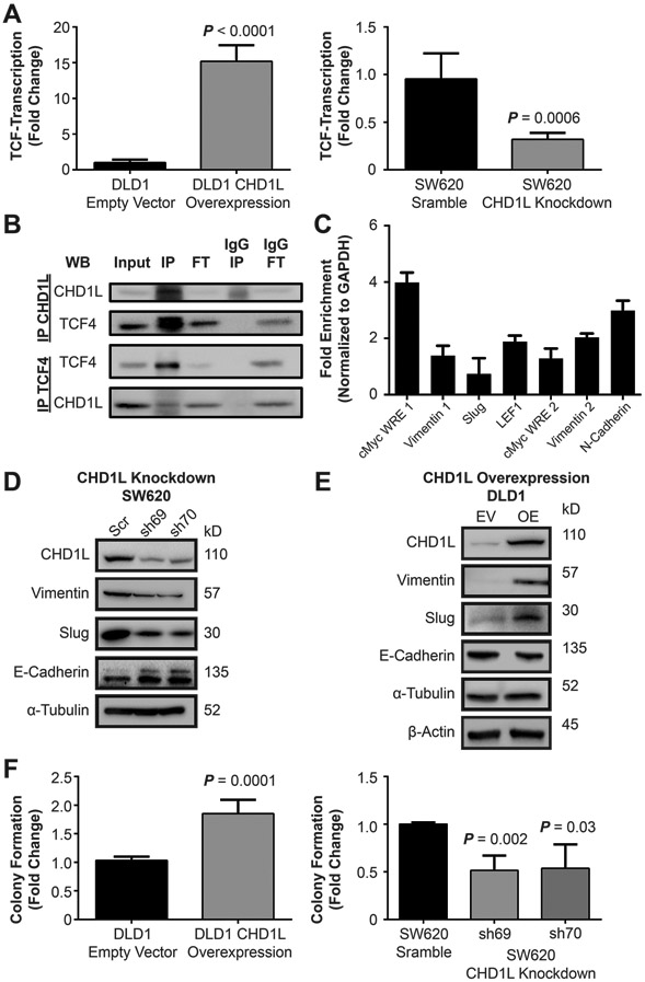
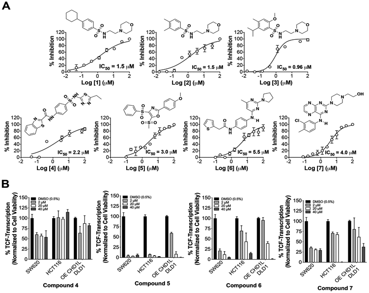
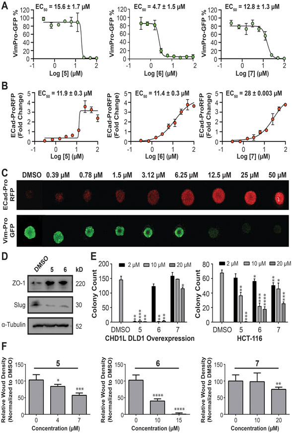
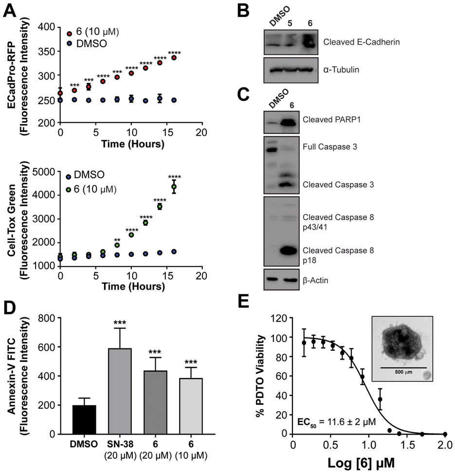
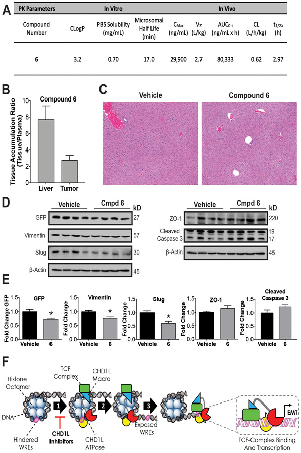
Since the discovery of CHD1L in 2008, it has emerged as an oncogene implicated in the pathology and poor prognosis of a variety of cancers, including gastrointestinal cancers. However, a mechanistic understanding of CHD1L as a driver of colorectal cancer has been limited. Until now, there have been no reported inhibitors of CHD1L, also limiting its development as a molecular target. We sought to characterize the clinicopathologic link between CHD1L and colorectal cancer, determine the mechanism(s) by which CHD1L drives malignant colorectal cancer, and discover the first inhibitors with potential for novel treatments for colorectal cancer. The clinicopathologic characteristics associated with CHD1L expression were evaluated using microarray data from 585 patients with colorectal cancer. Further analysis of microarray data indicated that CHD1L may function through the Wnt/TCF pathway. Thus, we conducted knockdown and overexpression studies with CHD1L to determine its role in Wnt/TCF-driven epithelial-to-mesenchymal transition (EMT). We performed high-throughput screening (HTS) to identify the first CHD1L inhibitors. The mechanism of action, antitumor efficacy, and drug-like properties of lead CHD1L inhibitors were determined using biochemical assays, cell models, tumor organoids, patient-derived tumor organoids, and in vivo pharmacokinetics and pharmacodynamics. Lead CHD1L inhibitors display potent in vitro antitumor activity by reversing TCF-driven EMT. The best lead CHD1L inhibitor possesses drug-like properties in pharmacokinetic/pharmacodynamic mouse models. This work validates CHD1L as a druggable target and establishes a novel therapeutic strategy for the treatment of colorectal cancer.


Conflict of interest statement

Disclosure of Conflict of Interest: The authors declare no conflicts of interest.

Figures

Figure 1:
Figure 1:. Clinicopathological characteristics associated with CHD1L expression.
(A) OS of CHD1L expression in CRC patients, P = 0.0167. (B) OS of CHD1L expression in Stage II/III patient cohort, P = 0.0191. (C) Differential expression of CHD1L in CRC by stage, P = 0.028. (D) Differential expression of CHD1L among N staging in CRC patients, P = 0.013 (N1), 0.05 (N2) relative to N0. (E) Differential expression of CHD1L among CRC molecular subtypes, P < 0.001. (F) Expression of CHD1L between CRC patients with normal and deficient DNA mismatch repair, P < 0.001.
Figure 2:
Figure 2:. CHD1L mediates TCF-transcription in CRC.
(A) Overexpression of CHD1L in DLD1 cells shows an increase in TCF-transcription using the TOPflash TCF-transcription reporter assay (P < 0.0001), knockdown of CHD1L in SW620 cells using shRNA decreases TCF-transcription (P = 0.0006). Mean value of fold change from three independent experiments ± s.d. are shown. (B) Co-immunoprecipitation of CHD1L with TCF4 from SW620 cells, IP (immunoprecipitation), FT (flow-through). (C) Chromatin immunoprecipitation of CHD1L with WNT response element DNA promoter sites in SW620 cells. (D) Evaluation attenuated gene expression of EMT associated genes with SW620CHD1L-KD cells by Western blot. (E) Evaluation of induction of EMT gene expression in DLD1CHD1L-OE cells by Western blot. (F) Evaluation of CSC colony formation in DLD1CHD1L-OE, (P = 0.0001) and SW620CHD1L-KD (P = 0.002, 0.03). Mean values of fold change are from three independent experiments ± s.d. Representative colony images are shown in the supplemental information.
Figure 3:
Figure 3:. Validation of CHD1L inhibitors identified from HTS.
(A) cat-CHD1L ATPase IC50 dose responses with hits 1-7. Mean IC50 values are calculated from three independent experiments and representative graphs are shown. (B) SW620, HCT116, and DLD1CHD1L-OE cells with TOPflash reporter were used to measure inhibition of TCF-transcription using 3 doses over 24 h.
Figure 4:
Figure 4:. CHD1L inhibitors reverse EMT and the malignant phenotype in CRC.
Dose responses for CHD1L inhibitors that modulate EMT measured by high-content imaging of (A) downregulation of VimPro-GFP reporter and (B) Upregulation of EcadPro-RFP reporter. Data curves were fit using Log [CHD1L inhibitor] vs. variable slope (4 parameters) in GraphPad Prism. Mean EC50 values ± SEM are calculated from three independent experiments (C) Representative images exhibiting reversion of EMT by compound 6 in SW620 tumor organoids measured by EMT reporter assays. (D) Western blot analysis showing protein expression changes of additional EMT biomarkers slug and ZO1. (E) CSC stemness measured by clonogenic colony formation after pretreatment with CHD1L inhibitors in DLD1CHD1L-OE and HCT116 cells. (F) Inhibition of invasive potential of HCT116 cells after treatment of CHD1L inhibitors. Welch’s t-test statistical analysis was used to determine significance, where * = P ≤ 0.05, ** = P ≤ 0.01, *** = P ≤ 0.001, **** = P ≤ 0.0001.
Figure 5:
Figure 5:. Lead compound 6 induces apoptosis in CRC cell lines and PDTOs
(A) Time course evaluation of the induction E-cadherin expression using Ecad-ProRFP reporter assay and cytotoxicity using Cell-Tox Green. (B) Western blot analysis of induction of cleaved E-cadherin after treatment of 5 and 6 for 48 hours in CRC organoids. (C) Western blot analysis of induction of caspase dependent apoptosis cadherin after treatment of 6 for 48 hours in CRC organoids. (D) Annexin V-FITC staining analysis of apoptosis after treatment of SN-38 and 6 for 12 hours (E) Cytotoxicity of 6 in PDTO CRC102 using CellTiter-Blue. Mean EC50 values ± s.d. are calculated from six independent experiments and representative graph is shown with inset of a representative PDTO. Welch’s t-test statistical analysis was used to determine significance, where * = P ≤ 0.05, ** = P ≤ 0.01, *** = P ≤ 0.001, **** = P ≤ 0.0001.
Figure 6:
Figure 6:. In vivo pharmacology and proposed mechanism of action.
(A) Summary of compound 6 in vivo and in vitro pharmacokinetic parameters. The consensus LogP (CLogP) values were obtained using the SwissADME web tools.(50) Compound 6 was administered by i.p. injection to athymic nude mice QD for 5 days to measure (B) Accumulation in SW620 xenograft tumors and (C) Histopathological assessment of liver toxicity. Representative photomicrograph sections (5x magnification) of liver in both vehicle and compound 6 treated animals. The images demonstrate normal hepatic cord and lobule architecture, with no evidence of hepatocyte degeneration, necrosis, hyperplasia, or parenchymal inflammation. (D) Western blots from xenograft tumors for vehicle and 6 were done to measure PD effects on EMT. (E) Densitometry of blot intensity showing mean value and SEM of four tumor xenograft samples. (F) Proposed mechanism of action of CHD1L mediated TCF-transcription where CHD1L is activated through binding TCF-complex members. (1) Once activated, CHD1L is directed to hindered WREs localized on chromatin. (2) Chromatin remodeling and DNA translocation occurs exposing WRE sites. (3) TCF-complex binds to exposed WREs facilitated by CHD1L, promoting EMT genes and other genes associated with mCRC. CHD1L ATPase inhibitors effectively prevent step 1, leading to the reversion of EMT and other malignant properties of CRC. Welch’s t-test statistical analysis was used to determine significance, where * = P ≤ 0.05

References

    1. Lorch Y, Maier-Davis B, Kornberg RD. Mechanism of chromatin remodeling. Proc Natl Acad Sci U S A 2010;107(8):3458–62 doi 10.1073/pnas.1000398107. - DOI - PMC - PubMed
    1. Kumar R, Li DQ, Muller S, Knapp S. Epigenomic regulation of oncogenesis by chromatin remodeling. Oncogene 2016;35(34):4423–36 doi 10.1038/onc.2015.513. - DOI - PubMed
    1. Swygert SG, Peterson CL. Chromatin dynamics: interplay between remodeling enzymes and histone modifications. Biochim Biophys Acta 2014;1839(8):728–36 doi 10.1016/j.bbagrm.2014.02.013. - DOI - PMC - PubMed
    1. Erdel F, Krug J, Langst G, Rippe K. Targeting chromatin remodelers: signals and search mechanisms. Biochim Biophys Acta 2011;1809(9):497–508 doi 10.1016/j.bbagrm.2011.06.005. - DOI - PubMed
    1. Brownlee PM, Meisenberg C, Downs JA. The SWI/SNF chromatin remodelling complex: Its role in maintaining genome stability and preventing tumourigenesis. DNA Repair (Amst) 2015;32:127–33 doi 10.1016/j.dnarep.2015.04.023. - DOI - PubMed

Publication types

MeSH terms