Review
. 2020 Jul 24;295(30):10340-10367.
doi: 10.1074/jbc.REV120.011473. Epub 2020 Jun 4.

Role of the lipid bilayer in outer membrane protein folding in Gram-negative bacteria

Affiliations
Review

Role of the lipid bilayer in outer membrane protein folding in Gram-negative bacteria

Jim E Horne et al. J Biol Chem. .

Abstract

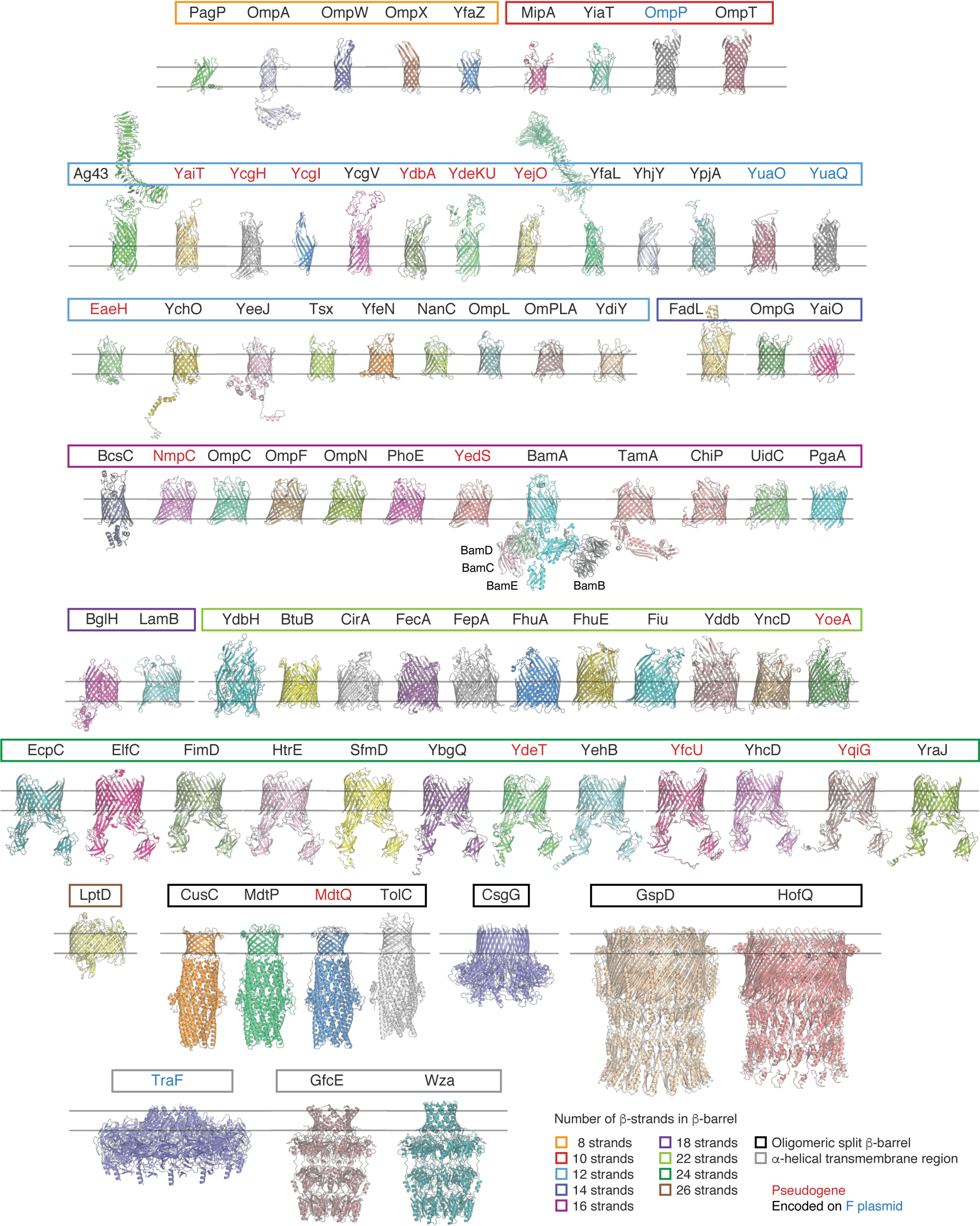
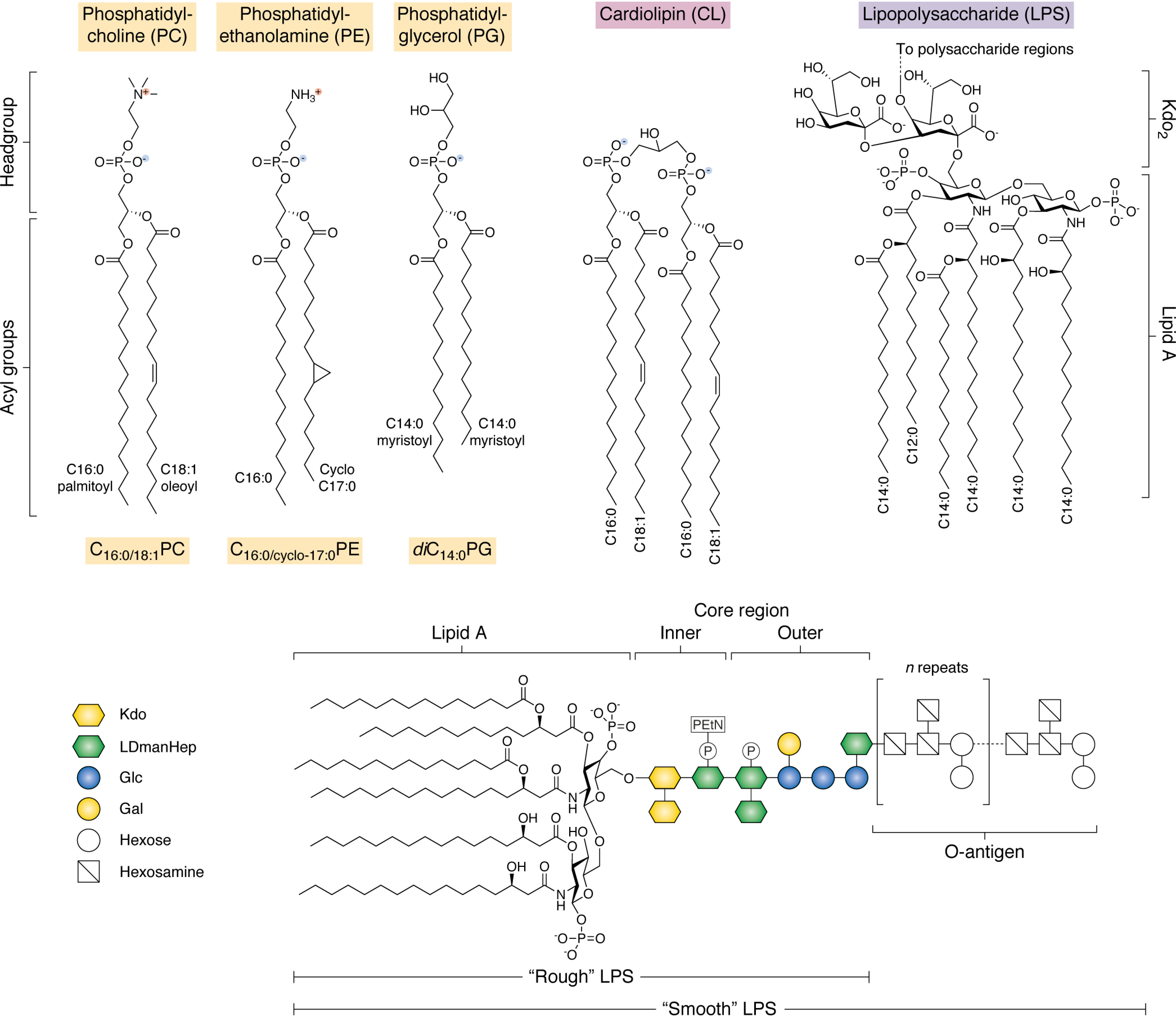
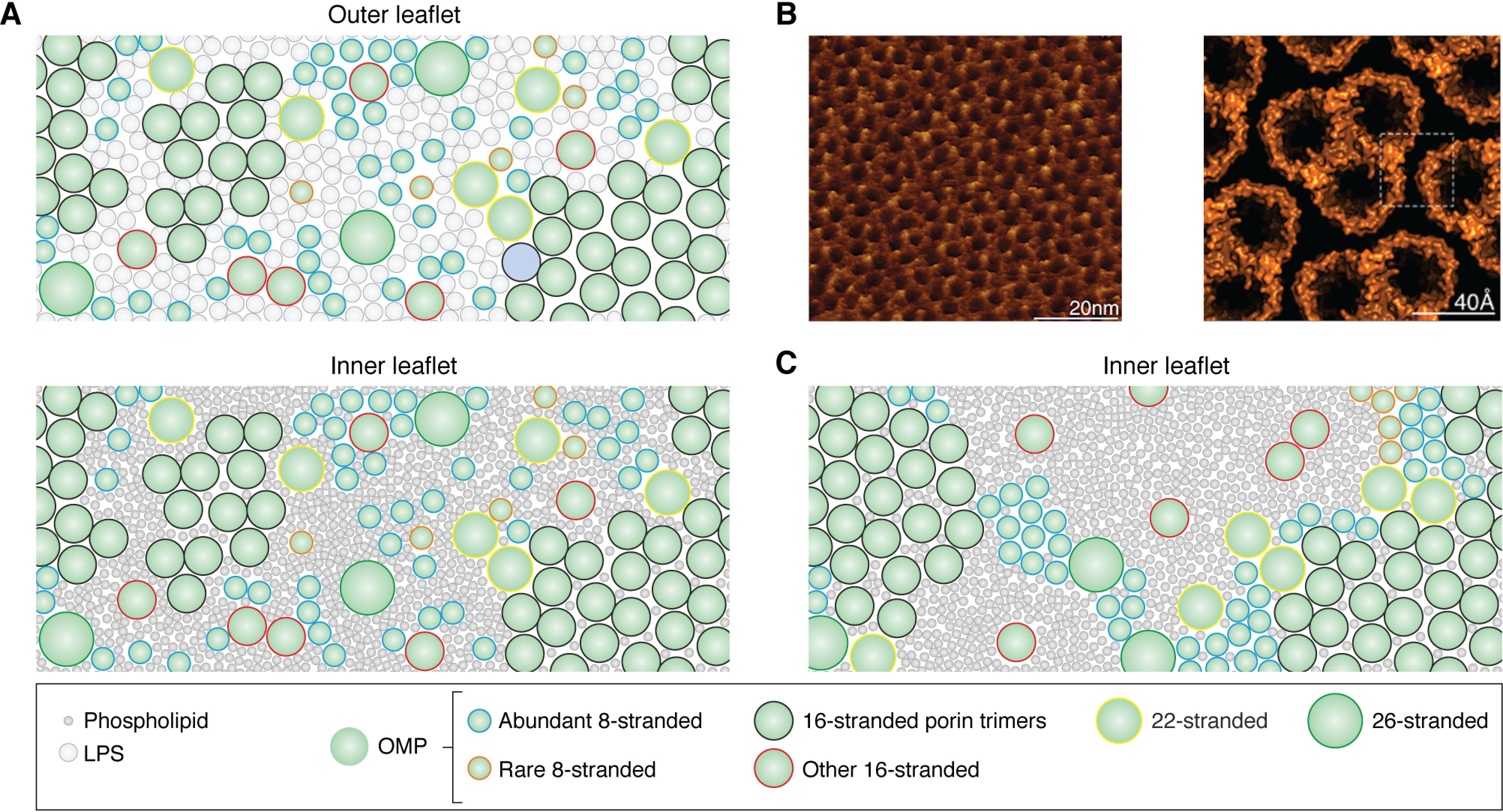
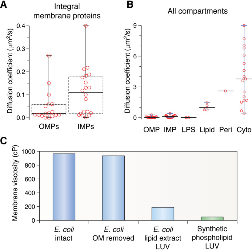
β-Barrel outer membrane proteins (OMPs) represent the major proteinaceous component of the outer membrane (OM) of Gram-negative bacteria. These proteins perform key roles in cell structure and morphology, nutrient acquisition, colonization and invasion, and protection against external toxic threats such as antibiotics. To become functional, OMPs must fold and insert into a crowded and asymmetric OM that lacks much freely accessible lipid. This feat is accomplished in the absence of an external energy source and is thought to be driven by the high thermodynamic stability of folded OMPs in the OM. With such a stable fold, the challenge that bacteria face in assembling OMPs into the OM is how to overcome the initial energy barrier of membrane insertion. In this review, we highlight the roles of the lipid environment and the OM in modulating the OMP-folding landscape and discuss the factors that guide folding in vitro and in vivo We particularly focus on the composition, architecture, and physical properties of the OM and how an understanding of the folding properties of OMPs in vitro can help explain the challenges they encounter during folding in vivo Current models of OMP biogenesis in the cellular environment are still in flux, but the stakes for improving the accuracy of these models are high. OMP folding is an essential process in all Gram-negative bacteria, and considering the looming crisis of widespread microbial drug resistance it is an attractive target. To bring down this vital OMP-supported barrier to antibiotics, we must first understand how bacterial cells build it.

Keywords: BAM complex; Gram-negative bacteria; OMPome; antibiotic resistance; disorderase; folding kinetics; lipid; lipid membrane; membrane bilayer; membrane protein; membrane protein folding; outer membrane; protein folding; protein-lipid interactions; β-barrel assembly machinery (BAM) complex.


Conflict of interest statement

Conflict of interest—The authors declare that they have no conflicts of interest with the contents of this article.

Figures

Figure 1.
Figure 1.
Structures of transmembrane proteins found in the OM of E. coli K-12 MG1655. A list of all known and predicted transmembrane proteins in the OM of E. coli K-12 strain MG1655 was manually curated, creating the “OMP-ome.” The Protein Data Bank was then searched for solved structures of these proteins or close homologues. Where no high-resolution solved 3D structures were available, homology models were generated using the I-TASSER server (RRID:SCR_014627) (396). For two proteins, NfrA (the N4 bacteriophage receptor), and FlgH (the flagellar L-ring protein), no homology models could be generated. Predictions for YaiO, YcgI, YdbH, and YhjY generated deformed or broken barrels (possibly due to a lack of homology to existing structures), but their predictions are displayed to indicate their approximate structure. Extracellular domains of autotransporters have only been shown where accurate models could be built or crystal structures were available. OMPs are grouped here by the number of β-strands and then by protein family. The non-OMP subunits of the BAM complex are labeled below the central BamA subunit. Protein names are in red if they represent pseudogenes (inactivated by mutation in this strain) and blue if they are encoded on the F plasmid. The color of the box surrounding the protein names represents the number of β-strands in the β-barrel. Light Orange, 8; red, 10; light blue, 12; violet, 14; pink, 16; purple, 18; light green, 22; dark green, 24; brown, 26; black, oligomeric split β-barrel; gray, α-helical transmembrane region. Structures were aligned with each other by their β-barrel domains and rendered individually in PyMOL 2.X (Schrödinger, LLC). A list of the proteins with their associated family and PDB code can be found in Table S1.
Figure 2.
Figure 2.
Common lipid types found in bacterial outer membranes and/or used in in vitro studies of OMP folding. Top, schematic of the generic structure of phospholipids and LPS. Bacterial lipids can be conceptualized as having two “domains”: a polar headgroup and a hydrophobic acyl tail region. In phospholipids, the acyl tails are connected by an ester linkage to a phosphate group and a variable headgroup region. PC and PE are zwitterionic, whereas PG carries a net negative charge. Note that PC lipids are not commonly found in bacterial membranes but are often used for OMP folding-studies in vitro due to their net neutral charge and propensity to form bilayers. Cardiolipin comprises two acyl tail regions connected by phosphate groups via a glycerol linkage and carries a net double negative charge. LPS is found exclusively in the OM of Gram-negative bacteria and varies considerably between species in both the number and length of acyl tails in the lipid A region and the sugar composition in the polysaccharide region (shown below). Here the most common structure of lipid A-Kdo2 for E. coli K-12 LPS is shown in full. Bottom, the architecture of a generic LPS is shown. The lipid A and core region are consistent with LPS found in E. coli K-12; however, this strain does not naturally produce an O-antigen, whereas many environmental and clinical strains do. Strains lacking the O-antigen region are said to contain “rough” LPS, and this can further be divided into subtypes dependent on truncations in the core region. The most extreme of these that is still viable at 37 °C under laboratory growth conditions is “deep rough” LPS, containing only lipid A-Kdo2. The O-antigen region is highly variable within species and can contain as many as 40 glycan repeats. Kdo, keto-deoxyoctulosonate; LDmanHep, l-glycero-d-manno-heptose; Glc, glucose; Gal, galactose; P, phosphate group; PEtN, phosphorylethanolamine.
Figure 3.
Figure 3.
Model depicting the structural organization of the E. coli OM. A schematic displays the degree of crowding in the OM. A, view of an imagined OM showing the dense packing of different size OMPs in monomers, dimers, and trimers interspersed with LPS in the outer leaflet (top) and phospholipids in the inner leaflet (bottom). Phospholipids are represented as dark gray circles with a diameter proportional to the headgroup size of PE/PG and LPS as light gray circles with a diameter proportional to the size of lipid A. Different OMPs are represented as idealized circles with diameters proportional to their number of strands. Blue outline, abundant 8-stranded; orange outline, rare 8-stranded; black, 16-stranded porin trimers; red, other 16-stranded; yellow, 22-stranded; green, 26-stranded. The overall LPR in this schematic is ∼9:1, with ∼2 LPS and ∼7 phospholipids per OMP, consistent with estimates for the LPR of the E. coli OM. B, left, high-resolution AFM image of OM extracts from Roseobacter dentrificans imaged from the periplasmic side showing a dense lattice of porin trimers. Right, atomic model of the packing of porin trimers derived from the AFM data. Reproduced with permission from Jarosławski et al. (156). This research was originally published in Molecular Microbiology. Jarosławski, S., Duquesne, K., Sturgis, J. N., and Scheuring, S. High-resolution architecture of the outer membrane of the Gram-negative bacteria Roseobacter denitrificans. Molecular Microbiology 2009; 74:1211–1222. © Wiley–Blackwell. C, view of an imagined OM with the same LPR as in A but assuming a more extreme clustering of most OMPs. Only the inner leaflet is shown. Despite having the same LPR values, the buried surface area of the clustered OMPs frees up more lipid to form larger bulk lipid domains.
Figure 4.
Figure 4.
Comparison of physical properties of bacterial membranes. A, box plots showing the range of diffusion coefficients reported for OMPs and IMPs (see “A crowded environment”). Boxes show interquartile range calculated by the Tukey method with the median indicated as a boldface horizontal line. Whiskers show the minimum and maximum values. B, comparison of the diffusion coefficients of membrane proteins with other components of bacteria. Whiskers are only shown for components that have three or more values reported in the literature. All values are reported from in vivo studies. LPS, diffusion of LPS molecules in S. typhimurium. Lipid, diffusion rate of a fluorescent lipid reporter probe in E. coli membranes. Peri, diffusion of soluble protein in the E. coli periplasm. Cyto, diffusion of soluble protein in the E. coli cytoplasm. C, viscosities of different membrane environments as measured by the use of fluorescent BODIPY C10 lipid reporter probes. E. coli data are from Mika et al. (175), and synthetic phospholipid data are from Wu et al. (225). BODIPY C10 specifically incorporates into the IM of E. coli, and removal of the OM minimally affects the measured viscosity. Synthetic phospholipid 200 nm LUVs were comprised of DLPC, DMPC, POPC, or DOPC.
Figure 5.
Figure 5.
Physical and mechanical properties of a lipid bilayer. A, top, the phase of a lipid bilayer depends on the temperature, with the lipids being in an ordered (gel) phase below the Tm and in a (liquid) disordered phase above the Tm. At the transition temperature, frustration between packing of regions of gel and liquid phase causes defects to occur at these boundaries. Bottom, a typical differential scanning calorimetry curve illustrating the thermal response of a DMPC (diC14:0PC) bilayer with the regions of each phase colored as above. B, the hydrophobic thickness of a membrane depends on the lipid acyl chain length. However, when an OMP becomes embedded in a lipid bilayer, the membrane responds by trying to “match” the hydrophobic thickness of the bilayer to that of the protein to minimize the energetic penalty of exposing polar lipid headgroups to a hydrophobic OMP surface or hydrophobic acyl tails to polar OMP loops. C, mixtures of lipids can separate, forming “rafts” or domains dependent on the physical conditions and lipid type. CL has a high propensity for negative curvature and has been shown to be enriched at cell poles and division sites where the membrane constricts. CL has also been observed to bind to membrane protein complexes such as the BAM complex (397) and cluster under patches of LPS in MD simulations (209, 210), suggesting that it may help stabilize bilayer packing defects (which might be induced by LPS) and stabilize regions of large hydrophobic mismatch (e.g. around embedded proteins such as BamA). D, schematic describing stored curvature elastic stress and how this depends on lipid type. Adapted from Booth and Curnow (398). This research was originally published in Current Opinion in Structural Biology. Booth, P. J., and Curnow, P. Folding scene investigation: membrane proteins. Current Opinion in Structural Biology 2009; 19:8–13. © Elsevier. Attractive and repulsive interactions driven by the packing of lipids create a pressure differential along the normal of the membrane that must be overcome to deform or alter lipid packing. Incorporation of lipids that have a tendency toward negative curvature (due to the relative size of the headgroup versus the acyl chain (e.g. PE lipids)) into a bilayer formed from lipids with a neutral or low curvature tendency (e.g. PC lipids) generates a stress force within the bilayer due to the opposing tendencies for bilayer formation of these lipids.
Figure 6.
Figure 6.
The structure and architecture of OmpA. The 8-stranded β-barrel OmpA has been used for many studies of OMP folding in vitro. It comprises a transmembrane β-barrel domain and a soluble periplasmic peptidoglycan-binding domain. Neighboring β-strands are connected on their extracellular side by a long disordered “loop” and on the periplasmic side by a short “turn.” OMP β-strands are usually numbered from the N terminus (NT) to the C terminus (CT), and the C-terminal β-strand often contains a conserved motif of Gly-X-X-Ar-X-Ar (where Ar represents any aromatic residue), indicated in purple on OmpA, thought to be important for recognition by BAM (386, 399). Many OMPs contain an enrichment in aromatic Trp and Tyr residues in their β-barrel domain, particularly at the interfacial region between the lipid headgroups (approximate position indicated by the gray line) and acyl tails (approximate position indicated by the gray box) known as an “aromatic girdle.” Trp (red) and Tyr (blue) residues found in the β-barrel domain of OmpA are indicated above. This model of OmpA was created in PyMol 2.X (Schrödinger, LLC) by fusing the NMR structures of the E. coli OmpA β-barrel (PDB code 1G90) (400) and its periplasmic domain (PDB code 2MQE) (401).
Figure 7.
Figure 7.
Mechanisms of OMP folding in vitro and in vivo. A, Mechanism of spontaneous OMP folding in vitro as described for OmpA. In vitro studies have shed light on the folding pathway of model OMPs, particularly OmpA, after dilution out of high concentrations of denaturant in the presence of a lipid bilayer. Adapted from Danoff and Fleming (351). This research was originally published in Biochemistry. Danoff, E. J., and Fleming, K. G. Membrane defects accelerate outer membrane β-barrel protein folding. Biochemistry 2015; 54:97–99. © American Chemical Society. 1, immediately after dilution out of denaturant, the chain undergoes hydrophobic collapse; 2, the polypeptide chain then binds to the surface of a membrane; 3, the nascent OMP then begins to form secondary structure as it brings together neighboring β-strands to form β-hairpins while still mostly exposed to the aqueous environment; 4, these β-hairpins associate and begin to insert into the acyl tail region of the membrane; 5, the tertiary structure of the barrel is complete with the final step likely being a slower equilibration of side chains and extrusion of hydrophilic loops from the barrel lumen. B, proposed mechanisms of BAM-catalyzed folding of OMPs in vivo. The nascent OMP is shown in red or purple, BamA is shown in green, and BamD is shown in yellow (other subunits have been omitted for clarity). 1, BamA-assisted. Substrate OMPs are delivered to BAM or directly to the membrane by periplasmic chaperones. These nascent OMPs then fold spontaneously into a region of destabilized membrane in front of the lateral gate of BamA, essentially following the same pathway as described in A. 2, BamA-budding. Binding/recognition of the OMP occurs on BAM before β-strands are added a β-hairpin at a time between β1 and β16 of BamA, forming a semisymmetric hybrid-barrel intermediate. Once all β-hairpins are added, this folded OMP then buds off from BamA. 3, BamA-swing/elongation. Binding/recognition of the OMP occurs on BAM and folding starts with templating of the C-terminal β-strand of a nascent OMP against β1 of BamA. Folding proceeds in the periplasm through the stepwise addition of more β-strands. Once all β-strands have been added, a conformational change in BamA “swings” the folded β-barrel into the membrane. 4, BamA lumen–catalyzed. This model begins as described in 3 with templating against BamA β1. However, formation of further β-strands is catalyzed against the lumen wall of BamA with a conserved motif in loop 6 of BamA (not shown) possibly stabilizing this interaction. In all of these models, BamD (yellow) may play an important role in substrate recognition and/or the conformational cycle.
Figure 8.
Figure 8.
Conformations of the BamA lipid-facing lateral gate. Shown are example structures of E. coli BamA adopting different conformations around the location of the β1–β16 seam/gate. BamA has been observed in both gate open and closed states with the open state observed in the presence of other BAM subunits but not in structures of BamA in isolation. Furthermore, in all structures of the full BAM complex, β16 of BamA adopts a kinked conformation at a highly conserved glycine (Gly807) in both the open (PDB code 5EKQ; BamACDE) (368) and closed (PDB code 5D0O; BamABCDE) (357) states of the gate. Residues Asn427–Gly433 (β1) are highlighted in light blue, residues Phe802–Trp810 (β16) are indicated in orange, and the kink is further highlighted with spheres (Ile806–Trp810). This kink is also observed in structures of BamA from Salmonella enterica (PDB code 5OR1) (370) and Neisseria gonorrhoeae (PDB code 4K3B) (355) (not shown) and in the BamA homologue, TamA, which also plays a role in OMP assembly (PDB codes 4N74 and 4C00) (387) (not shown). BamA with a closed gate and no kink has been observed in isolation (PDB code 4N75; BamAΔ1–427) (366) and in a hybrid BamA containing a C-terminal 9-residue extension (colored yellow) comprised of part of turn 3 and β7 from OmpX, which may represent a mimic of an OMP-BamA folding intermediate (PDB code 6FSU) (371). Structures are represented in an asymmetric bilayer with a mixture of phospholipids with 14-18 carbon acyl chains (shown in violet) in the inner leaflet and E. coli rough LPS in the outer leaflet (acyl chains in white). Note the different hydrophobic thickness between each leaflet. Asymmetric bilayer was built using the GNOMM server (402).

Similar articles

Cited by

References

    1. Prilipov A., Phale P. S., Koebnik R., Widmer C., and Rosenbusch J. P. (1998) Identification and characterization of two quiescent porin genes, nmpC and ompN, in Escherichia coli BE. J. Bacteriol. 180, 3388–3392 10.1128/JB.180.13.3388-3392.1998 - DOI - PMC - PubMed
    1. Fàbrega A., Rosner J. L., Martin R. G., Solé M., and Vila J. (2012) SoxS-dependent coregulation of ompN and ydbK in a multidrug-resistant Escherichia coli strain. FEMS Microbiol. Lett. 332, 61–67 10.1111/j.1574-6968.2012.02577.x - DOI - PubMed
    1. Li G.-W., Burkhardt D., Gross C., and Weissman J. S. (2014) Quantifying absolute protein synthesis rates reveals principles underlying allocation of cellular resources. Cell 157, 624–635 10.1016/j.cell.2014.02.033 - DOI - PMC - PubMed
    1. Soufi B., Krug K., Harst A., and Macek B. (2015) Characterization of the E. coli proteome and its modifications during growth and ethanol stress. Front. Microbiol. 6, 103 10.3389/fmicb.2015.00103 - DOI - PMC - PubMed
    1. Henning U., Höhn B., and Sonntag I. (1973) Cell envelope and shape of Escherichia coli K12: the ghost membrane. Eur. J. Biochem. 39, 27–36 10.1111/j.1432-1033.1973.tb03099.x - DOI - PubMed

Publication types

MeSH terms

Substances