. 2020 Jul 8;40(28):5413-5430.
doi: 10.1523/JNEUROSCI.2730-19.2020. Epub 2020 Jun 4.

Modular and Distinct Plexin-A4/FARP2/Rac1 Signaling Controls Dendrite Morphogenesis

Affiliations

Modular and Distinct Plexin-A4/FARP2/Rac1 Signaling Controls Dendrite Morphogenesis

Victor Danelon et al. J Neurosci. .

Abstract

Diverse neuronal populations with distinct cellular morphologies coordinate the complex function of the nervous system. Establishment of distinct neuronal morphologies critically depends on signaling pathways that control axonal and dendritic development. The Sema3A-Nrp1/PlxnA4 signaling pathway promotes cortical neuron basal dendrite arborization but also repels axons. However, the downstream signaling components underlying these disparate functions of Sema3A signaling are unclear. Using the novel PlxnA4KRK-AAA knock-in male and female mice, generated by CRISPR/cas9, we show here that the KRK motif in the PlxnA4 cytoplasmic domain is required for Sema3A-mediated cortical neuron dendritic elaboration but is dispensable for inhibitory axon guidance. The RhoGEF FARP2, which binds to the KRK motif, shows identical functional specificity as the KRK motif in the PlxnA4 receptor. We find that Sema3A activates the small GTPase Rac1, and that Rac1 activity is required for dendrite elaboration but not axon growth cone collapse. This work identifies a novel Sema3A-Nrp1/PlxnA4/FARP2/Rac1 signaling pathway that specifically controls dendritic morphogenesis but is dispensable for repulsive guidance events. Overall, our results demonstrate that the divergent signaling output from multifunctional receptor complexes critically depends on distinct signaling motifs, highlighting the modular nature of guidance cue receptors and its potential to regulate diverse cellular responses.SIGNIFICANCE STATEMENT The proper formation of axonal and dendritic morphologies is crucial for the precise wiring of the nervous system that ultimately leads to the generation of complex functions in an organism. The Semaphorin3A-Neuropilin1/Plexin-A4 signaling pathway has been shown to have multiple key roles in neurodevelopment, from axon repulsion to dendrite elaboration. This study demonstrates that three specific amino acids, the KRK motif within the Plexin-A4 receptor cytoplasmic domain, are required to coordinate the downstream signaling molecules to promote Sema3A-mediated cortical neuron dendritic elaboration, but not inhibitory axon guidance. Our results unravel a novel Semaphorin3A-Plexin-A4 downstream signaling pathway and shed light on how the disparate functions of axon guidance and dendritic morphogenesis are accomplished by the same extracellular ligand in vivo.

Keywords: Semaphorin signaling; axonal repulsion; dendritic branching; guidance receptors; neural development.


Figures

Figure 1.
Figure 1.
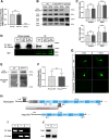
Generation of the PlxnA4KRK-AAA mutant mouse line and the Farp2 KO mouse line. A, qRT-PCR analysis revealed unchanged PlxnA4 mRNA transcript levels in E13.5 PlxnA4KRK-AAA heads compared with WT littermate controls. PlxnA4 KO heads were processed as well as a negative control. Quantification was performed using a relative quantification analysis (ΔΔCt) and presented as mean ± SEM. ***p < 0.001 (Student's t test). B, C, Western blot analysis of E13.5 DRGs and cortices extracted from PlxnA4KRK-AAA and WT littermate controls revealed similar Plexin-A4 and Nrp1 protein levels in both genotypes. Heads of E13.5 PlxnA4 KO were used as a negative control. Data are mean ± SEM. Statistical analysis: Student's t test. D, PlxnA4KRK-AAA, WT littermates, and PlxnA4 KO cortices were subjected to anti-Nrp1 immunoprecipitation and probed using a Plexin-A4 antibody. No change in Nrp1-Plexin-A4 association was detected in the PlxnA4KRK-AAA mutant compared with WT. E, F, 7 DIV cortical neurons from E13.5 PlxnA4KRK-AAA and WT littermate embryos were subjected to a cell-surface biotinylation assay, followed by Western blot probing for Plexin-A4. The total mass of biotinylated proteins in the range of ∼120-260 kDa, detected and quantified using streptavidin-HRP, was used as loading control. Data are mean ± SEM; N.S., Non Significant. G, Primary cortical neurons of PlxnA4KRK-AAA, WT littermates, and PlxnA4 KO embryos were immunostained in culture using a Plexin-A4 antibody. Plexin-A4 protein levels and its spatial pattern in the PlxnA4KRK-AAA mutant were comparable with the WT, whereas virtually no Plexin-A4 was detected in the negative control KO. Scale bar, 25 µm. H, Schematic representation of the Farp2 locus on chromosome 1 (GRCm38-mm10) and the CRISPR-Cas9 KO design. Two sgRNAs flanking an 18,167 bp sequence were used: sgRNA 1 targeted a region upstream of the Farp2 gene promoter, and sgRNA 2 targeted a sequence in intron 2. P1 and P2 (black triangles) represent primers used for detection of the KO allele in a PCR analysis of genomic DNA, targeting sequences outside of the deletion area (upstream of sgRNA1 and downstream of sgRNA2, respectively). Similarly, P3 and P4 (gray triangles) represent primers used for detection of the WT allele and were targeted at intron 1. RT 1 and 2 (blue triangles) represent primers used for reverse transcription analysis of Farp2 gene expression levels; RT 1 targeted a sequence comprised of the 3′ of exon 4 and 5′ of exon 5, and RT 2 targeted exon 6. I, PCR analysis of tail genomic DNA obtained from three E13.5 embryos of a Farp2 Het x Het cross, using primers 1-4 (P1-P4). Lengths of the WT and KO alleles are 551 and 271 bp, respectively. +/+, WT; +/−, Farp2 heterozygous; −/−, Farp2 KO. J, Reverse transcription analysis of WT (+/+) and KO (−/−) cDNA using either Farp2 primers RT 1-2 (top), or Actin β (Actb) primers as internal control (bottom). G, H, Successful KO of the Farp2 gene and its transcripts.
Figure 2.
Figure 2.
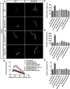
PlxnA4KRK-AAA and Farp2 KO DRG axons show intact Sema3A-dependent responses in vitro. A, DRG explants from WT, PlxnA4KRK-AAA, and Farp2 KO E13.5 embryos were grown for 48 h, treated with 0.1, 0.5, or 1 nm AP-Sema3A (only 1 nm is shown) or control conditioned media and stained using phalloidin-rhodamine to visualize growth cone collapse. Arrows indicate intact growth cones. Arrowheads indicate collapsed growth cones. B, C, Quantification of collapse response as a mean percentage of collapsed growth cones out of the total ± SEM. Statistical analysis: two-way ANOVA with post hoc Tukey test. Scale bar, 50 µm. D, DRG explants from E13.5 PlxnA4KRK-AAA and WT littermates or Farp2 KO and WT littermates were cocultured in collagen droplets for 48 h with a COS1 aggregate either secreting myc-Sema3A (dashed circle) or expressing control PAY1-GFP (green). Cultures were visualized using anti-tubulin Class III immunostaining. E, Quantification of axonal repulsion using the proximal/distal (P/D) ratio, as indicated in the schematic representation of the collagen axonal repulsion assay in D. Data are mean ± SEM. Statistical analysis: Student's t test; N.S., Non Significant. Scale bar, 500 µm.
Figure 3.
Figure 3.
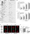
Both PlxnA4KRK-AAA and Farp2 KO neurons are nonresponsive to Sema3A-induced cortical neuron dendrite outgrowth in vitro. A, Representative confocal micrographs of dissociated primary cortical neurons obtained from WT, PlxnA4 KO, PlxnA4KRK-AAA, and Farp2 KO E13.5 embryos. The neurons were treated with AP or Sema3A 5 nm for 24 h. B-E, Sholl analysis quantified dendritic growth and branching in WT, PlxnA4 KO, PlxnA4KRK-AAA, and Farp2 KO cortical neurons (B), measuring the number of dendritic intersections, the total dendritic length (C), the DCI (D), and the number of dendritic tips (E). Data are mean ± SEM from three independent cultures of each genotype. ***p < 0.001 (two-way ANOVA with post hoc Tukey test). Scale bar, 25 µm.
Figure 4.
Figure 4.
Both PlxnA4KRK-AAA and Farp2 KO cortical neurons are nonresponsive, even to higher concentrations of Sema3A. A, Representative confocal micrographs of dissociated primary cortical neurons obtained from WT, PlxnA4KRK-AAA, and Farp2 KO E13.5 embryos. The WT neurons were treated with AP or Sema3A 5 nm, and the PlxnA4KRK-AAA and Farp2 KO neurons were treated with AP or Sema3A 10 nm for 24 h. B–E, Sholl analysis quantified dendritic growth and branching in WT, PlxnA4KRK-AAA, and Farp2 KO cortical neurons (B), measuring the number of dendritic intersections, the total dendritic length (C), the DCI (D), and the number of dendritic tips (E). Data are mean ± SEM from three independent cultures of each genotype. ***p < 0.001 (two-way ANOVA with post hoc Tukey test). Scale bar, 25 µm.
Figure 5.
Figure 5.
Reduced basal dendritic arborization in layer 5 cortical neurons from PlxnA4KRK-AAA and Farp2 KO adult animals. A, Golgi-stained images of brain (2- to 3-month-old) sections of WT, PlxnA4 KO, PlxnA4KRK-AAA, Farp2 KO, and PlxnA4+/KRK-AAA/Farp2+/−. B, Sholl analysis quantification revealed reduced and altered branching patterns of basal dendrites from layer V cortical neurons in PlxnA4 KO, PlxnA4KRK-AAA, Farp2 KO, and PlxnA4+/KRK-AAA/Farp2+/− compared with WT and to PlxnA4+/KRK-AAA or Farp2+/− littermates. C-E, Quantifications of total dendritic length (C), DCI (D), and dendritic tips (E) in all genotype analyzed. Data are mean ± SEM. *p < 0.05 (one-way ANOVA followed by post hoc Tukey test). F, G, Representative images of the layer 5 cortical neurons and Sholl analysis obtained from a WT, PlxnA4KRK-AAA, and PlxnA4+/KRK-AAA/Farp2+/− Thy1-GFP mice. Statistical analysis: one-way ANOVA followed by post hoc Tukey test. Data are mean ± SEM; n = 3 brains/genotype. *p < 0.05, statistical difference between WT and PlxnA4KRK-AAA mutant or PlxnA4 KO or Farp2 KO in B and G. #p < 0.05, statistical difference between WT and PlxnA4KRK-AAA/+/Farp2+/− or PlxnA4 KO in B and G. Scale bars: A, F, 25 µm.
Figure 6.
Figure 6.
PlxnA4KRK-AAA and Farp2 KO animals show normal axonal projections in the PNS and CNS in vivo. A, WT, PlxnA4KRK-AAA, Farp2 KO, or PlxnA4 KO E12.5 embryos were immunostained using a Neurofilament antibody to visualize cutaneous sensory axons pattern. PlxnA4KRK-AAA and Farp2 KO embryos were comparable with the WT, whereas the PlxnA4 KO exhibited an expected hyperinnervation, including a previously described discrete phenotype (white arrows). Scale bar, 500 µm. B, WT, PlxnA4KRK-AAA, Farp2 KO, or PlxnA4 KO E13.5 embryos were immunostained using a TH antibody to visualize sympathetic axons. PlxnA4KRK-AAA and Farp2 KO embryos were comparable with the WT, whereas the PlxnA4 KO exhibited an expected medial protrusion of sympathetic axons (black arrows). Scale bar, 250 µm. C, WT, PlxnA4KRK-AAA, Farp2 KO, or PlxnA4 KO P30 brains were stained using hematoxylin. PlxnA4KRK-AAA and Farp2 KO embryos were comparable with the WT, whereas the PlxnA4 KO exhibited an expected disrupted formation of the anterior commissure (white arrows). Scale bar, 500 µm.
Figure 7.
Figure 7.
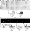
Inhibition of Rac signaling abolishes Sema3A-mediated cortical neuron dendrite elaboration but does not hinder Sema3A-dependent growth cone collapse of WT or PlxnA4KRK-AAA DRG axons in vitro. A, DRG explants from WT and PlxnA4KRK-AAA littermates E13.5 embryos were grown for 48 h, treated for 30 min with the pan-Rac inhibitor EHT 1864 at a concentration of 5 μm, 10 μm, or media alone as control. Then, 1 nm Sema3A or control conditioned media was added for 30 min, followed by fixation and phalloidin-rhodamine staining for assessment of growth cone collapse. Black arrows indicate intact growth cones. Arrowheads indicate collapsed growth cones. B, C, Quantification of collapse response as a mean percentage of collapsed growth cones out of the total ± SEM in WT and PlxnA4KRK-AAA axons, respectively; N.S., Non Significant. Scale bar, 50 µm. D, Representative confocal micrographs of dissociated primary cortical neurons obtained from WT E13.5 embryos. The neurons were treated with 5 nm AP, 5 nm Sema3A, 5 nm Sema3A + 2.5 μm EHT, 5 nm Sema3A + 5 μm EHT, and 5 nm Sema3A + 10 μm EHT. E-H, Sholl analysis of dendritic intersections (E), total dendritic length (F), the DCI (G), and number of dendritic tips (H) for all treatment conditions described above. Data are mean ± SEM from three independent cultures. ***p < 0.001 (two-way ANOVA with post hoc Tukey test). Scale bars: A, D, 25 µm.
Figure 8.
Figure 8.
Sema3A-dependent dendritic arborization relies on the FARP2 GEF and downstream Rac1 GTPase activation. A, Representative confocal micrographs of dissociated primary neurons obtained from Farp2 KO animals. The neurons were treated with 5 nm AP, treated with 5 nm Sema3A, transfected with Farp2-HA cDNA and treated with 5 nm AP, transfected with Farp2-HA cDNA and treated with 5 nm Sema3A and transfected with Farp2-HA cDNA, treated with 5 nm Sema3A + 2.5 uM EHT for 24 h. B, Sholl analysis of dendritic intersections in the five different groups. Data are mean ± SEM from three independent cultures. **, #p < 0.01 (two-way ANOVA with post hoc Tukey test). **Statistical difference between Farp2 KO+Farp2-HA and Farp2 KO+Farp2- HA+Sema3A. #Statistical difference between Farp2 KO+Farp2-HA+Sema3A and Farp2 KO+Farp2-HA+Sema3A + EHT. Scale bars: A, 25 µm.
Figure 9.
Figure 9.
Sema3A increases Rac activation in cortical neurons and requires the presence of Rac1 for dendrite elaboration. A, Rac1-specific siRNA decrease Rac1 total protein levels. B, Quantification of Rac1 levels from three independent experiments. *p < 0.05 (Student's t test). C, Representative confocal micrographs of dissociated primary cortical neurons obtained from WT E13.5 embryos transfected either with scramble siRNA or Rac1-specific siRNA. D, Sholl analysis of dendritic intersections. E, Total dendritic length. F, DCI. G, Number of dendritic tips for all treatment conditions described above. Data are mean ± SEM from three independent cultures. **p < 0.01 (two-way ANOVA with post hoc Tukey test). Scale bars: C, 25 µm. H, Pulldown assay showing the effect of 30 min stimulation of Sema3A on Rac1 activation in WT neurons, in PlxnA4KRK-AAA, and in Farp2 KO neurons. I, Quantification of Rac1-GTP fold change from three independent experiments. *p < 0.05 (one-way ANOVA).
Figure 10.
Figure 10.
Differential requirement for the KRK motif and its downstream signaling effector FARP2 in Plexin-A4-mediated cellular responses. A, Sema3A-Nrp1/Plexin-A4 signaling promotes basal dendrite elaboration in deep-layer pyramidal cortical neurons on one hand and growth cone collapse and axonal repulsion on the other. B, Substitution of the KRK motif of Plexin-A4 to AAA, ablation of the Plexin-A4-binding effector, the Rac1 GEF FARP2, or inhibition of Rac1-specifically abrogate dendrite elaboration but not growth cone collapse and axonal repulsion.

Similar articles

Cited by

References

    1. Anzo M, Sekine S, Makihara S, Chao K, Miura M, Chihara T (2017) Dendritic Eph organizes dendrodendritic segregation in discrete olfactory map formation in Drosophila. Genes Dev 31:1054–1065. 10.1101/gad.297424.117 - DOI - PMC - PubMed
    1. Barnes AP, Polleux F (2009) Establishment of axon-dendrite polarity in developing neurons. Annu Rev Neurosci 32:347–381. 10.1146/annurev.neuro.31.060407.125536 - DOI - PMC - PubMed
    1. Bradford MM. (1976) A rapid and sensitive method for the quantitation of microgram quantities of protein utilizing the principle of protein-dye binding. Anal Biochem 72:248–254. 10.1016/0003-2697(76)90527-3 - DOI - PubMed
    1. Castellani V, Chedotal A, Schachner M, Faivre-Sarrailh C, Rougon G (2000) Analysis of the L1-deficient mouse phenotype reveals cross-talk between Sema3A and L1 signaling pathways in axonal guidance. Neuron 27:237–249. 10.1016/S0896-6273(00)00033-7 - DOI - PubMed
    1. Chari R, Mali P, Moosburner M, Church GM (2015) Unraveling CRISPR-Cas9 genome engineering parameters via a library-on-library approach. Nat Methods 12:823–826. 10.1038/nmeth.3473 - DOI - PMC - PubMed

Publication types