. 2020 Jun 4;11(6):423.
doi: 10.1038/s41419-020-2637-3.

Integrated analysis of the molecular pathogenesis of FDXR-associated disease

Affiliations

Integrated analysis of the molecular pathogenesis of FDXR-associated disease

Jesse D Slone et al. Cell Death Dis. .

Abstract

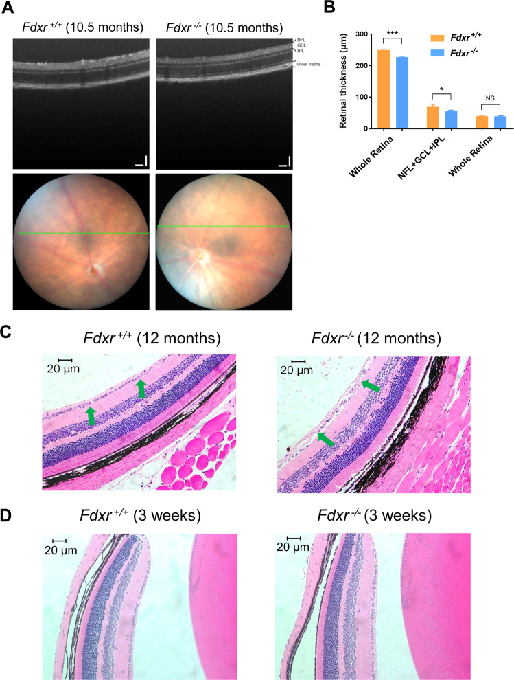
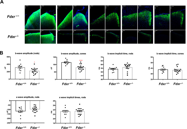
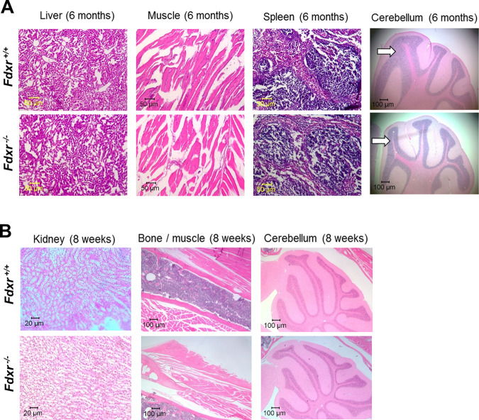
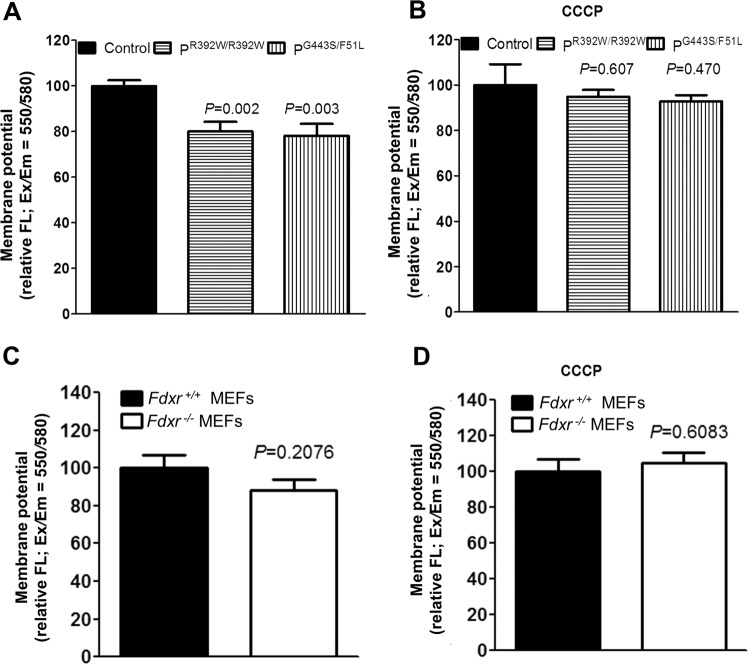
The mitochondrial flavoprotein ferredoxin reductase (FDXR) is required for biogenesis of iron-sulfur clusters and for steroidogenesis. Iron-sulfur (Fe-S) clusters are ubiquitous cofactors essential to various cellular processes, and an increasing number of disorders are associated with disruptions in the synthesis of Fe-S clusters. Our previous studies have demonstrated that hypomorphic mutations in FDXR cause a novel mitochondriopathy and optic atrophy in humans and mice, attributed in part to reduced function of the electron transport chain (ETC) as well as elevated production of reactive oxygen species (ROS). Inflammation and peripheral neuropathy are also hallmarks of this disease. In this paper, we demonstrate that FDXR mutation leads to significant optic transport defects that are likely to underlie optic atrophy, a major clinical presentation in FDXR patients, as well as a neurodegenerative loss of cells in the central nervous system (CNS). Molecular analysis indicates that FDXR mutation also leads to mitochondrial iron overload and an associated depolarization of the mitochondrial membrane, further supporting the hypothesis that FDXR mutations cause neurodegeneration by affecting FDXR's critical role in iron homeostasis.


Conflict of interest statement

The authors declare that they have no conflict of interest.

Figures

Fig. 1
Fig. 1. Fdxr-mediated optic atrophy and RGC loss.
a Optic disc and OCT scanning of P120 mice. The green line in optic disc images indicates the position of OCT scanning. Scale bars: 100 μm. b Quantification of retinal thickness from OCT scanning images of retinas from Fdxr+/+ and Fdxr−/− mice (*P < 0.05, **P < 0.01, ***P < 0.001); n = 14 retinal images derived from two animals, per genotype). c H&E-stained sagittal sections through the optic disc show that the number of retinal ganglion cells (RGCs) in the Ganglion Cell Layer (green arrows) are markedly reduced in H&E sections of eyes from 12-month-old Fdxr/− mutant mice, as compared with Fdxr+/+ control mice. d H&E staining of retinal sections from mice at 3 weeks of age indicates no difference in number of RGCs between Fdxr+/+ and Fdxr/− animals.
Fig. 2
Fig. 2. Fdxr mutation causes functional defects in retinal neurons.
a Anterograde axonal transport of mouse retinal ganglion cells (RGCs) was measured in the mouse eyes. Seven-month-old Fdxr+/+ control and Fdxr−/− mutant mice were anesthetized with an inhalation of 2.5% isoflurane. Brains were cryo-sectioned through the superior colliculi with a thickness of 50 µm. Transport of CTB-Fluor 488 from the eye to the superior colliculi in the brain was greatly reduced in the Fdxr−/− mutant mice, as compared with the transport in the Fdxr+/+ control mice. Scale bars: 100 μm. b Electroretinography was performed in 4-month-old Fdxr+/+ and Fdxr−/− mouse eyes. After light stimulation, both ocular photoreceptor rods and cones showed decreased b-wave amplitude in the Fdxr/ mutants relative to control mice.
Fig. 3
Fig. 3. Pathology assessment of tissues from Fdxr mutant mice.
a H&E staining of Fdxr mutant mouse tissues from the liver, muscle, spleen, and brain/cerebellum from 6-month-old Fdxr−/− mice as compared with Fdxr+/+ control mice. The cerebellar granular layers for the mutant and control samples are indicated with white arrows. In general, the cerebellar granular layer of the mutant sample shows a reduced volume (relative to the other cerebellar layers) than what is observed in the control sample. b H&E staining of tissues from the kidneys, bone, and muscle, and cerebellum from 2-month-old Fdxr/ mice as compared with Fdxr+/+ control mice.
Fig. 4
Fig. 4. Gait abnormalities and peripheral nerve conduction defects of FdxrR389Q/R389Q mutant mice relative to wild-type (WT) mice.
Significant impairments in rear-gait dynamics (slower swing times and shorter time in stance position) were observed in Fdxr mutants relative to WT controls independent of sex (b, d), whereas front-gait dynamics appeared unaffected (a, c). Altered rear-gait dynamics in FdxrR389Q/R389Q were associated with abnormal hindpaw position and significantly impaired sciatic function as measured by pawprint angle (e) and impaired hindlimb base support as measured by increased hindpaw width (f). To confirm the underlying neurophysiology of these behavioral defects, CMAP recordings were taken from sciatic nerves of Fdxr+/+ (black) and Fdxr−/− (red) mice at 2 months of age (g). Arrows indicate onset of electrical stimulation of the sciatic nerve during recording. Conduction velocities (CVs) were measured from recorded waves using a 2 mA (h) or 4 mA (i) electrical stimulation of the sciatic nerve (n = 16 for Fdxr+/+ and Fdxr/− mice; P < 0.001). Data are representative of two independent experiments.
Fig. 5
Fig. 5. FDXR mutation regulates iron metabolism.
Iron levels (Fe2+ plus Fe3+) were measured by QuantiChrom iron assay in extracts from control and patient fibroblasts carrying FDXRR392W/R392W or FDXRG443S/F51L mutations (a, c), and in extracts from Fdxr−/− mutant mice and Fdxr+/+ littermates (b, d). Iron levels in mitochondrial extracts (a, b) were reduced in FDXR mutant cells and tissues. In contrast, cytoplasmic extracts showed no significant differences between FDXR mutant cells/tissues and their corresponding controls (c, d). e Fdxr deficiency leads to iron overload at the age of 10.5 months in mice. Tissues from Fdxr/− mutant mice and Fdxr+/+ littermates were stained with Prussian blue to measure iron accumulation. The results are shown for the liver, heart, muscle, kidney, and spleen. f Prussian blue staining was also carried on various brain tissues from Fdxr−/− and Fdxr+/+ mice. The results show increased iron levels in the cerebellum, cortex, lateral ventricle, thalamus, and olfactory bulb.
Fig. 6
Fig. 6. FDXR mutation leads to a reduction in mitochondrial membrane potential.
a The mitochondrial membrane potential (ΔΨm) was measured in patient fibroblasts with FDXRR392W/R392W or FDXRG443S/F51L mutations and control fibroblasts, using a TMRE-based assay system. ΔΨm was determined according to the relative fluorescence intensity at Ex/Em = 550/580, in the absence of 10 µM of carbonyl cyanide 3-chlorophenylhydrazone (CCCP). b Mitochondrial membrane potential of the cell lines from panel a, as measured in the presence of 10 µM of carbonyl cyanide 3-chlorophenylhydrazone (CCCP), is shown as a control. c Mitochondrial membrane potential, as determined in mouse embryonic fibroblasts (MEFs) from Fdxr−/− mutant mice and Fdxr+/+ control littermates. d Mitochondrial membrane potential of the cell lines from panel d, as measured in the presence of 10 µM of CCCP, is shown as a positive control. The average of three to five determinations is shown for each cell line.

References

    1. Wang J, Pantopoulos K. Regulation of cellular iron metabolism. Biochemical J. 2011;434:365–381. doi: 10.1042/BJ20101825. - DOI - PMC - PubMed
    1. Campuzano V, et al. Friedreich’s ataxia: autosomal recessive disease caused by an intronic GAA triplet repeat expansion. Science. 1996;271:1423–1427. doi: 10.1126/science.271.5254.1423. - DOI - PubMed
    1. Rotig A, et al. Aconitase and mitochondrial iron-sulphur protein deficiency in Friedreich ataxia. Nat. Genet. 1997;17:215–217. doi: 10.1038/ng1097-215. - DOI - PubMed
    1. Cavadini P, O’Neill HA, Benada O, Isaya G. Assembly and iron-binding properties of human frataxin, the protein deficient in Friedreich ataxia. Hum. Mol. Genet. 2002;11:217–227. doi: 10.1093/hmg/11.3.217. - DOI - PubMed
    1. Lodi R, et al. Deficit of in vivo mitochondrial ATP production in patients with Friedreich ataxia. Proc. Natl Acad. Sci. USA. 1999;96:11492–11495. doi: 10.1073/pnas.96.20.11492. - DOI - PMC - PubMed

Publication types

MeSH terms