. 2020 Jun;582(7811):283-288.
doi: 10.1038/s41586-020-2323-8. Epub 2020 May 20.

poly(UG)-tailed RNAs in genome protection and epigenetic inheritance

Affiliations

poly(UG)-tailed RNAs in genome protection and epigenetic inheritance

Aditi Shukla et al. Nature. 2020 Jun.

Erratum in

Abstract

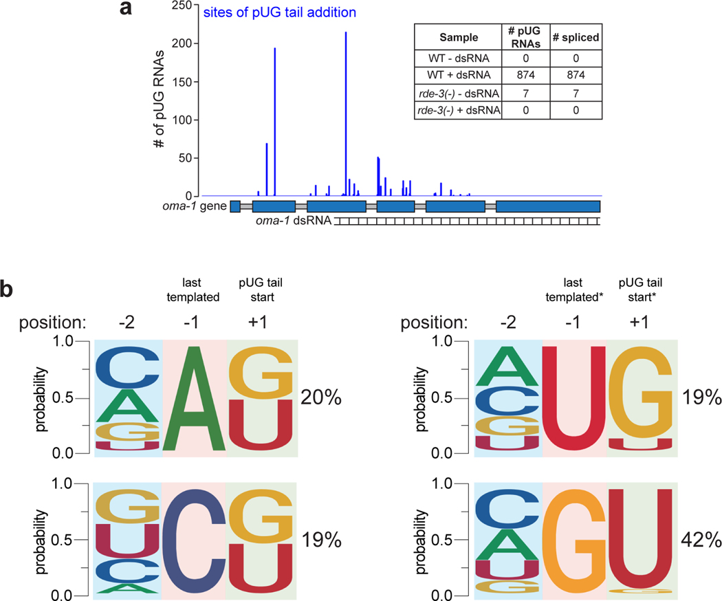
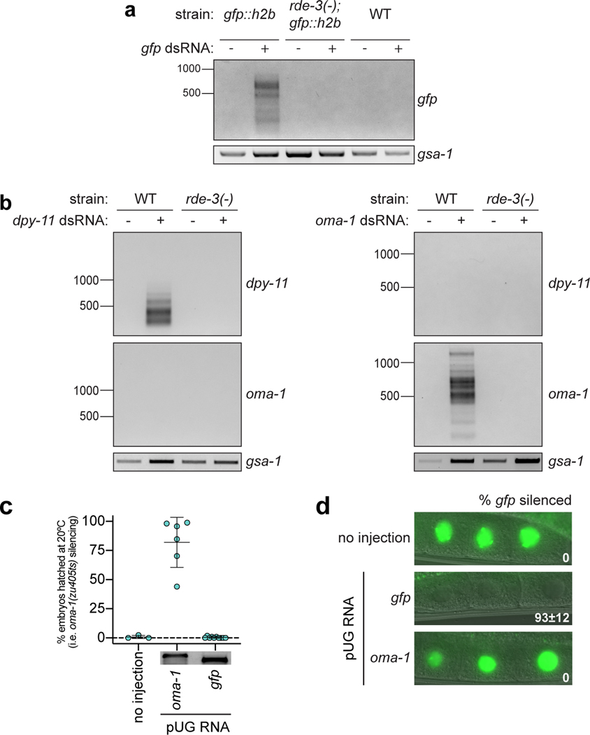
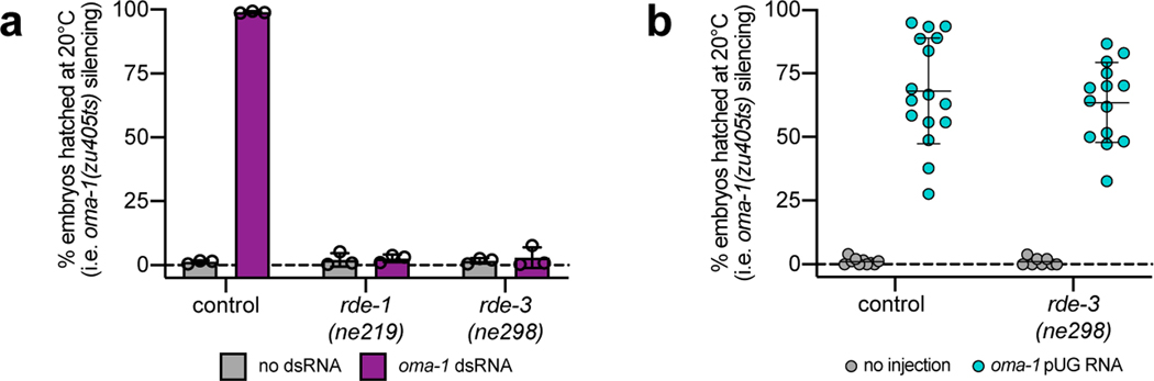
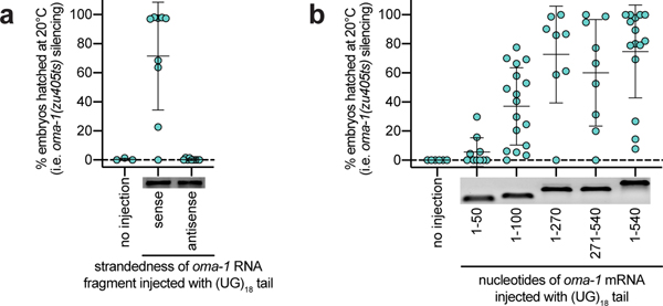
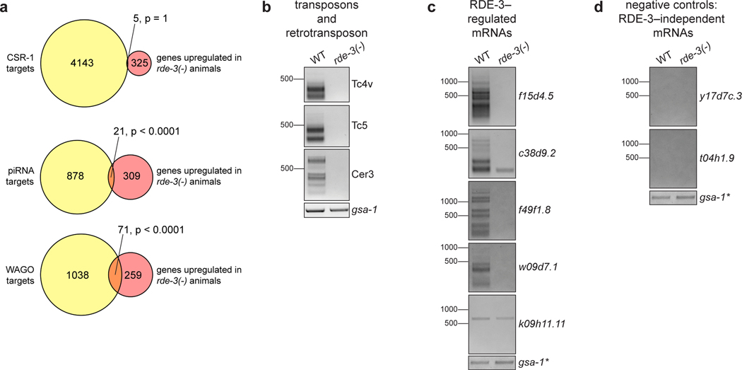
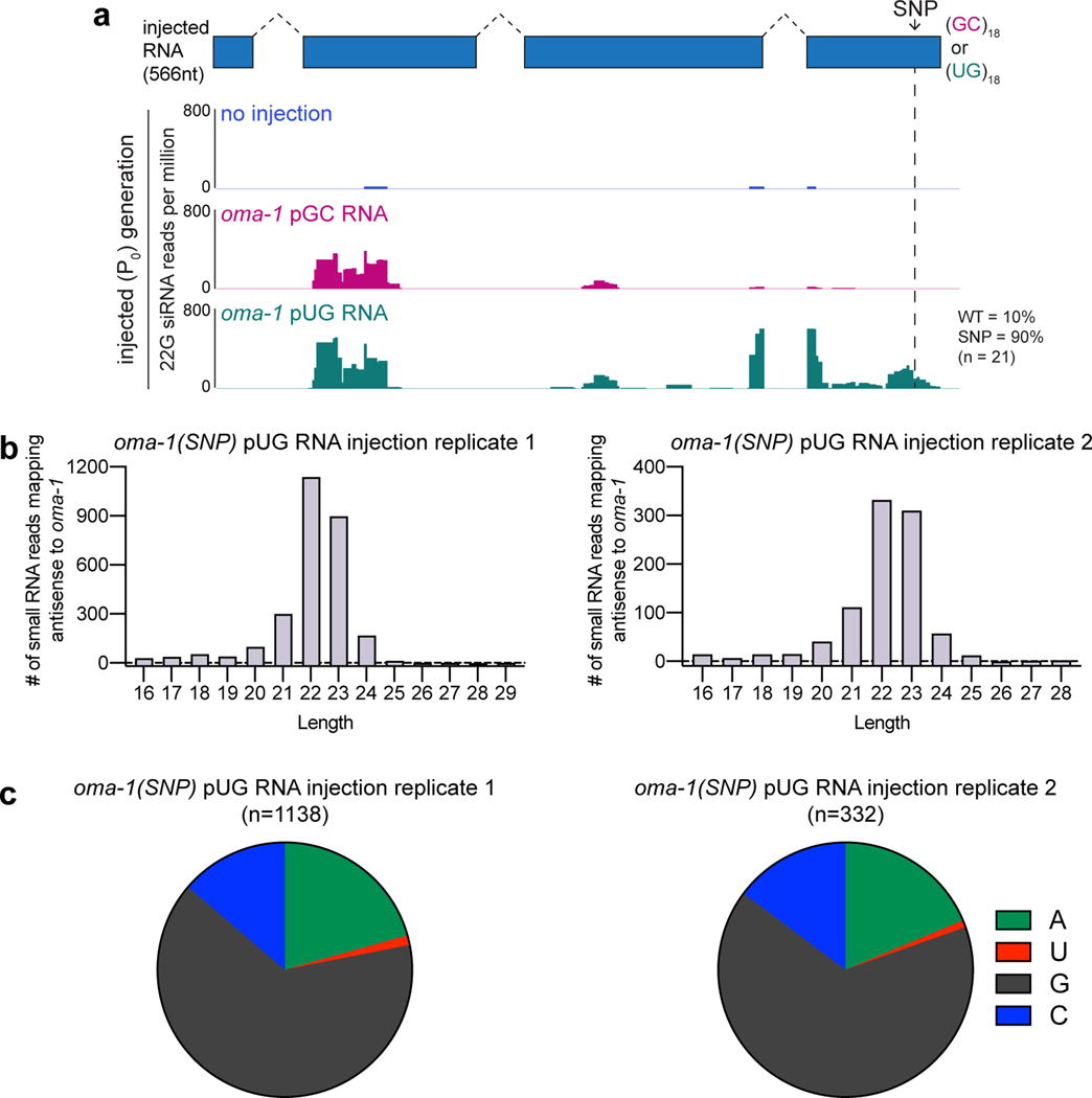
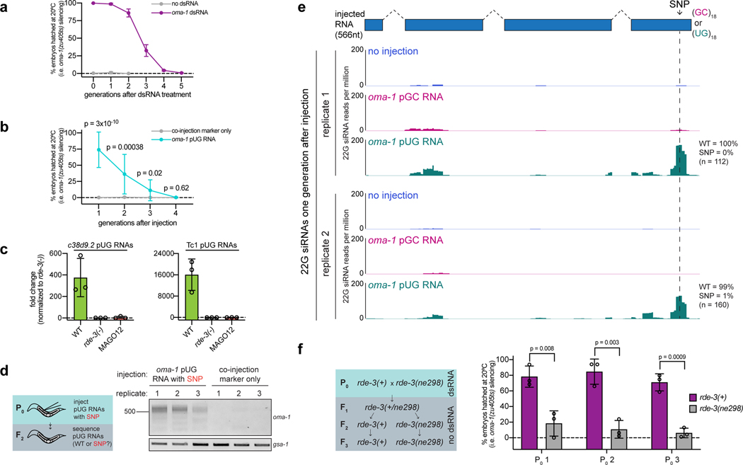
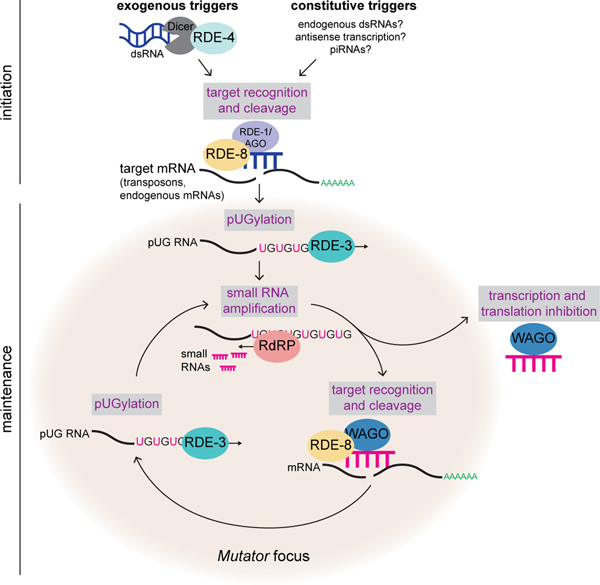
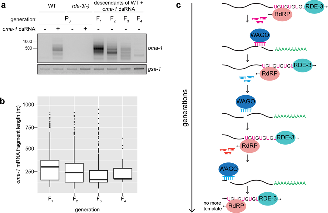
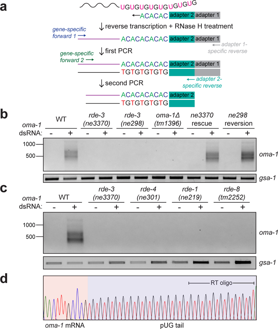
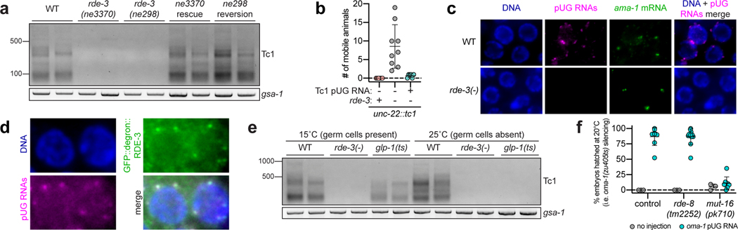
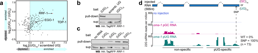
Mobile genetic elements threaten genome integrity in all organisms. RDE-3 (also known as MUT-2) is a ribonucleotidyltransferase that is required for transposon silencing and RNA interference in Caenorhabditis elegans1-4. When tethered to RNAs in heterologous expression systems, RDE-3 can add long stretches of alternating non-templated uridine (U) and guanosine (G) ribonucleotides to the 3' termini of these RNAs (designated poly(UG) or pUG tails)5. Here we show that, in its natural context in C. elegans, RDE-3 adds pUG tails to targets of RNA interference, as well as to transposon RNAs. RNA fragments attached to pUG tails with more than 16 perfectly alternating 3' U and G nucleotides become gene-silencing agents. pUG tails promote gene silencing by recruiting RNA-dependent RNA polymerases, which use pUG-tailed RNAs (pUG RNAs) as templates to synthesize small interfering RNAs (siRNAs). Our results show that cycles of pUG RNA-templated siRNA synthesis and siRNA-directed pUG RNA biogenesis underlie double-stranded-RNA-directed transgenerational epigenetic inheritance in the C. elegans germline. We speculate that this pUG RNA-siRNA silencing loop enables parents to inoculate progeny against the expression of unwanted or parasitic genetic elements.


Conflict of interest statement

Competing interests

M.W. has a patent (US20160145666A1) through Wisconsin Alumni Research Foundation (Madison, WI) for methods, kits, and compositions of matter relating to poly(UG) polymerases.

Figures

Extended Data Fig. 1.
Extended Data Fig. 1.. Analysis of oma-1 pUGylation sites.
a, Illumina MiSeq was performed (n=1 biological experiment) on oma-1 pUG PCR products derived from WT and rde-3(−) animals, +/− oma-1 dsRNA. # of sequenced pUG RNAs (y-axis) mapping to each pUGylation site (x-axis) is shown. Inset: total number of sequenced oma-1 pUG RNAs from indicated samples and total number of these sequenced pUG RNAs in which the oma-1 sequence was spliced. b, MiSeq-sequenced oma-1 pUG RNAs were sorted into four groups based on the nucleotide (nt) at the last templated position (−1) of the oma-1 mRNA. The % of oma-1 pUG RNAs (MiSeq reads) with each nt in the −1 position is shown. Logo analysis was then performed on each of the four groups to determine the probability of finding each nucleotide at the first position of the pUG tail (+1), as well as at the second-to-last templated nucleotide of oma-1 (−2). This analysis showed that if the last templated nucleotide of the oma-1 mRNA fragment was an A or a C, then RDE-3 was equally likely to add a U or a G as the first nucleotide of an elongating pUG tail. If, however, the last templated nt was a U or G, then RDE-3 preferentially added a G or U, respectively, as the first nt in an elongating pUG tail. *Note: To perform the analyses in this figure, we assumed that if a U or G could have been genomically encoded, then it was. If, instead, RDE-3 added the U or G shown in the −1 position as the first nucleotide of the pUG tail, then these data show that the second nucleotide that RDE-3 prefers to add is a G after a U or a U after a G. CCA-adding rNT enzymes modify the 3’ termini of tRNAs with non-templated CCA nts. The mechanism by which these enzymes add non-templated nonhomopolymeric stretches of nts is thought to involve allosteric regulation of the nt binding pocket by the 3’ nt of a substrate tRNA. A similar mechanism may explain how RDE-3 can add pUG tails to its mRNA substrates. For instance, when the 3’ nt of an RDE-3 substrate is a U, the rNTP binding pocket of RDE-3 might adopt a structure that preferentially binds G and vice versa when 3’ nt of an RDE-3 substrate is a G. Such a model could explain how a single rNT enzyme adds perfectly alternating U and G nts to RNA substrates. There are also alternative models for how RDE-3 might add pUG tails to an RNA. These include: 1) the existence a poly(AC) nucleic acid template used by RDE-3 during pUG tail synthesis, 2) the existence of one or more rNTs that cooperate with RDE-3 to produce pUG tails, or 3) the possibility that RDE-3 binds and incorporates UG or GU dinucleotides. We disfavor the first two possibilities as these models are difficult to reconcile with the observation that RDE-3 adds UG repeats to tethered RNAs in yeast or in Xenopus oocytes. The third proposed model may be true, but because our sequencing shows that pUG tails can initiate with either a U or G (this figure, Supplementary Table 1), then RDE-3 would need to be able to bind both UG and GU dinucleotides. Determining the mechanism by which RDE-3 adds pUG tails will likely involve structural studies and/or in vitro pUGylation assays using recombinant RDE-3 protein.
Extended Data Fig. 2.
Extended Data Fig. 2.. RNAi-triggered pUGylation and pUG RNA–directed gene silencing are general and sequence-specific.
a,gfp::h2b, rde-3(−); gfp::h2b and WT (no gfp::h2b) animals were fed E.coli expressing either empty vector control or gfp dsRNA. b, WT and rde-3(−) animals were fed E.coli expressing empty vector control and either oma-1 or dpy-11 dsRNA. For a and b, gfp, dpy-11 and oma-1 pUG RNAs were detected using the assay outlined in Fig. 1a. Data is representative of 3 biologically independent experiments. c, rde-1(ne219); oma-1(zu405ts) animals were injected with either an oma-1 (n=6) or gfp (n=10) pUG RNA. n=3 for no injection. % embryos hatched was scored for the progeny of injected animals. Inset: injected RNAs run on a 2% agarose gel to assess RNA integrity. Error bars: s.d. of the mean. d, rde-1(ne219); gfp::h2b animals were injected with either an oma-1 or gfp pUG RNA (n=10 for both, 3 for no injection). Mean % progeny with gfp::h2b silenced is indicated ± standard deviation (s.d.). For c and d, all pUG tails were 36nt in length.
Extended Data Fig. 3.
Extended Data Fig. 3.. RDE-3–mediated pUGylation is necessary for RNAi.
a, Animals of the indicated genotypes (all harboring the oma-1(zu405ts) mutation) were treated +/− oma-1 dsRNA. For each experiment, % embryos hatched was scored at 20°C and averaged for 6 individual animals per treatment for each genotype. rde-1(ne219) mutants, which cannot respond to dsRNA, serve as a control for this experiment. Error bars: s.d. of the mean for 3 biologically independent experiments. b, Control or rde-3(ne298) animals (all rde-1(ne219); oma-1(zu405ts) background) were injected with oma-1 pUG RNAs and % embryos hatched was scored at 20°C. n=10 noninjected and 16 injected animals for control. n=8 noninjected and 14 injected animals for rde-3(ne298). Error bars: s.d. of the mean.
Extended Data Fig. 4.
Extended Data Fig. 4.. pUG tails must be appended to sense RNAs of >50nt for functionality.
rde-1(ne219); oma-1(zu405ts) animals were injected with: a, an oma-1 pUG RNA consisting of the sense or antisense strand of the same 541nt long oma-1 mRNA fragment (beginning at the aug) with a 36nt 3’ pUG tail (n=9 for both; n=3 for no injection). b, oma-1 pUG RNAs consisting of oma-1 mRNA fragments of varying lengths (with position 1 starting at the aug of the oma-1 mRNA sequence) all appended to a 36nt pUG tail. n=6 (no injection), 10 (1–50), 17 (1–100), 8 (1–270), 9 (271–541) and 15 (1–541). For a and b, % embryonic arrest was scored at 20°C. Error bars: s.d. of the mean.
Extended Data Fig. 5.
Extended Data Fig. 5.. Endogenous targets of pUGylation in C. elegans.
a, mRNAs upregulated in rde-3(−) mutants (Supplementary Table 2) were compared to published lists of: (1) RNAs targeted by CSR-1bound endo-siRNAs, (2) piRNA-targeted mRNAs (based on predictive and experimental approaches), and (3) WAGO-class mRNAs. p-values were generated using a one-sided Fisher’s exact test. This analysis showed statistically significant overlap between the mRNAs upregulated in rde-3(−) mutants and both piRNAs targets and WAGO-class mRNAs. b-d, Total RNA was extracted from WT or rde-3(−) animals. The assay outlined in Fig. 1a was used to detect pUG RNAs for b, two DNA transposons (Tc4v and Tc5) and a retrotransposon (Cer3) that were significantly upregulated in rde-3(−) animals; c, predicted protein-coding mRNAs that were significantly upregulated in rde-3(−) animals; and d, two randomly selected mRNAs whose expression does not change in rde-3(−) mutants. Data is representative of 3 biologically independent experiments. *Note: the same RT samples were used for panels c and d and, therefore, the gsa-1 loading control is the same for both panels.
Extended Data Fig. 6.
Extended Data Fig. 6.. Mutator foci likely coordinate pUG RNA biogenesis within germ cells.
a,dpy-11 and oma-1 pUG PCR (Fig. 1a) were performed on total RNA from glp-1(q224/ts) animals grown at 15°C (germ cells present) or 25°C (<99% of germ cells), +/− oma-1 and dpy-11 dsRNA. Data is representative of 2 biologically independent experiments. *Note: the samples in a are the same as those used in Fig. 3e and, therefore, the gsa-1 loading control is the same. b, oma-1 pUG PCR was performed on total RNA extracted from wild-type, rde-3(−), and mut-16(pk710) animals, +/− oma-1 dsRNA. Data is representative of 4 biologically independent experiments. c, qRT-PCR was used to quantify levels of oma-1 pUG RNAs in wild-type, rde-3(−), and mut-16(pk710) animals, +/− oma-1 dsRNA. Data is represented as fold change in the levels of oma-1 pUG RNAs +/− oma-1 dsRNA (y-axis) for each strain (x-axis). n=3 biologically independent samples per treatment for each strain. Error bars: s.d. of the mean. d, qRT-PCR was used to quantify levels of Tc1 pUG RNAs in wild-type, rde-3(−), and mut-16(pk710) animals. Note: the RNA samples used for d are the same as those used in c, except that the data for +/− oma-1 dsRNA samples were pooled for each strain. n=6 biologically independent samples for each strain. Error bars: s.d. of the mean. The analyses in c and d showed that mut-16 mutant animals produced more oma-1, but fewer Tc1, pUG RNAs, than wild-type animals. The increased levels of oma-1 pUG RNAs in mut-16(pk710) animals was also suggested by the gel in b. Together, these data suggest that Mutator foci likely have an important role in coordinating pUG RNA biogenesis in germ cells, as pUG RNA levels become misregulated in mut-16(pk710) mutants.
Extended Data Fig. 7.
Extended Data Fig. 7.. pUG RNAs are templates for RdRPs.
a, A biological replicate of the experiment shown in Fig. 4d was performed. oma-1(SNP) pUG or pGC RNAs were injected into rde-1(ne219); oma-1(zu405ts) germlines. SNP location is indicated with the dotted line. Injected animals were collected 1–4 hours after injection, total RNA was isolated and small RNAs (20–30nt) were sequenced. Distribution of 22G siRNAs mapping antisense to oma-1 is shown, with 22G siRNA reads normalized to reads per million total reads. oma-1 pUG (but not pGC) RNA injection triggered 22G siRNA production near the site of the pUG tail (“pUG-specific” 22G siRNAs). For unknown reasons, both pUG and pGC RNA injections triggered small RNA production ≅400bp 5’ of either tail. b, Length distribution of small RNA reads mapping antisense to oma-1 is shown for small RNAs sequenced after oma-1(SNP) pUG RNA injections (Fig. 4d and a). c, Proportion of 22nt long small RNAs mapping antisense to oma-1 containing 5’ adenine, uracil, guanine, or cytosine is shown.
Extended Data Fig. 8.
Extended Data Fig. 8.. De novo pUGylation events in progeny are required for TEI.
a,oma-1(zu405ts) animals were fed bacteria expressing empty vector control or oma-1 dsRNA and % embryos hatched at 20°C was scored for 6 generations. Error bars represent s.d. of the mean of three biologically independent experiments. For each experiment, % embryos hatched at 20°C was averaged for 6 individual animals per treatment for each genotype. b, rde-1(ne219); oma-1(zu405ts) animals were injected with co-injection marker alone (n=12) or co-injection marker + oma-1 pUG RNA (n=19) and % embryos hatched at 20°C was scored for four generations in lineages of animals established from injected parents (see Methods for details of experimental setup). Error bars: s.d. of the mean. p-values: two-tailed unpaired Student’s t-test. c, c38d9.2 and Tc1 pUG RNA expression quantified in embryos harvested from wild-type, rde-3(−) or MAGO12 animals using qRT-PCR. Fold change normalized to rde-3(−). Each point (n) represents a biologically independent replicate, n=3 independent replicates/strain. Error bars: s.d. of the mean. d, Same experiment as Fig. 5d. rde-1(ne219); oma-1(zu405ts) animals were injected with an oma-1(SNP) pUG RNA or with co-injection marker only. Co-injection marker-expressing F1 progeny were picked and allowed to lay their F2 broods. oma-1 pUG PCR was performed on total RNA from F2 progeny. Shown is data from three biological replicates. e, Two biological replicates of small RNAs sequenced from the progeny of rde-1(ne219); oma-1(zu405ts) animals injected with oma-1(SNP) pUG or pGC RNAs are shown. Dotted line indicates the location of the SNP incorporated into oma-1. Distribution of 22G siRNAs mapping antisense to oma-1 is shown, with 22G siRNA reads normalized to reads per million total reads. In Fig. 4d and Extended Data Fig. 7a, small RNAs were sequenced 1–4 hours after injection and 100% of 22G siRNAs antisense to the region of the engineered SNP in oma-1 were found to encode the complement of the SNP. Shown here, <1% of 22G siRNAs from progeny of injected animals encoded the SNP complement. Note: siRNAs mapping near the pUG tail were observed only after oma-1(SNP) pUG RNA injection (pUG-specific siRNAs). For unknown reasons, both oma-1(SNP) pUG and pGC RNAs triggered small RNA production 5’ of the pUG-specific siRNAs. It is possible that these siRNAs were triggered by systems that respond to foreign RNAs, such as the piRNA system. Further work will be needed to ascertain the etiology of these siRNAs. f, Same experiment as Fig. 5e. oma-1(zu405ts) hermaphrodites were fed oma-1 dsRNA and crossed to rde-3(ne298); oma-1(zu405ts) males. F2 progeny from this cross were genotyped for rde-3(ne298). WT and rde-3(ne298) homozygous F3 progeny were phenotyped for % embryonic arrest at 20°C. 3 biologically independent crosses (P0 1–3) were performed. Error bars: s.d. of the mean. p-values: two-tailed unpaired Student’s t-tests.
Extended Data Fig. 9.
Extended Data Fig. 9.. Working model for pUG RNA/siRNA cycling during RNAi.
Initiation: exogenous and constitutive (i.e. genomically encoded such as dsRNA, piRNAs) triggers direct RDE-3 to pUGylate RNAs previously fragmented by factors in the RNAi pathway. Maintenance: pUG RNAs are templates for 2° siRNA synthesis by RdRPs. Argonaute proteins (termed WAGOs) bind 2° siRNAs and: 1) target homologous RNAs for transcriptional and translational silencing,,,, as well as 2) direct the cleavage and de novo pUGylation of additional mRNAs. In this way, cycles of pUG RNA-based siRNA production and siRNA-directed mRNA pUGylation maintain silencing over time and across generations. This model shows germline perinuclear condensates termed Mutator foci as the likely sites of pUG RNA biogenesis in germ cells for several reasons. RDE-3 localizes to Mutator foci and we show in Fig. 3d that endogenous pUG RNAs localize to Mutator foci. The fact that enzyme and enzyme product both localize to Mutator foci suggests that Mutator foci may be sites of RNA pUGylation. In addition, while pUG RNAs are still made in mut-16 mutants (Extended Data Fig. 6b-d), which lack Mutator foci, the levels of both dsRNA-triggered and endogenous pUG RNAs are misregulated. Thus, while RDE-3 still has enzymatic activity in the absence of Mutator foci, these perinuclear condensates are likely coordinating target recognition and pUGylation in wild-type animals. Indeed, both the endonuclease RDE-8, which cleaves mRNAs targeted by dsRNA, and the RdRP RRF-117 also localize to Mutator foci, further suggesting that pUG RNA/siRNA cycling occurs in Mutator foci. Previous studies have shown that animals lacking RDE-3 still produce some 22G endo- siRNAs, including 22G siRNAs that associate with the Argonaute CSR-1 and whose biogenesis depends upon the RdRP EGO-1,. Thus, EGO-1 may also produce some 22G siRNAs via a pUG RNA-independent mechanism. A previous study showed that, in rrf-1 mutants that lack germlines, sel-1 RNAi causes a small fraction of sel-1 mRNA fragments to be uridylated in a largely RDE-3–dependent manner in the soma. This data suggests that, in somatic tissues, RDE-3 may add non-templated Us to the 3’ termini of mRNA fragments generated during RNAi. It was proposed that this uridylation may be important for turnover or decay of RNAi targets. Our work, combined with this earlier data about RDE-3–dependent uridylation, suggests two models. First, RDE-3 may possess two distinct catalytic activities: uridylation and pUGylation. According to this model, RDE-3 might add Us or UGs depending on context (e.g. cell/tissue-type or developmental timing). Alternatively, the mRNA uridylation observed in the soma could depend upon RDE-3 and the pUGylation system, but may be mediated by another, currently unknown, poly(U) polymerase.
Extended Data Fig. 10.
Extended Data Fig. 10.. pUG RNA shortening may act as a brake on TEI.
a, The gel shown is the same as in Fig. 5a, except that oma-1 pUG RNAs from the P0 generation are included for WT and rde-3(−) animals. Data is representative of 3 biologically independent experiments. b, oma-1 pUG RNA reads from MiSeq (n=1 biological experiment) were mapped to oma-1 and the length of the oma-1 mRNA portion of each pUG RNA read was determined (y-axis). Shown is a Box and Whisker plot representing the interquartile range (IQR, box) and median (line in the box) of lengths at the indicated generations after dsRNA treatment. The y-axis starts at the aug of the oma-1 mRNA. The whiskers extend to values below and above 1.5*IQR from the first and third quartiles, respectively. Data beyond the end of the whiskers are outliers and plotted as points. These data support the gel in a, showing that pUG RNAs get shorter in each generation during RNAi-triggered TEI. c, A “ratchet” model to explain pUG RNA shortening. pUG RNA shortening may be due to the 3’→5’ directionality of RdRPs, which, during the maintenance phase of pUG/siRNA cycling (see model in Extended Data Fig. 9), causes each turn of the pUG/siRNA cycle to trigger cleavage and pUGylation of target mRNAs at sites more 5’ than in the previous cycle. Eventually, pUG RNAs are too short to act as RdRP templates, cycling cannot be maintained and silencing ends. Additional support for the ratchet model comes from Fig. 5c, which shows that RNAi-triggered pUG RNAs are longer in MAGO12 mutant animals than in wild-type animals. Note: the P0 generation animals in Fig. 5c were exposed to dsRNA continuously from embryos to adulthood, when they were harvested. These longer pUG RNAs are likely due to continued initiation of pUGylation triggered by the exogenously provided dsRNA without downstream pUG/siRNA cycling. In the absence of this cycling, pUG RNA shortening does not occur. Finally, a number of recent studies in C. elegans have reported transgenerational inheritance of acquired traits, which lasts 3–4 generations. As shown in a, the expression of oma-1 RNAi–directed pUG RNAs also perdures for 3–4 generations. These shared generational timescales of inheritance hint that the inheritance of acquired traits in C. elegans may be mediated by pUG RNAs whose generational “half-life” is limited to 3–4 generations due to the built-in brake on TEI provided by pUG RNA shortening.
Figure 1.
Figure 1.. pUG tails are added to mRNA fragments in vivo.
a, Assay to detect gene-specific pUG RNAs. Note: (AC)9 RT oligo can anneal anywhere along the pUG tail. b, oma-1 pUG PCR on total RNA isolated from animals of indicated genotypes, +/− oma-1 dsRNA (RNAi). rde-3 mutants were rescued as described in Main text and Methods. gsa-1, which has an 18nt long genomically encoded pUG repeat in its 3’UTR, is a loading control. Wild-type (WT) vs. rde-3(ne3370) and WT vs. rde-3(ne298) data is representative of >10 and 2 biologically independent experiments, respectively. c, oma-1 pUG PCR on total RNA from animals of indicated genotypes, +/− oma-1 dsRNA. Data is representative of 3 biologically independent experiments. d, Sanger sequencing chromatogram (red=T, black=G, blue=C, green=A) of an oma-1 pUG PCR product.
Figure 2.
Figure 2.. pUG tails convert inert RNA fragments into agents of gene silencing.
a-c, To control for potential dsRNA contamination in in vitro transcription reactions, RNAs were injected into rde-1(ne219) mutants, which cannot respond to dsRNA. a, Fluorescence micrographs showing −1 to −3 oocytes of adult progeny of rde-1(ne219); gfp::h2b animals injected in the germline with in vitro transcribed RNAs consisting of the first 369nt of gfp mRNA with the indicated 3’ terminal repeats. Mean % progeny with gfp::h2b silenced is indicated ± standard deviation (s.d.). # of injected animals (n) = 3 (no injection); 9 [gfp dsRNA, (AU)18]; 10 [no tail, (GC)18, (AC)18]; and 16 [(UG)18]. b-c, oma-1(zu405ts) animals lay arrested embryos at 20°C unless oma-1(zu405ts) is silenced. Adult rde-1(ne219); oma-1(zu405ts) animals were injected in the germline with in vitro transcribed RNAs consisting of the first 541nt of oma-1 mRNA with b, indicated 3’ terminal repeats or c, varying 3’ pUG tail lengths; different 3’ UG repeat sequences; or with (UG)18 on the 3’ end, 5’ end or in the middle of the oma-1 mRNA. For all oma-1 pUG RNA injection data, each point represents % hatched embryos laid by 5 progeny derived from one injected animal at 20°C (see Methods). Error bars: s.d. of the mean. Insets: injected RNAs run on 2% agarose gel to assess RNA integrity. For b, n=6 (no injection); 10 (oma-1 dsRNA, no tail); 12 [(UG)18]; 9 [(GC)18]; and 8 [(AU)18, (AC)18]. For c, n=9 [no injection #1, (U18G18), (UUGG)9]; 10 [(UG)0, 1, 8, 14, scrambled UGs, 3’ (UG)18, internal (UG)18]; 8 [(UG)5, 5’ (UG)18]; 12 [(UG)18 #1]; 5 [(UG)18 #2]; 11 [(UG)40]; 6 (no injection #2) and 3 (no injection #3).
Figure 3.
Figure 3.. Endogenous pUG RNAs exist and localize to germline Mutator foci.
a, Tc1 pUG PCR (Fig. 1a) on total RNA from two replicates of indicated genotypes (rescue/reversion as in Fig. 1b). WT vs. rde-3(ne3370) and WT vs. rde-3(ne298) is representative of >5 and 2 biologically independent experiments, respectively. b, 18 and 22 rde-3(−); unc-22::tc1 animals were injected with Tc1 pUG RNA + co-injection marker or co-injection marker alone, respectively. Each data point (n) represents # of mobile progeny (indicating Tc1 mobilized from unc-22) laid by 25 randomly pooled co-injection marker–expressing progeny derived from injected animals (see Methods). n=9 for co-injection marker only, 6 for Tc1 pUG RNA + co-injection marker, 6 for noninjected rde-3(+); unc-22::tc1. Error bars: s.d. of the mean. c-d, Fluorescence micrographs of adult pachytene stage germ cell nuclei. DNA stained with 4’,6-diamidino-2-phenylindole (DAPI, blue). Data is representative of 3 biologically independent experiments. c, RNA FISH to detect pUG RNAs on germlines dissected from WT or rde-3(−) animals using (AC)9 DNA oligo conjugated to Alexa 647 (magenta). Positive control: RNA FISH to detect ama-1 mRNA (green). d, pUG RNA FISH (magenta) combined with immunofluorescence to detect GFP::degron::RDE-3 (green). e, Tc1 pUG PCR on total RNA isolated from replicates of glp-1(q224/ts) animals grown at 15°C (germ cells present) or 25°C (<99% of germ cells). Data is representative of 2 biologically independent experiments. f, Control, rde-8(tm2252) or mut-16(pk710) animals (all rde-1(ne219); oma-1(zu405ts) background) were injected with oma-1 pUG RNAs (n=9, 12 and 8, respectively) and % embryos hatched scored at 20°C. n=3 for all no injection. Error bars: s.d. of the mean.
Figure 4.
Figure 4.. pUG RNAs are templates for RdRPs.
a, LC-MS/MS was performed on proteins that bound to 5’ biotinylated RNA oligos [(UG)5, (UG)18 or 36 scrambled UGs] conjugated to streptavidin beads. Shown is a scatter plot of log2-transformed fold enrichment in (UG)18 vs. scrambled UG pull-down (x-axis) and (UG)18 vs. (UG)5 pull-down (y-axis). Proteins enriched ≥2-fold in (UG)18 vs. beads-only pull-down are plotted. b-c, Indicated 5’ biotinylated RNA oligos were conjugated to streptavidin beads and incubated with extracts from animals expressing HA::tagRFP::RRF-1. Bead-bound material (pull-down) and supernatant (sup) were subjected to ɑ-HA immunoblotting. Data is representative of 2 biologically independent experiments. d, rde-1(ne219); oma-1(zu405ts) animals were injected with SNP-containing (dotted line) oma-1 (oma-1(SNP)) pUG or pGC-tailed RNAs and collected 1–4 hours later; small RNAs (20–30nts) were sequenced. Distribution of 22G siRNAs mapping antisense to oma-1 is shown, with 22G siRNA reads normalized to reads per million total reads. pUG-specific: 22G siRNAs observed only after oma-1(SNP) pUG RNA injection; non-specific: 22G siRNAs observed after oma-1(SNP) pUG and pGC RNA injections. See Extended Data Fig. 7 for biological replicate and details about sequenced small RNAs.
Figure 5.
Figure 5.. pUG RNA/siRNA cycles drive heritable gene silencing.
a,oma-1 pUG PCR performed on total RNA from descendants of oma-1 dsRNA-treated animals. b, rde-1(ne219); gfp::h2b animals were injected with gfp pUG RNA, and gfp expression was monitored for six generations. n=3 (no injection), 9 (gfp pUG RNA). Error bars: s.d. of the mean. c, oma-1 pUG PCR performed on total RNA from oma-1 dsRNA-treated (P0) animals of indicated genotypes and their progeny (F1). Note: pUG RNAs appear longer in MAGO12 animals (Extended Data Fig. 10). d, pUG RNAs were Sanger sequenced from F2 progeny of rde-1(ne219); oma-1(zu405ts) animals injected with oma-1(SNP) pUG RNA. e, oma-1(zu405ts) hermaphrodites were fed oma-1 dsRNA and crossed to rde-3(ne298); oma-1(zu405ts) males (3 biologically independent crosses). oma-1 pUG PCR was performed on total RNA from rde-3(+) or rde-3(ne298) F3 progeny. a, c, d, e. Data is representative of 3 biologically independent experiments.

Comment in

References

Main references

    1. Collins J, Saari B. & Anderson P. Activation of a transposable element in the germ line but not the soma of Caenorhabditis elegans. Nature 328, 726–728 (1987). - PubMed
    1. Ketting RF, Haverkamp THA, van Luenen HGAM & Plasterk RHA mut-7 of C. elegans, Required for Transposon Silencing and RNA Interference, Is a Homolog of Werner Syndrome Helicase and RNaseD. Cell 99, 133–141 (1999). - PubMed
    1. Tabara H. et al. The rde-1 gene, RNA interference, and transposon silencing in C. elegans. Cell 99, 123–132 (1999). - PubMed
    1. Chen C-CG et al. A member of the polymerase beta nucleotidyltransferase superfamily is required for RNA interference in C. elegans. Curr. Biol 15, 378–383 (2005). - PubMed
    1. Preston MA et al. Unbiased screen of RNA tailing activities reveals a poly(UG) polymerase. Nat. Methods 16, 437–445 (2019). - PMC - PubMed

Additional references

    1. Brenner S. The genetics of Caenorhabditis elegans. Genetics 77, 71–94 (1974). - PMC - PubMed
    1. Smith T, Heger A. & Sudbery I. UMI-tools: modeling sequencing errors in Unique Molecular Identifiers to improve quantification accuracy. Genome Res. 27, 491–499 (2017). - PMC - PubMed
    1. Martin M. Cutadapt removes adapter sequences from high-throughput sequencing reads. EMBnet.journal 17, 10–12 (2011).
    1. Dobin A. et al. STAR: ultrafast universal RNA-seq aligner. Bioinformatics 29, 15–21 (2013). - PMC - PubMed
    1. Li H. et al. The Sequence Alignment/Map format and SAMtools. Bioinformatics 25, 2078–2079 (2009). - PMC - PubMed

Publication types

MeSH terms