Myeloid Cell-Derived Arginase in Cancer Immune Response
- PMID: 32499785
- PMCID: PMC7242730
- DOI: 10.3389/fimmu.2020.00938
Myeloid Cell-Derived Arginase in Cancer Immune Response
Abstract
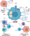
Amino acid metabolism is a critical regulator of the immune response, and its modulating becomes a promising approach in various forms of immunotherapy. Insufficient concentrations of essential amino acids restrict T-cells activation and proliferation. However, only arginases, that degrade L-arginine, as well as enzymes that hydrolyze L-tryptophan are substantially increased in cancer. Two arginase isoforms, ARG1 and ARG2, have been found to be present in tumors and their increased activity usually correlates with more advanced disease and worse clinical prognosis. Nearly all types of myeloid cells were reported to produce arginases and the increased numbers of various populations of myeloid-derived suppressor cells and macrophages correlate with inferior clinical outcomes of cancer patients. Here, we describe the role of arginases produced by myeloid cells in regulating various populations of immune cells, discuss molecular mechanisms of immunoregulatory processes involving L-arginine metabolism and outline therapeutic approaches to mitigate the negative effects of arginases on antitumor immune response. Development of potent arginase inhibitors, with improved pharmacokinetic properties, may lead to the elaboration of novel therapeutic strategies based on targeting immunoregulatory pathways controlled by L-arginine degradation.
Keywords: T lymphocyte; T-cell metabolism; arginase; arginine; immunosuppression; immunotherapy; tumor immunology.
Copyright © 2020 Grzywa, Sosnowska, Matryba, Rydzynska, Jasinski, Nowis and Golab.
Figures


Similar articles
-
Inhibition of arginase by CB-1158 blocks myeloid cell-mediated immune suppression in the tumor microenvironment.J Immunother Cancer. 2017 Dec 19;5(1):101. doi: 10.1186/s40425-017-0308-4. J Immunother Cancer. 2017. PMID: 29254508 Free PMC article.
-
Pathophysiology of Arginases in Cancer and Efforts in Their Pharmacological Inhibition.Int J Mol Sci. 2024 Sep 10;25(18):9782. doi: 10.3390/ijms25189782. Int J Mol Sci. 2024. PMID: 39337272 Free PMC article. Review.
-
Metabolomic reprogramming of the tumor microenvironment by dual arginase inhibitor OATD-02 boosts anticancer immunity.Sci Rep. 2025 May 28;15(1):18741. doi: 10.1038/s41598-025-03446-1. Sci Rep. 2025. PMID: 40437024 Free PMC article.
-
Metabolism of L-arginine by myeloid-derived suppressor cells in cancer: mechanisms of T cell suppression and therapeutic perspectives.Immunol Invest. 2012;41(6-7):614-34. doi: 10.3109/08820139.2012.680634. Immunol Invest. 2012. PMID: 23017138 Free PMC article. Review.
-
Biochemistry, pharmacology, and in vivo function of arginases.Pharmacol Rev. 2025 Jan;77(1):100015. doi: 10.1124/pharmrev.124.001271. Epub 2024 Nov 22. Pharmacol Rev. 2025. PMID: 39952693 Free PMC article. Review.
Cited by
-
Impact of obesity‑associated myeloid‑derived suppressor cells on cancer risk and progression (Review).Int J Oncol. 2024 Aug;65(2):79. doi: 10.3892/ijo.2024.5667. Epub 2024 Jun 28. Int J Oncol. 2024. PMID: 38940351 Free PMC article. Review.
-
Metabolic barriers to cancer immunotherapy.Nat Rev Immunol. 2021 Dec;21(12):785-797. doi: 10.1038/s41577-021-00541-y. Epub 2021 Apr 29. Nat Rev Immunol. 2021. PMID: 33927375 Free PMC article. Review.
-
Glutamine metabolism inhibition has dual immunomodulatory and antibacterial activities against Mycobacterium tuberculosis.Nat Commun. 2023 Nov 16;14(1):7427. doi: 10.1038/s41467-023-43304-0. Nat Commun. 2023. PMID: 37973991 Free PMC article.
-
Dysbiosis of skin microbiome and gut microbiome in melanoma progression.BMC Microbiol. 2022 Feb 25;22(1):63. doi: 10.1186/s12866-022-02458-5. BMC Microbiol. 2022. PMID: 35216552 Free PMC article.
-
Reinventing the human tuberculosis (TB) granuloma: Learning from the cancer field.Front Immunol. 2022 Dec 15;13:1059725. doi: 10.3389/fimmu.2022.1059725. eCollection 2022. Front Immunol. 2022. PMID: 36591229 Free PMC article. Review.
References
Publication types
MeSH terms
Substances
LinkOut - more resources
Full Text Sources
Medical
Research Materials
Miscellaneous

