Review
. 2020 May 15:11:940.
doi: 10.3389/fimmu.2020.00940. eCollection 2020.

The Tumor Microenvironment: A Milieu Hindering and Obstructing Antitumor Immune Responses

Affiliations
Review

The Tumor Microenvironment: A Milieu Hindering and Obstructing Antitumor Immune Responses

Alireza Labani-Motlagh et al. Front Immunol. .

Abstract

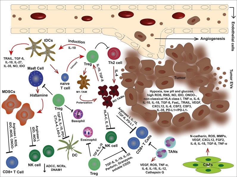
The success of cancer immunotherapy relies on the knowledge of the tumor microenvironment and the immune evasion mechanisms in which the tumor, stroma, and infiltrating immune cells function in a complex network. The potential barriers that profoundly challenge the overall clinical outcome of promising therapies need to be fully identified and counteracted. Although cancer immunotherapy has increasingly been applied, we are far from understanding how to utilize different strategies in the best way and how to combine therapeutic options to optimize clinical benefit. This review intends to give a contemporary and detailed overview of the different roles of immune cells, exosomes, and molecules acting in the tumor microenvironment and how they relate to immune activation and escape. Further, current and novel immunotherapeutic options will be discussed.

Keywords: antitumor; immune cells; immune response; immunosuppression; tumor; tumor microenvironment.


Figures

Figure 1
Figure 1
An overview of tumor microenvironment. In the immunosuppressive TME, malignant cells debilitate the antitumor immune responses through secretion of offensive and detrimental molecules, collaboration with cancer-associated stromal cells, and exploit immune scape mechanisms to outwit the immune cells. Tumor cells alter their milieu by lowering pH and glucose but high production of VEGF, non-classical HLA class I, death ligands such as FasL and TRAIL, anti-inflammatory cytokines, and metabolites such as IDO, ROS, RNS, ONOO, and NO. These molecules can not only inhibit the immune cells but also elicit the stroma cells and facilitate tumor development. The cancer-associated stroma cells favor tumor cells by suppressing the immune responses and even induce each other. Tregs are capable to inhibit effector immune cells, eosinophils, basophils, and mast cells. Mast cells themselves induce MDSCs by releasing histamine. Tregs also stimulate tDC via IL-10 and impose M1-TAM polarization into M2-type. In turn, M2-TAMs eliminate effector cells via non-classical HLA class I, arginase I, IL-10, TGF-β, and PD-L1. In addition, MDSCs hinder effector cells by releasing arginase I, and metabolites such as IDO, ROS, ONOO, and iNOS. TANs are other players that eliminate CD8+ T cells. The condition becomes more complicated with CAFs that promote angiogenesis, tumor growth, and invasion.
Figure 2
Figure 2
Tumor-cell escape and NK cell-mediated cytotoxicity. Tumor cell-released immunosuppressive exosomes expressing surface NKG2DLs impair the NK cell-mediated recognition and cytotoxicity. The exsosomes released by tumor cells internally carry the DNAM-1 ligands therefore they are not capable to bind the DNAM-1, leaving this activating receptor free to bind to its correlated ligands on tumor cells and kill them through apoptosis due to releasing perforin and granzyme B. EOC, epithelial ovarian cancer.
Figure 3
Figure 3
The effect of exosomes in a niche of tumor microenvironment. Different cell types and cancer cells crosstalk in tumor via EVs. In the TME, cancer-associated stroma cells promote tumor progression via exosomes. Tumor cell-derived exosomes inhibit NK cells, and T cells, elicit MDSC expansion and Treg suppressive function, stimulate angiogenesis, and metastasis, and polarize macrophages and neutrophils into TAMs and TANs, respectively. Malignant cells also receive support via exosomes released by cancer-associated stromal cells. However, exosomes released by NK cells induce tumor cell apoptosis through cytotoxicity function.

References

    1. Swann JB, Smyth MJ. Immune surveillance of tumors. J Clin Investig. (2007) 117:1137–46. 10.1172/JCI31405 - DOI - PMC - PubMed
    1. Gül N, Babes L, Siegmund K, Korthouwer R, Bögels M, Braster R, et al. . Macrophages eliminate circulating tumor cells after monoclonal antibody therapy. J Clin Investig. (2014) 124:812–23. 10.1172/JCI66776 - DOI - PMC - PubMed
    1. Martin JH, Edwards SW. Changes in mechanisms of monocyte/macrophage-mediated cytotoxicity during culture. Reactive oxygen intermediates are involved in monocyte-mediated cytotoxicity, whereas reactive nitrogen intermediates are employed by macrophages in tumor cell killing. J Immunol. (1993) 150:3478–86. - PubMed
    1. Urban JL, Shepard HM, Rothstein JL, Sugarman BJ, Schreiber H. Tumor necrosis factor: a potent effector molecule for tumor cell killing by activated macrophages. Proc Natl Acad Sci USA. (1986) 83:5233–37. 10.1073/pnas.83.14.5233 - DOI - PMC - PubMed
    1. Sektioglu IM, Carretero R, Bulbuc N, Bald T, Tuting T, Rudensky AY, et al. . Basophils promote tumor rejection via chemotaxis and infiltration of CD8+ T cells. Cancer Res. (2017) 77:291–302. 10.1158/0008-5472.CAN-16-0993 - DOI - PubMed

LinkOut - more resources