. 2020 Apr 8;11(5):2447-2460.
doi: 10.1364/BOE.385729. eCollection 2020 May 1.

Robotic microscopy for everyone: the OpenFlexure microscope

Affiliations

Robotic microscopy for everyone: the OpenFlexure microscope

Joel T Collins et al. Biomed Opt Express. .

Abstract

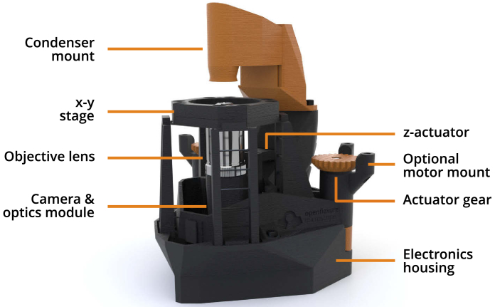
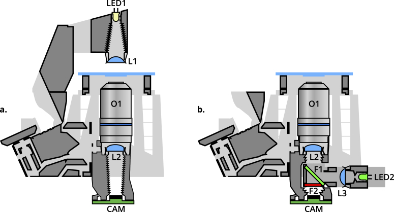
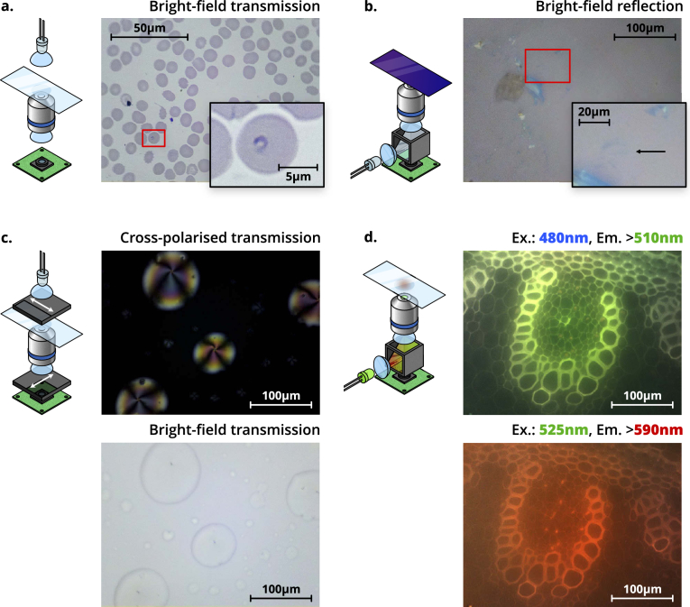
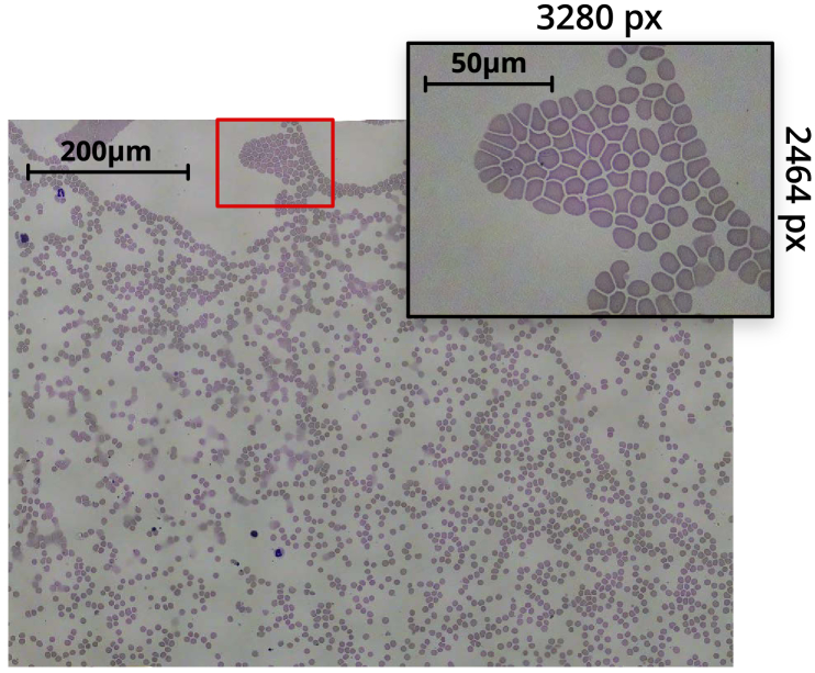
Optical microscopes are an essential tool for both the detection of disease in clinics, and for scientific analysis. However, in much of the world access to high-performance microscopy is limited by both the upfront cost and maintenance cost of the equipment. Here we present an open-source, 3D-printed, and fully-automated laboratory microscope, with motorised sample positioning and focus control. The microscope is highly customisable, with a number of options readily available including trans- and epi- illumination, polarisation contrast imaging, and epi-florescence imaging. The OpenFlexure microscope has been designed to enable low-volume manufacturing and maintenance by local personnel, vastly increasing accessibility. We have produced over 100 microscopes in Tanzania and Kenya for educational, scientific, and clinical applications, demonstrating that local manufacturing can be a viable alternative to international supply chains that can often be costly, slow, and unreliable.


Conflict of interest statement

The authors declare no conflicts of interest.

Figures

Fig. 1.
Fig. 1.
Overview of the OpenFlexure Microscope design, in transmission bright-field configuration. The condenser mount houses an illumination LED and a plastic condenser lens, while the optics module sits below the stage and houses an objective lens, tube lens, and camera. The entire optics module is attached to the z-actuator, providing variable focus. Both the optics module and x-y stage are controlled by actuator gears at the back of the microscope, optionally driven by stepper motors. A detachable electronics housing stores optional electronic parts, such as motor controllers and a Raspberry Pi, for automation.
Fig. 2.
Fig. 2.
Cross-section schematics of trans- (a.) and epi- (b.) illumination configurations. LED1 provides transmission illumination, with a condenser lens L1. LED2 and lens L3 provide epi-illumination, connected to a removable filter cube housing two filters, F1 at 45° and F2. These filters can be removed or replaced by beamsplitters or polarizers to enable bright-field, fluorescence, or polarization-contrast epi-illuminated imaging. In both configurations, a standard RMS objective O1 and tube lens L2 image the sample onto the camera sensor CAM.
Fig. 3.
Fig. 3.
Schematics (left) and images (right) of four different imaging modalities possible using the OFM. a. Trans-illumination bright-field imaging of a Giemsa-stained thin blood smear, obtained with a 100×, 1.25NA oil immersion objective. The inset shows a magnified section of the image, highlighting a ring-form trophozoite of Plasmodium falciparum. b. Epi-illumination bright-field imaging of a group of thin graphene flakes, obtained with a 40×, 0.65NA dry objective. The inset shows a magnified section of the image, highlighting a resolvable tri-layer graphene flake (contrast has been digitally enhanced for clarity by increasing brightness and gamma). c. Polarisation-contrast trans-illumination image of 5CB liquid crystal droplets. A bright-field image is shown below for comparison. Both images were obtained with a 40×, 0.65NA dry objective, and depict the same region of the sample. Arrows on the polarisers denote the transmission axis. d. Fluorescence images of unstained Lily of the Valley (convallaria majalis) rhizome, at two different wavelengths. The excitation wavelength (“Ex.”), and minimum emission wavelength imaged (“Em.”) are shown above the respective panels. Both images were obtained with a 40×, 0.65NA dry objective, and depict the same region of the sample. Both the colour of illumination LED, and the filters in the filter cube, will change depending on the application.
Fig. 4.
Fig. 4.
Tiled scan image of a Giemsa-stained thin blood smear, obtained with a 100×, 1.25NA oil immersion objective. The inset highlights an individual 8-megapixel image from the scan. The composite image was obtained from a 10×10 grid of captures. After accounting for image overlap and skewing, and cropping out edges of the composite with missing sections, the resulting image is 14920 px ×11270 px (170 megapixel).

References

    1. Wollman A. J. M., Nudd R., Hedlund E. G., Leake M. C., “From Animaculum to single molecules: 300 years of the light microscope,” Open Biol. 5(4), 150019 (2015).10.1098/rsob.150019 - DOI - PMC - PubMed
    1. Yadav P., “Health product supply chains in developing countries: Diagnosis of the root causes of underperformance and an agenda for reform,” Heal. Syst. & Reform 1(2), 142–154 (2015).10.4161/23288604.2014.968005 - DOI - PubMed
    1. Malkin R. A., “Barriers for medical devices for the developing world,” Expert Rev. Med. Devices 4(6), 759–763 (2007).10.1586/17434440.4.6.759 - DOI - PubMed
    1. Malkin R., Keane A., “Evidence-based approach to the maintenance of laboratory and medical equipment in resource-poor settings,” Med. Biol. Eng. Comput. 48(7), 721–726 (2010).10.1007/s11517-010-0630-1 - DOI - PubMed
    1. Maia Chagas A., “Haves and have nots must find a better way: The case for open scientific hardware,” PLoS Biol. 16(9), e3000014 (2018).10.1371/journal.pbio.3000014 - DOI - PMC - PubMed

LinkOut - more resources