Review
. 2020 Sep;69(9):1673-1697.
doi: 10.1007/s00262-020-02616-6. Epub 2020 Jun 4.

Pro-tumorigenic functions of macrophages at the primary, invasive and metastatic tumor site

Affiliations
Review

Pro-tumorigenic functions of macrophages at the primary, invasive and metastatic tumor site

Elaheh Nasrollahzadeh et al. Cancer Immunol Immunother. 2020 Sep.

Abstract

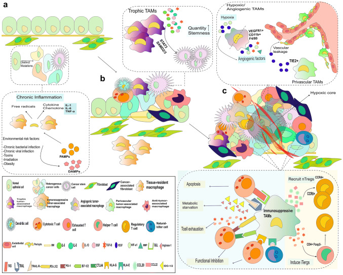
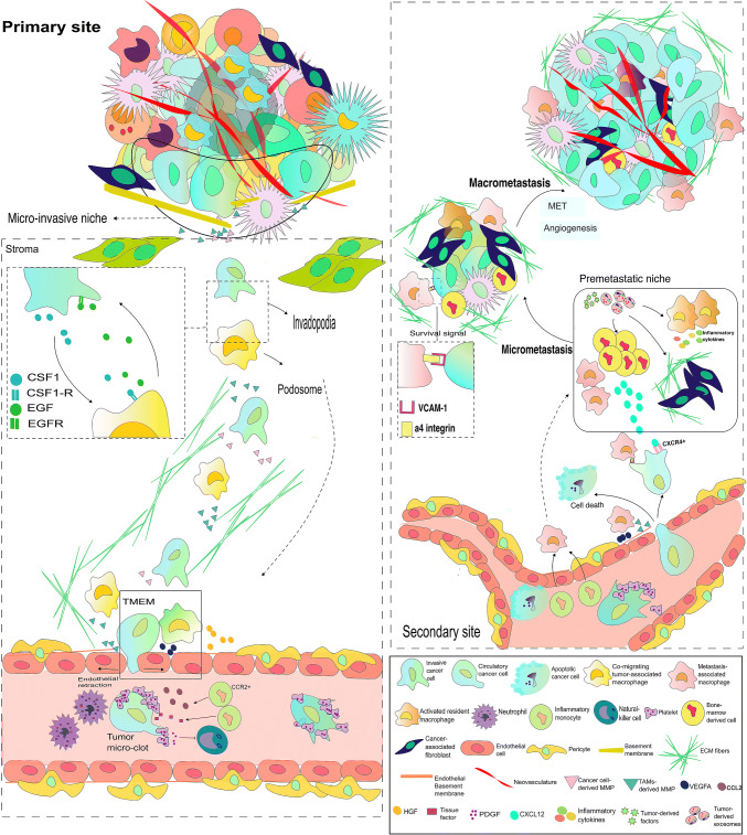
The tumor microenvironment (TME) not only facilitates cancer progression from the early formation to distant metastasis, but also it differs itself from time to time alongside the tumor evolution. Tumor-associated macrophages (TAMs), whether as pre-existing tissue-resident macrophages or recruited monocytes, are an inseparable part of this microenvironment. As their parents are broadly classified into a dichotomic, simplistic M1 and M2 subtypes, TAMs also exert paradoxical and diverse phenotypes as they are settled in different regions of TME and receive different microenvironmental signals. Briefly, M1 macrophages induce an inflammatory precancerous niche and flame the early oncogenic mutations, whereas their M2 counterparts are reprogrammed to release various growth factors and providing an immunosuppressive state in TME as long as abetting hypoxic cancer cells to set up a new vasculature. Further, they mediate stromal micro-invasion and co-migrate with invasive cancer cells to invade the vascular wall and neural sheath, while another subtype of TAMs prepares suitable niches much earlier than metastatic cells arrive at the target tissues. Accordingly, at the neoplastic transformation, during the benign-to-malignant transition and through the metastatic cascade, macrophages are involved in shaping the primary, micro-invasive and pre-metastatic TMEs. Whether their behavioral plasticity is derived from distinct genotypes or is fueled by microenvironmental cues, it could define these cells as remarkably interesting therapeutic targets.

Keywords: Metastasis; Primary tumor; Pro-tumorigenic functions; Therapeutic targets; Tumor microenvironment; Tumor-associated macrophages.


Conflict of interest statement

The authors declare that they have no conflict of interest.

Figures

Fig. 1
Fig. 1
Macrophages before and after neoplastic transformation. Cancer risk factors like chronic infections, radiation and obesity seem to be engaged to an unresolving inflammatory condition. Tissue resident macrophages behave as M1-like macrophages and detect DAMPs/PAMPs as signs of inflammation and release free radicals and inflammatory cytokines/chemokines like IL-1, IL-6 and TNF-a in the normal inflamed tissues. These oxidative stresses are mutagen and create a pre-cancerous niche around the normal cells. a In carcinomas, the normal epithelium is exposed to this pre-cancerous niche and could undergo different mutations and probably earn neoplastic traits. The present inflammatory cytokines also stimulate neoplastic cells to express more oncogenic materials. As cancer initiates the early TME is also created. b Various BMDCs are recruited and educated as pro-tumor collaborators. Among these, different subsets of M2-like TAMs are involved. Trophic TAMs produce growth factors and cytokines that nourish bulk tumor cells and CSCs. Later, as cancer progress, the early TME evolves to a complex ecosystem. c Immunosuppressive TAMs directly promote immune evasion via threading the cytotoxic T cells viability or functionality by expressing different inhibitory ligands, leading to an increasing population of dead or exhausted T cells. Additionally, these TAMs also indirectly affect cytotoxic T cells through recruiting natural Tregs or inducing Th cells to express Foxp3, a Tregs marker. Plenty of growth factors and successful immune evasion are associated with a period of fast tumor growth. However, gradually the lack of nutritions, low oxygen pressure and acidosis overcome the cancer cells innate tendency for endless proliferation and limit tumor growth. The hypoxic core drive various pro-angiogenic factors. HIF-a1 and VEGF stimulate angiogenic TAMs to produce more amounts of pro-angiogenic factors to stimulate angiogenic buds formation from existing blood vessels. Perivascular-TAMs are settled in the perivascular niche to incite the leaky nature of new micro-vasculature. It is concluded that different subset of macrophages are involved in the cancer initiation and progression by participating in creation of precancerous niche, early TME and later evolve TME
Fig. 2
Fig. 2
TAMs lead aggressive cancer cells departure toward the far metastatic sites. The crowded hypoxic tumor core might be the first point of stromal invasion since the hunger select more aggressive cancer cells to survive and migrate toward the tumor borders, near the normal stroma. At the border, they are assisted with various BMDCs particularly macrophages to create a micro-invasive niche where TAMs prompt cancer cells to undergo EMT, at least partially (shown with blue-green cells). Hereafter, these cells are anchorage-independent, motile and highly invasive. Invasive cancer cells and accompanied TAMs release huge amounts of MMPs and other ECM remodeling enzymes to detach from the tumor stroma, breach the BM and invade the beneath normal stroma. Parallel to this, invasive cells and TAMs secret and send factors and exosomes to their desirable sites at targeted organs to recruit BMDCs, activate resident macrophages and fibroblasts to form an inflammatory niche and educate them to serve future metastatic cells. lnvasive cells owe their directional streaming to TAMs-derived chemotactic gradient of EGF. Mechanistically, co-migrating TAMs induce formation of plasma membrane protrusions in the invasive cells that is called invadopodia and act as their degrading feet. Further, ECs-derived HGF and collagen railways (shown as green lines) also enhance the chance of their vessel directed movement. By the blood vessel, at TMEM, perivascular-TAMs open the gateways to streaming cells mainly via secreting VEGF and loosening the endothelial junctions. In the circulation, various threats from the physical shear stress to immune attacks surround CTCs. Platelets can bind to wandering CTCs and form micro-clots and also release PDGF to protect them against both physical and immune threats. Micro-clots produced CCL2 and tissue factor (TF) recruit inflammatory CCR2+ monocytes. Cancer cells journey in the circulation could have different endings: death, entrapment in the small capillaries or arrest at vasculature of pre-metastatic sites. In the third condition, the recruited monocytes extravasate at the PMNs, turn to MAMs and ease the extravasation of arrested CTCs by a mechanism similar to the intravasation. Early as they are extravasated, there is still a high risk of death. However, MAMs-VCAM1 interaction with cancer cells a4integrin and activation of Ezrin-PI3K/Akt pathway is effective in promoting their survival. A little far from the point of arrest, BMDCs in the PMN release CXCL12 to pull the survived CCR4+cancer cells toward the PMN, where they can seed and form the micro-metastatic lesion. The low speed of proliferation in the partially mesenchymal cells and gradual inducing hypoxia are two main rate limiting steps in forming the macrometastasis. However, a population of educated CD11b+ Ly6high monocytic cells can induce EMT reversion in a process called MET. The condition go worth, toward the overt metastasis, if MAMs, CAFs and other BMDCs flip the angiogenic switch and provide new source of nutritions for the micrometastatic foci
Fig. 3
Fig. 3
Tumor-associated endoneural macrophages stimulate cancer cells for neural tracking. Once a neoplastic lesion is established, it produces various cytokines and induce an inflammatory condition in the surrounding environment. Specifically, about the adjacent nerves, this inflammatory environment causes the nerouinflammation as a kind of nerve injury that is later followed by a neural regenerative response. The injured nerves release high amounts of CCL2 to recruit BM-derived monocytes. The recruited monocytes turn to endoneural macrophages. a They are activated to secret a neural growth factor (GDNF) in response to tumor-derived signals. GDNF interacts with GFRa1 and mostly affects the injured nerve in a paracrine loop and causes axonogenesis (regenerative response). b Further, the GDNF gradient can pull cancer cells toward the axons, protrude their membranes to form invadopodia and induce expression of MMPs. c & d All these result in enhancing the cancer cells chance in tracking the nerve and invading the neural sheath

References

    1. Kitamura T, Qian BZ, Pollard JW. Immune cell promotion of metastasis. Nat Rev Immunol. 2015;15(2):73–86. doi: 10.1038/nri3789. - DOI - PMC - PubMed
    1. Mantovani A. Cancer: inflaming metastasis. Nature. 2009;457(7225):36. doi: 10.1038/457036b. - DOI - PubMed
    1. Hanahan D, Weinberg RA. Hallmarks of cancer: the next generation. Cell. 2011;144(5):646–674. doi: 10.1016/j.cell.2011.02.013. - DOI - PubMed
    1. de Visser KE, Eichten A, Coussens LM. Paradoxical roles of the immune system during cancer development. Nat Rev Cancer. 2006;6(1):24–37. doi: 10.1038/nrc1782. - DOI - PubMed
    1. McAllister SS, Weinberg RA. The tumour-induced systemic environment as a critical regulator of cancer progression and metastasis. Nat Cell Biol. 2014;16(8):717–727. doi: 10.1038/ncb3015. - DOI - PMC - PubMed