Case Reports
. 2020 Sep;162(9):2055-2059.
doi: 10.1007/s00701-020-04431-5. Epub 2020 Jun 4.

Bilateral thoracic disc herniation with abdominal wall paresis: a case report

Affiliations
Case Reports

Bilateral thoracic disc herniation with abdominal wall paresis: a case report

Vicki Marie Butenschoen et al. Acta Neurochir (Wien). 2020 Sep.

Erratum in

Abstract

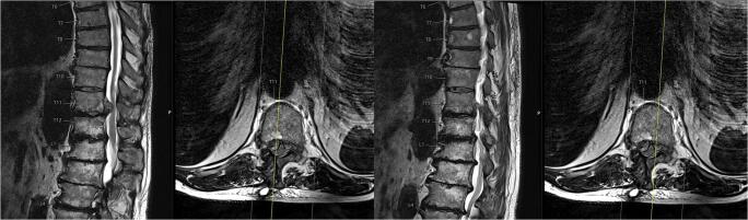
We present a rare case of a patient initially presenting with unilateral abdominal wall bulging and radicular pain caused by a lateral disc herniation at Th11/12, later suffering from a hernia recurrence with bilateral disc prolapse and motor deficits. The patient underwent sequesterectomy via a right hemilaminectomy at Th11, and after 8 weeks, a bilateral sequesterectomy with semirigid fusion Th11/12 was performed. Unilateral motor deficits at the thoracic level have been discussed in case reports; a bilateral disc protrusion with abdominal wall bulging occurring as a recurrent disc herniation has never been described before.

Keywords: Disc herniation; Motor deficit; Thoracic spine.


Figures

Fig. 1
Fig. 1
Initial presentation of the patient with abdominal wall bulging right-sided
Fig. 2
Fig. 2
The abdominal CT scan excludes the suspected diagnosis of an abdominal wall herniation
Fig. 3
Fig. 3
The MRI scan of the thoracolumbar spine T2 weighted axial and sagittal presents a right-sided disc herniation TH 11/12
Fig. 4
Fig. 4
In the clinical follow-up 8 weeks after the first operation, the patient presented with bilateral abdominal wall bulging
Fig. 5
Fig. 5
MRI of the thoracolumbar spine T2 weighted 8 weeks after initial presentation with clinical bilateral thoracic abdominal wall bulging and a bilateral relapse of disc herniation Th11/12
Fig. 6
Fig. 6
Intraoperative findings: A discectomy (a) and dynamic fusion (b) was performed
Fig. 7
Fig. 7
Postoperative X-ray in lateral (a) and anterior-posterior imaging (b)

References

    1. Baranto A, Börjesson M, Danielsson B, Hellström M, Swärd L. Acute chest pain in a top soccer player due to thoracic disc herniation. Spine (Phila Pa 1976) 2009;34:E359–E362. doi: 10.1097/BRS.0b013e3181a16d41. - DOI - PubMed
    1. Court C, Mansour E, Bouthors C. Thoracic disc herniation: surgical treatment. Orthop Traumatol Surg Res. 2018;104:S31–S40. doi: 10.1016/j.otsr.2017.04.022. - DOI - PubMed
    1. Dakwar E, Le TV, Baaj AA, Le AX, Smith WD, Akbarnia BA, Uribe JS. Abdominal wall paresis as a complication of minimally invasive lateral transpsoas interbody fusion. Neurosurg Focus. 2011;31:E18. doi: 10.3171/2011.7.FOCUS11164. - DOI - PubMed
    1. Dower A, Chatterji R, Swart A, Winder MJ. Surgical management of recurrent lumbar disc herniation and the role of fusion. J Clin Neurosci. 2016;23:44–50. doi: 10.1016/j.jocn.2015.04.024. - DOI - PubMed
    1. Drazin D, Ugiliweneza B, Al-Khouja L, Yang D, Johnson P, Kim T, Boakye M. Treatment of recurrent disc herniation: a systematic review. Cureus. 2016;8:e622. doi: 10.7759/cureus.622. - DOI - PMC - PubMed

Publication types

MeSH terms

Supplementary concepts