

Chest CT features of coronavirus disease 2019 (COVID-19) pneumonia: key points for radiologists
- PMID: 32500509
- PMCID: PMC7270744
- DOI: 10.1007/s11547-020-01237-4
Chest CT features of coronavirus disease 2019 (COVID-19) pneumonia: key points for radiologists
Abstract
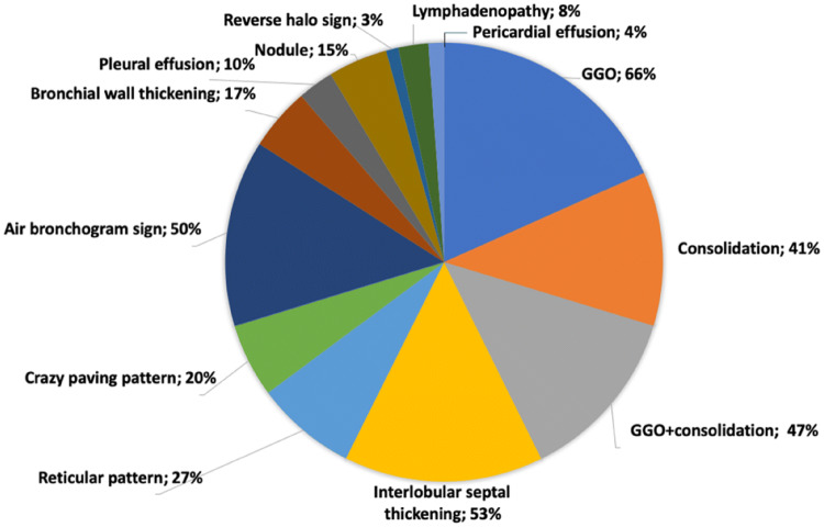
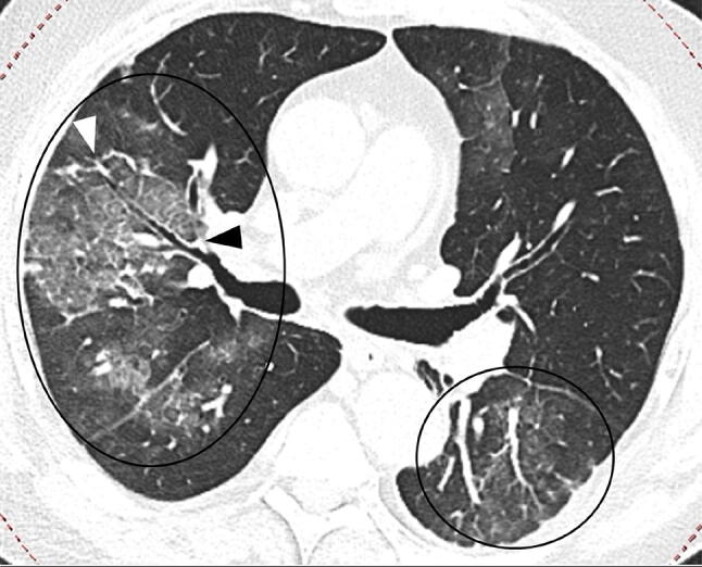
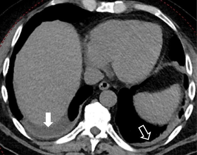
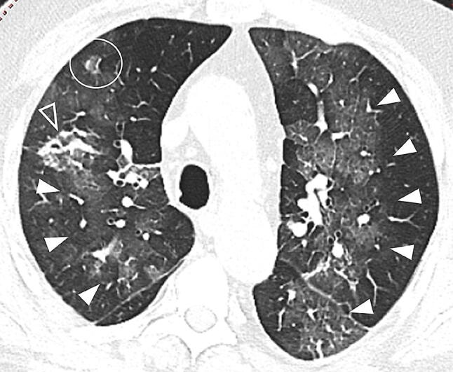
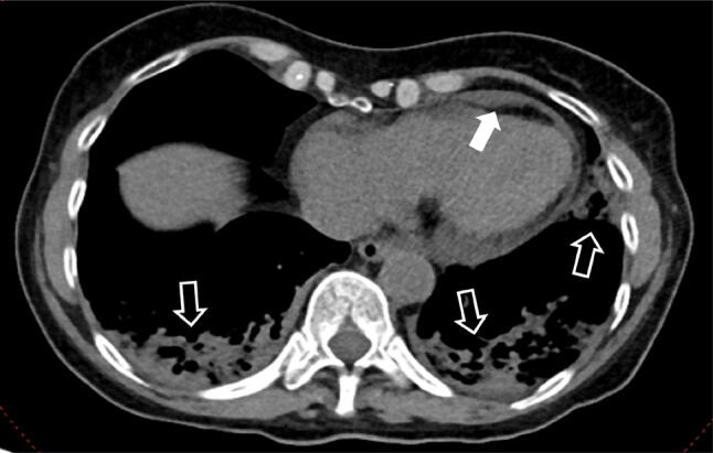
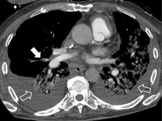
COVID-19 is an emerging infection caused by a novel coronavirus that is moving so rapidly that on 30 January 2020 the World Health Organization declared the outbreak a Public Health Emergency of International Concern and on 11 March 2020 as a pandemic. An early diagnosis of COVID-19 is crucial for disease treatment and control of the disease spread. Real-time reverse-transcription polymerase chain reaction (RT-PCR) demonstrated a low sensibility; therefore chest computed tomography (CT) plays a pivotal role not only in the early detection and diagnosis, especially for false negative RT-PCR tests, but also in monitoring the clinical course and in evaluating the disease severity. This paper reports the CT findings with some hints on the temporal changes over the course of the disease: the CT hallmarks of COVID-19 are bilateral distribution of ground glass opacities with or without consolidation in the posterior and peripheral lung, but the predominant findings in later phases include consolidations, linear opacities, "crazy-paving" pattern, "reversed halo" sign and vascular enlargement. The CT findings of COVID-19 overlap with the CT findings of other diseases, in particular the viral pneumonia including influenza viruses, parainfluenza virus, adenovirus, respiratory syncytial virus, rhinovirus, human metapneumovirus, etc. There are differences as well as similarities in the CT features of COVID-19 compared with those of the severe acute respiratory syndrome. The aim of this article is to review the typical and atypical CT findings in COVID-19 patients in order to help radiologists and clinicians to become more familiar with the disease.
Keywords: Chest CT; Consolidation; Coronavirus pneumonia; Crazy-paving pattern; Ground glass opacities; Lungs; Reticular pattern.
Conflict of interest statement
The authors declare that they have no conflict of interest.
Figures

Similar articles
-
Imaging and clinical features of patients with 2019 novel coronavirus SARS-CoV-2.Eur J Nucl Med Mol Imaging. 2020 May;47(5):1275-1280. doi: 10.1007/s00259-020-04735-9. Epub 2020 Feb 28. Eur J Nucl Med Mol Imaging. 2020. PMID: 32107577 Free PMC article.
-
Chest CT manifestations of new coronavirus disease 2019 (COVID-19): a pictorial review.Eur Radiol. 2020 Aug;30(8):4381-4389. doi: 10.1007/s00330-020-06801-0. Epub 2020 Mar 19. Eur Radiol. 2020. PMID: 32193638 Free PMC article. Review.
-
The role of imaging in 2019 novel coronavirus pneumonia (COVID-19).Eur Radiol. 2020 Sep;30(9):4874-4882. doi: 10.1007/s00330-020-06827-4. Epub 2020 Apr 15. Eur Radiol. 2020. PMID: 32296940 Free PMC article. Review.
-
Coronavirus Disease 2019 (COVID-19): Role of Chest CT in Diagnosis and Management.AJR Am J Roentgenol. 2020 Jun;214(6):1280-1286. doi: 10.2214/AJR.20.22954. Epub 2020 Mar 4. AJR Am J Roentgenol. 2020. PMID: 32130038
-
Similarities and Differences of Early Pulmonary CT Features of Pneumonia Caused by SARS-CoV-2, SARS-CoV and MERS-CoV: Comparison Based on a Systemic Review.Chin Med Sci J. 2020 Sep 30;35(3):254-261. doi: 10.24920/003727. Chin Med Sci J. 2020. PMID: 32972503 Free PMC article.
Cited by
-
Prognostic peripheral blood biomarkers at ICU admission predict COVID-19 clinical outcomes.Front Immunol. 2022 Nov 14;13:1010216. doi: 10.3389/fimmu.2022.1010216. eCollection 2022. Front Immunol. 2022. PMID: 36451808 Free PMC article.
-
Heart Failure and Cardiomyopathies: CT and MR from Basics to Advanced Imaging.Diagnostics (Basel). 2022 Sep 23;12(10):2298. doi: 10.3390/diagnostics12102298. Diagnostics (Basel). 2022. PMID: 36291987 Free PMC article. Review.
-
Highlighting COVID-19: What the imaging exams show about the disease.World J Radiol. 2021 May 28;13(5):122-136. doi: 10.4329/wjr.v13.i5.122. World J Radiol. 2021. PMID: 34141092 Free PMC article. Review.
-
The design of a point of care FET biosensor to detect and screen COVID-19.Sci Rep. 2023 Mar 18;13(1):4485. doi: 10.1038/s41598-023-31679-5. Sci Rep. 2023. PMID: 36934198 Free PMC article.
-
Atypical Immunologic Manifestations of COVID-19: a Case Report and Narrative Review.SN Compr Clin Med. 2023;5(1):108. doi: 10.1007/s42399-023-01448-6. Epub 2023 Mar 18. SN Compr Clin Med. 2023. PMID: 36970579 Free PMC article.
