. 2020 Jul;45(7):531-533.
doi: 10.1097/RLU.0000000000003100.

Incidental CT Findings Suspicious for COVID-19-Associated Pneumonia on Nuclear Medicine Examinations: Recognition and Management Plan

Affiliations

Incidental CT Findings Suspicious for COVID-19-Associated Pneumonia on Nuclear Medicine Examinations: Recognition and Management Plan

Mark Tulchinsky et al. Clin Nucl Med. 2020 Jul.

Abstract

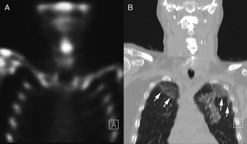
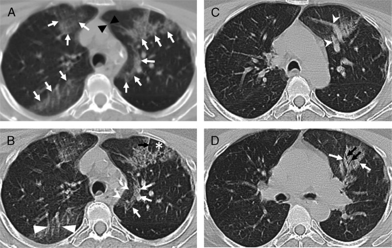
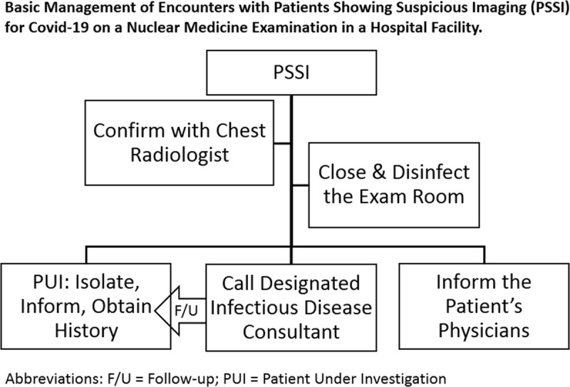
Some patients undergoing routine SPECT/CT and PET/CT examinations during the COVID-19 pandemic may incidentally reveal findings of COVID-19-associated pneumonia (C-19AP) on localizing CT. It is critical for nuclear medicine physicians to develop diagnostic skills for timely recognition of typical findings of C-19AP on a localizing CT. Furthermore, it is our responsibility to know the optimal practices for safely isolating and managing such patients while protecting the staff, other patients at the facility, family and/or friend accompanying the patients, and the public in general from risky exposure to COVID-19 sources. We offer several steps following an encounter suspicious of C-19AP.


Conflict of interest statement

Conflicts of interest and sources of funding: none declared.

Figures

FIGURE 1
FIGURE 1
Skeletal SPECT/CT through the mid chest, showing coronal skeletal SPECT (A) and the matching slice of the low-dose localizing CT (B) that is displayed in a lung window. There are large, peripheral GGOs (arrows) in both lungs.
FIGURE 2
FIGURE 2
Shown are axial slices of localizing low-dose CT obtained with a non-breathholding technique as part of SPECT/CT study that revealed findings commonly described in patients with C-19AP. A, The localizing, unenhanced, axial CT image is shown. Respiratory motion artifact is creating double shadows (black arrowheads) and blurring the fine detail. Nevertheless, multiple scattered areas of GGOs are noted in the peripheral distribution (white arrows), a few of which in the left lung demonstrate nodular consolidation. B, The same image was processed using the edge enhancement filter, which improved visualization of associated interlobular and intralobular septal thickening (black arrow) with central lobular sparing (asterisk). A semilunar-shaped reticular-nodular infiltrate (white arrows) is surrounding a GGO (white indented-base arrowheads), called a reversed halo sign. There is a noticeable vascular enlargement coursing through a GGO (white flat-base arrowheads). C, Another slice showing peripheral GGO containing vascular enlargement. D, A slice showing reticular-nodular opacity (white arrow) with an air-bronchogram (black arrows).
FIGURE 3
FIGURE 3
Schematic of basic steps in managing an encounter with a patient showing suspicious imaging for C-19AP.

References

    1. Ghebreyesus TA. WHO Director-General's opening remarks at the media briefing on COVID-19 - 11 March 2020. WHO Speaches. Available at: https://www.who.int/dg/speeches/detail/who-director-general-s-opening-re.... Accessed March 15, 2020.
    1. Bai HX, Hsieh B, Xiong Z, et al. Performance of radiologists in differentiating COVID-19 from viral pneumonia on chest CT. Radiology. 2020;200823. - PMC - PubMed
    1. Coronaviridae Study Group of the International Committee on Taxonomy of Viruses The species severe acute respiratory syndrome-related coronavirus: classifying 2019-nCoV and naming it SARS-CoV-2. Nat Microbiol. 2020;5:536–544. - PMC - PubMed
    1. Qin C, Liu F, Yen TC, et al. 18F-FDG PET/CT findings of COVID-19: a series of four highly suspected cases. Eur J Nucl Med Mol Imaging. 2020;47:1281–1286. - PMC - PubMed
    1. Zou S, Zhu X. FDG PET/CT of COVID-19. Radiology. 2020;200770. - PMC - PubMed

MeSH terms