. 2020 Jul 2;107(1):24-33.
doi: 10.1016/j.ajhg.2020.05.010. Epub 2020 Jun 4.

Homozygous Mutations in BTG4 Cause Zygotic Cleavage Failure and Female Infertility

Affiliations

Homozygous Mutations in BTG4 Cause Zygotic Cleavage Failure and Female Infertility

Wei Zheng et al. Am J Hum Genet. .

Abstract

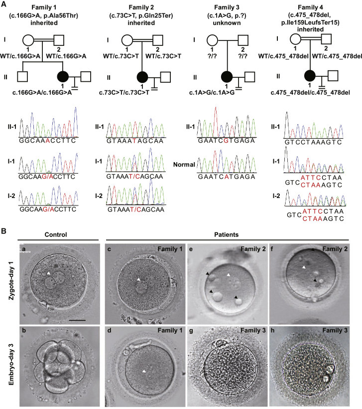
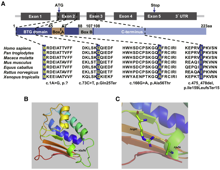
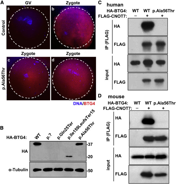
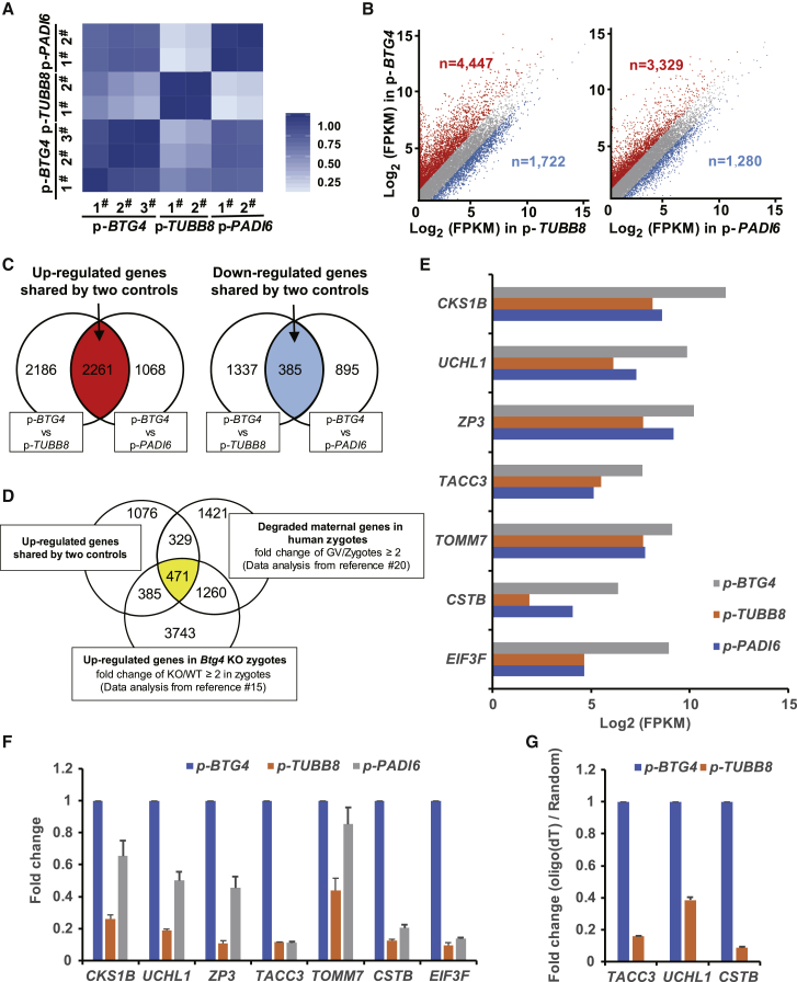
Zygotic cleavage failure (ZCF) is a unique early embryonic phenotype resulting in female infertility and recurrent failure of in vitro fertilization (IVF) and/or intracytoplasmic sperm injection (ICSI). With this phenotype, morphologically normal oocytes can be retrieved and successfully fertilized, but they fail to undergo cleavage. Until now, whether this phenotype has a Mendelian inheritance pattern and which underlying genetic factors play a role in its development remained to be elucidated. B cell translocation gene 4 (BTG4) is a key adaptor of the CCR4-NOT deadenylase complex, which is involved in maternal mRNA decay in mice, but no human diseases caused by mutations in BTG4 have previously been reported. Here, we identified four homozygous mutations in BTG4 (GenBank: NM_017589.4) that are responsible for the phenotype of ZCF, and we found they followed a recessive inheritance pattern. Three of them-c.73C>T (p.Gln25Ter), c.1A>G (p.?), and c.475_478del (p.Ile159LeufsTer15)-resulted in complete loss of full-length BTG4 protein. For c.166G>A (p.Ala56Thr), although the protein level and distribution of mutant BTG4 was not altered in zygotes from affected individuals or in HeLa cells, the interaction between BTG4 and CNOT7 was abolished. In vivo studies further demonstrated that the process of maternal mRNA decay was disrupted in the zygotes of the affected individuals, which provides a mechanistic explanation for the phenotype of ZCF. Thus, we provide evidence that ZCF is a Mendelian phenotype resulting from mutations in BTG4. These findings contribute to our understanding of the role of BTG4 in human early embryonic development and provide a genetic marker for female infertility.

Keywords: BTG4; female infertility; mutation; zygotic cleavage failure.


Conflict of interest statement

The authors declare no competing interests.

Figures

Figure 1
Figure 1
Pedigree-based Identification of the BTG4 Pathogenic Variants and the ZCF Phenotype (A) Genetic analysis in four families affected by ZCF. Black circles indicate the affected individuals. Sanger sequencing confirmation is shown below the pedigrees. The variants c.166G>A (p.Ala56Thr) (family 1), c.73C>T (p.Gln25Ter) (family 2), and c.475_478del (p.Ile159LeufsTer15) (family 4) in BTG4 were inherited from the parents of the affected individuals, but the inheritance pattern of family 3 was unknown due to lack of information about the parents (indicated by question marks). (B) The morphology of a control zygote and the affected individuals’ zygotes on day 1 and/or day 3. The white arrowheads indicate the pronuclei, and the black arrowheads indicate the small vacuoles. Scale bar = 20 μm.
Figure 2
Figure 2
Location of Variants, Amino Acid Conservation across Species, and Structural Implications of BTG4 Amino Acid Substitution. (A) Location of the variants in the gene and protein structure and the conservation of the mutated amino acids in seven different species. All four residues are highly conserved across species. (B) Overview of the predicted structure of the wild-type BTG4 protein. Arrow indicates the location of Ala56. (C) Magnified view of the predicted structure surrounding Ala56. The yellow dashed line represents the predicted hydrogen bond.
Figure 3
Figure 3
Localization and Level of the Mutant Protein in Oocytes and HeLa cells and the Effect of the c.166G>A (p.Ala56Thr) Variant on the BTG4-CNOT7 Interaction (A) Immunofluorescence of control and c.166G>A (p.Ala56Thr) GV oocytes and zygotes. BTG4 is present after the GV stage (A and B), and zygotes with the c.166G>A (p.Ala56Thr) variant exhibit similar BTG4 protein localization as the controls (C and D). (B) Immunoblot analysis of HeLa cell extracts after transfection with wild-type or mutant HA-tagged BTG4 expression plasmids. The wild-type protein and the c.166G>A (p.Ala56Thr) variant were both about 37 kDa, the truncated c.475_478del (p.Ile159LeufsTer15) variant was about 20 kDa, and the c.1A>G (p.?) and c.73C>T (p.Gln25Ter) variants showed no detectable bands. α-Tubulin was used as the internal control. (C) Co-IP assay showing the loss of the BTG4 and CNOT7 interaction by the c.166G>A (p.Ala56Thr) variant in humans. Cell lysates were immunoprecipitated with anti-FLAG antibody, and the resulting protein samples were separated by SDS-PAGE and analyzed via immunoblot using antibodies against the HA tag. (D) Co-IP assay showing the loss of the BTG4 and CNOT7 interaction by the c.166G>A (p.Ala56Thr) variant in mice. Cell lysates were immunoprecipitated with anti-FLAG antibody and analyzed via immunoblot using antibodies against HA.
Figure 4
Figure 4
The Pathogenic Variants in BTG4 Resulted in Impaired Maternal mRNA Decay in Zygotes with the c.166G>A (p.Ala56Thr) Variant (A) Heatmap of the Spearman correlation coefficients among zygotes with the three different variants: BTG4 (c.166G>A [p.Ala56Thr]), TUBB8 (c.922G>A [p.Gly308Ser]), and PADI6 (c.1521dup [p.Ser508GlnfsTer5]). (B) Scatterplot comparing transcripts between BTG4 variant (c.166G>A, p.Ala56Thr) and TUBB8 (c.922G>A [p.Gly308Ser]) and/or PADI6 (c.1521dup [p.Ser508GlnfsTer5]) variant zygotes. Transcripts that were increased or decreased by more than 2-fold in the BTG4 variant zygotes are highlighted in red or blue, respectively. (C) Venn diagrams showing the shared upregulated (red region pointed by arrow) or downregulated (blue region pointed by arrow) genes between the BTG4 variant (c.166G>A [p.Ala56Thr]) and the TUBB8 variant (c.922G>A [p.Gly308Ser]) and between the BTG4 variant and the PADI6 variant (c.1521dup [p.Ser508GlnfsTer5]). (D) Venn diagrams showing the overlap of upregulated genes in zygotes harboring the BTG4 variant (c.166G>A [p.Ala56Thr]) with the reported human (GV/Zygote ≥ 2) and mouse (Btg4 knockout [KO] mice/wild type [WT] mice ≥ 2 in zygotes) mRNA sequencing data. (E) RNA-seq results confirming the relative expression of representative genes. (F) qRT-PCR verification of the expression of representative genes relative to GAPDH in zygotes with variants in different genes. The error bar denotes SD. (G) qRT-PCR results obtained from oligo-dT versus random primer-mediated reverse transcription reactions; these results reflect changes in the poly(A) tail length of the given transcripts from the above representative genes. The error bar denotes SD.

References

    1. Mascarenhas M.N., Flaxman S.R., Boerma T., Vanderpoel S., Stevens G.A. National, regional, and global trends in infertility prevalence since 1990: a systematic analysis of 277 health surveys. PLoS Med. 2012;9:e1001356. - PMC - PubMed
    1. Hart R.J. Physiological aspects of female fertility: role of the environment, modern lifestyle, and genetics. Physiol. Rev. 2016;96:873–909. - PubMed
    1. Matzuk M.M., Lamb D.J. The biology of infertility: research advances and clinical challenges. Nat. Med. 2008;14:1197–1213. - PMC - PubMed
    1. Yatsenko S.A., Rajkovic A. Genetics of human female infertility. Biol. Reprod. 2019;101:549–566. - PMC - PubMed
    1. Chen B., Zhang Z., Sun X., Kuang Y., Mao X., Wang X., Yan Z., Li B., Xu Y., Yu M. Biallelic mutations in PATL2 cause female infertility characterized by oocyte maturation arrest. Am. J. Hum. Genet. 2017;101:609–615. - PMC - PubMed

Publication types