. 2020 Jun 11;181(6):1380-1394.e18.
doi: 10.1016/j.cell.2020.04.056. Epub 2020 Jun 4.

Rad54 Drives ATP Hydrolysis-Dependent DNA Sequence Alignment during Homologous Recombination

Affiliations

Rad54 Drives ATP Hydrolysis-Dependent DNA Sequence Alignment during Homologous Recombination

J Brooks Crickard et al. Cell. .

Abstract

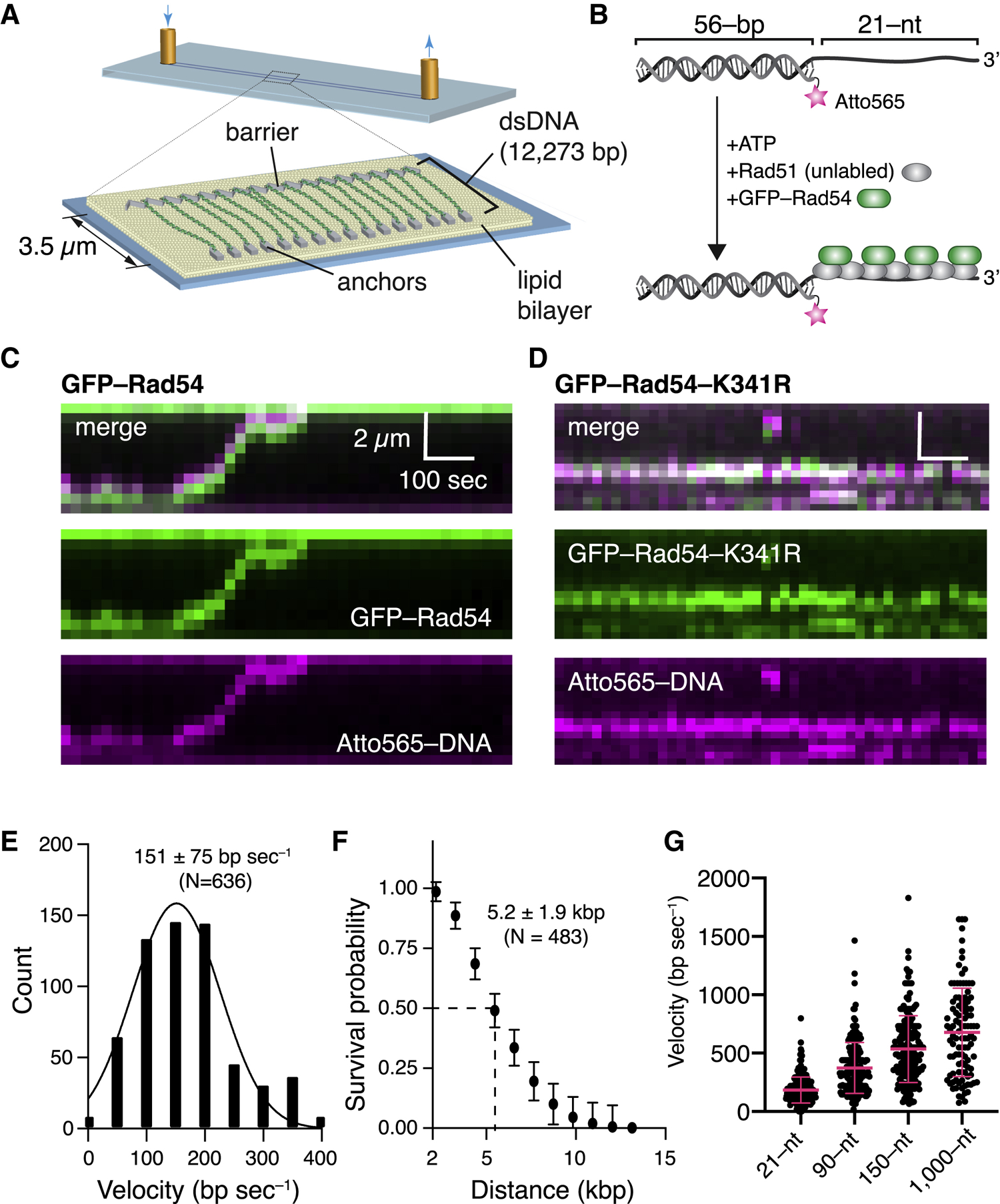
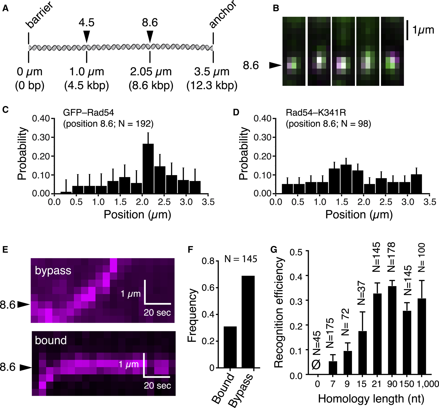
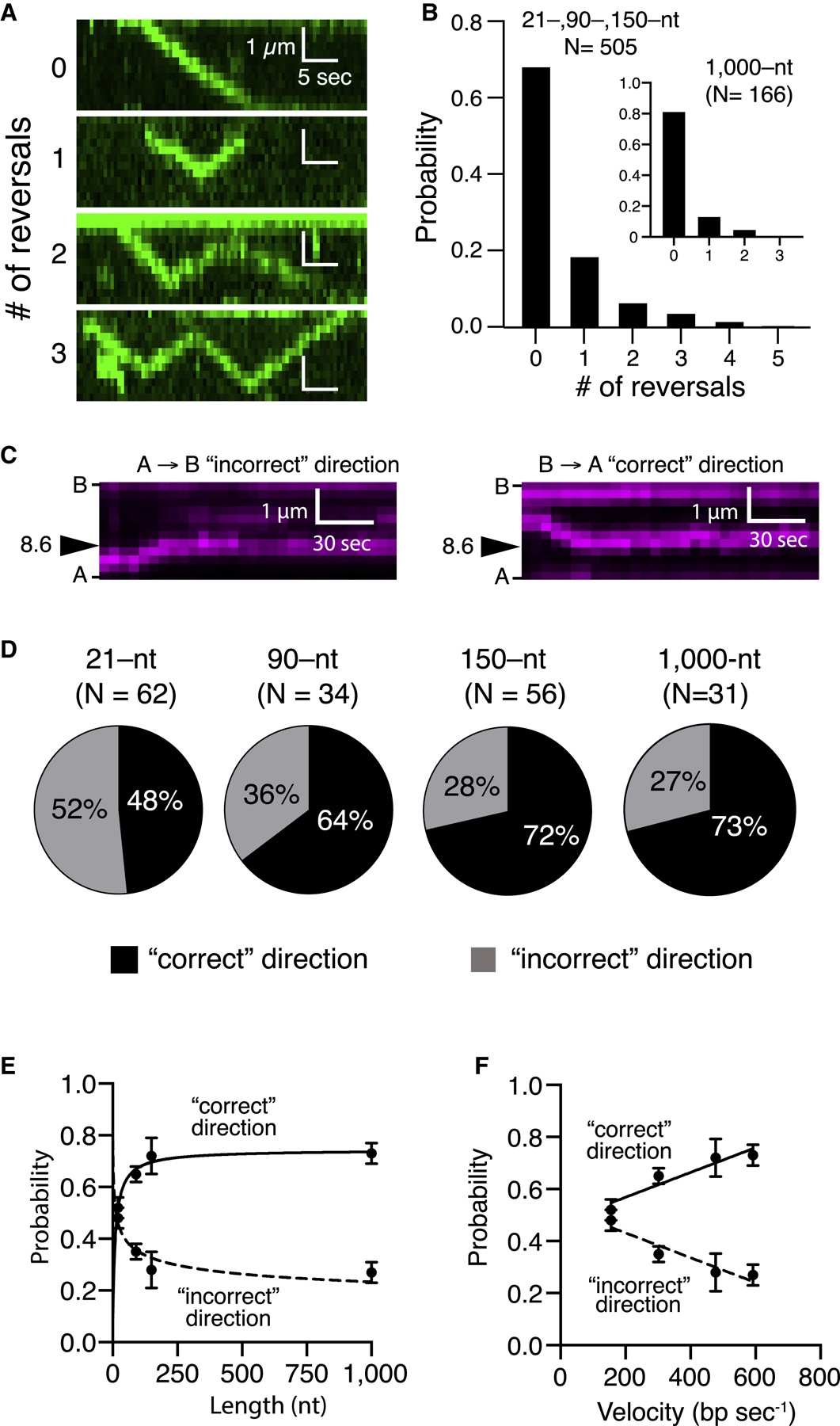
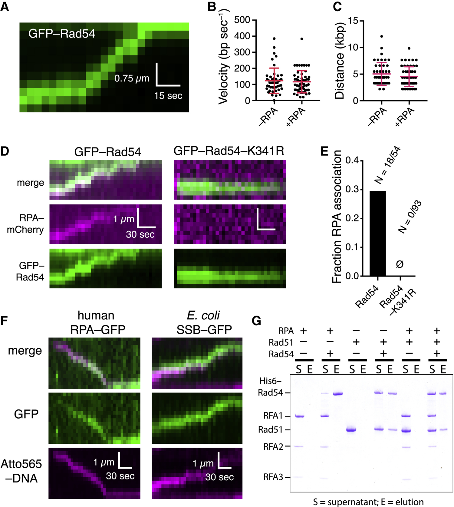
Homologous recombination (HR) helps maintain genome integrity, and HR defects give rise to disease, especially cancer. During HR, damaged DNA must be aligned with an undamaged template through a process referred to as the homology search. Despite decades of study, key aspects of this search remain undefined. Here, we use single-molecule imaging to demonstrate that Rad54, a conserved Snf2-like protein found in all eukaryotes, switches the search from the diffusion-based pathways characteristic of the basal HR machinery to an active process in which DNA sequences are aligned via an ATP-dependent molecular motor-driven mechanism. We further demonstrate that Rad54 disrupts the donor template strands, enabling the search to take place within a migrating DNA bubble-like structure that is bound by replication protein A (RPA). Our results reveal that Rad54, working together with RPA, fundamentally alters how DNA sequences are aligned during HR.

Keywords: DNA curtain; DNA double-strand break; Rad51; Rad54; homologous recombination; homology search; motor protein; replication protein A; single molecule.


Conflict of interest statement

Declaration of Interests The authors declare no competing interests.

Figures

Figure 1.
Figure 1.. Rad54 Acts as a Molecular Motor during the Homology Search
(A) DNA curtain assay. (B) PSC preparation. (C) Kymograph showing a PSC containing Rad51,GFP-Rad54 (green), and Atto565-DNA (magenta) translocating on the donor dsDNA. (D) Kymograph illustrating the behavior of a GFP-Rad54-K341R PSC. (E) Distribution of PSC translocation velocities(represents combined datasets for Rad54 and GFP-Rad54); the solid line is a Gaussian fit to the data. (F) Linear translocation distance graphed as survival probability; error bars (SD) were generated by bootstrapping (represents combined datasets for Rad54 and GFP-Rad54). (G) Distribution of PSC translocation velocities forthe tailed duplex DNA (21-nt ssDNA) and 90-, 150-, and 1,000-nt ssDNAs. Red lines represent mean and SD. In each case, the ssDNA is fully homologous to a specific target site on the donor dsDNA (Figure 2).
Figure 2.
Figure 2.. Target Recognition during the Homology Search
(A) Donor dsDNA schematic highlighting the locations of two different homologous targets. (B) Images of PSCs bound at target 8.6. (C) Binding distribution for PSCs containing Atto565-DNA homologous to target 8.6; error bars for all binding distributions (SD) were generated by bootstrapping. (D) Distribution of PSCs containing GFP-Rad54-K341R and Atto565-DNA. (E) Kymographs showing target recognition and target bypass. (F) Homology recognition and bypass for PSCs with 21 nt of homology. (G) Homology recognition efficiency for different lengths of homology; error bars represent SD of three independent experiments. Homology lengths of 0, 7, 9, 15, and 21 all correspond to the tailed duplex substrate with a 21-nt ssDNA overhang. The 90-, 150-, and 1,000-nt substrates were ssDNA molecules fully homologous to the 8.6 target site on the donor dsDNA.
Figure 3.
Figure 3.. Homology Recognition and Translocation Direction
(A) Kymographs illustrating examples of varying numbers of reversal events by the PSC (unlabeled Rad51, GFP-Rad54 [green], and unlabeled 150-nt ssDNA). (B) Frequency of observed translocation reversalevents; the main panel corresponds to pooled datasets for the tailed duplex (21-nt ssDNA), 90 nt and 150 nt, which were all similar in reversal characteristics, and the inset corresponds to the 1,000-nt ssDNA substrate. (C) Kymographs illustrating examples of homologoustarget recognition for translocation events occurring in either direction. B, barrier; A, anchor (Figure 1A; unlabeled Rad51, unlabeled Rad54, and Atto565-DNA [magenta]). (D) Relative fraction of first-passage recognition events occurring for PSC translocation in either direction for different-length PSC substrates. (E and F) Relative fraction of first-passage recognition events for either direction as a function of (E) PSC ssDNA length or (F) translocation velocity. The error bars represent the SD for three independent experiments. (E) and (F) represent different presentations and analyses of the same experimental dataset.
Figure 4.
Figure 4.. RPA Co-localizes with the PSC during the Homology Search
(A) Co-localization of RPA-GFP with the PSC. (B) Fraction of PSCs (translocating and stationary) that co-localize with RPA. (C) Fraction of PSCs that undergo translocationwith RPA co-localization. (D) First-passage recognition efficiency for different-length PSCs in the presence and absence of RPA; this dataset is not segregated for direction of approach. The error bars represent the SD of three independent experiments. (E) Fraction of first-passage recognition events forPSCs approaching the target site from the correct and incorrect directions for different-length PSCs in the presence and absence of RPA. The error bars represent the SD of three independent experiments. (F) Photobleaching analysis to count the number ofRPA molecules associated with the PSC. (G) RPA-GFP signal intensity for translocating andstationary PSCs and cumulative datasets. The red lines represent the mean and SD. (H) RPA-GFP signal intensity of different-length PSCs. The red lines represent the mean and SD.
Figure 5.
Figure 5.. Rad54 Alone Can Open dsDNA Strands during Translocation
(A) Kymograph showing GFP-Rad54 (green) translocation. (B) Comparison of translocation velocities of GFP-Rad54 only in the presence and absence of RPA; red lines represent mean and SD. (C) GFP-Rad54 processivity in the presence andabsence of RPA; red lines represent mean and SD. (D) Kymographs showing GFP-Rad54 or GFP-Rad54-K341R in the presence of RPA-mCherry. (E) Fraction of GFP-Rad54 and GFP-Rad54-K341R that co-localize with RPA-mCherry. (F) Kymographs showing human RPA and E. coli SSB tracking with the translocating S. cerevisiae PSC. (G) Pull-down assay showing that Rad54 and RPAdo not interact in solution.
Figure 6.
Figure 6.. Nucleosome Remodeling and Bypass during the Homology Search
(A) Schematic of nucleosome-bound donor dsDNA substrates labeled with Atto565-H2A or Alexa 488-H4. (B) Summary of different outcomes during PSC encounters with single nucleosomes for labeled H2A or labeled H4 with PSCs prepared with the tailed duplex (21-nt ssDNA) substrate. The bottom graph shows the combined labeled H2A and labeled H4 datasets. (C) Kymographs showing examples Atto565-H2A nucleosomes (magenta) being remodeled by PSCs labeled with GFP-Rad54 (green, left panels) and examples of Alexa 488-H4 nucleosomes (green) being remodeled by PSCs labeled with Atto565-labeled tailed duplex DNA (magenta, right panels). (D) Distributions of nucleosome remodeling events, sliding or eviction, for H2A-Atto565-labeled, H4-Alexa 488-labeled, and combined datasets. (E) Kymographs depicting nucleosomes (magenta) being bypassed by translocating PSCs (green) with or without evident PSC pausing at the nucleosome. (F) Fraction of bypass events where the PSCs pause during nucleosome bypass for nucleosomes labeled with H2A-Atto565, H4-Alexa 488, and combined datasets.
Figure 7.
Figure 7.. Model Describing the Influence of Rad54 on the Homology Search Mechanism
(A) Schematic depiction of the PSC linked to a donor dsDNA template via the binding activity of Rad54, where ATP-dependent forward progression of the complex is coupled to deformation of the DNA duplex, enabling RPA association, which, in turn, promotes homology recognition. (B) Model depicting rapid sampling of donor DNA by Rad51 ssDNA within the translocating PSC. In the cartoon schematic, Rad51 is not depicted for clarity, and Rad54 is shown bound at or near the 3′ end of the PSC ssDNA; similar principles may apply regardless of where Rad54 is located within the PCS. Additional details are in the main text.

References

    1. Adzuma K (1998). No sliding during homology search by RecA protein. J. Biol. Chem 273, 31565–31573. - PubMed
    1. Agarwal S, van Cappellen WA, Guénolé A, Eppink B, Linsen SE, Meijering E, Houtsmuller A, Kanaar R, and Essers J (2011). ATP-dependent and independent functions of Rad54 in genome maintenance. J. Cell Biol 192, 735–750. - PMC - PubMed
    1. Alexeev A, Mazin A, and Kowalczykowski SC (2003). Rad54 protein possesses chromatin-remodeling activity stimulated by the Rad51-ssDNA nucleoprotein filament. Nat. Struct. Biol 10, 182–186. - PubMed
    1. Alexiadis V, and Kadonaga JT (2002). Strand pairing by Rad54 and Rad51 is enhanced by chromatin. Genes Dev. 16, 2767–2771. - PMC - PubMed
    1. AlHaj Abed J, Erceg J, Goloborodko A, Nguyen SC, McCole RB, Saylor W, Fudenberg G, Lajoie BR, Dekker J, Mirny LA, and Wu CT (2019). Highly structured homolog pairing reflects functional organization of the Drosophila genome. Nat. Commun 10, 4485. - PMC - PubMed

Publication types

MeSH terms