. 2020 Jul 6;30(13):2495-2507.e7.
doi: 10.1016/j.cub.2020.04.071. Epub 2020 Jun 4.

Pathogenic Allodiploid Hybrids of Aspergillus Fungi

Affiliations

Pathogenic Allodiploid Hybrids of Aspergillus Fungi

Jacob L Steenwyk et al. Curr Biol. .

Abstract

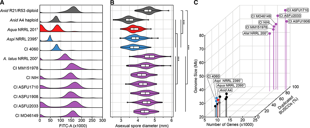
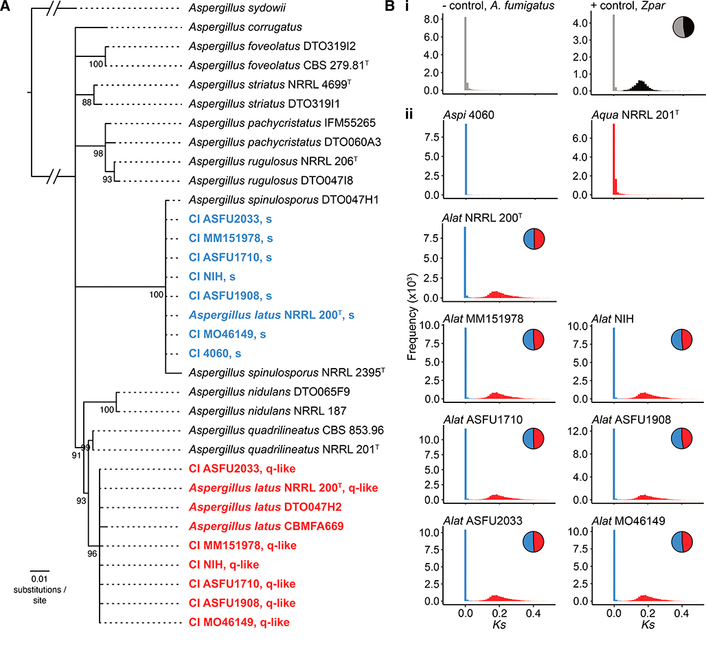
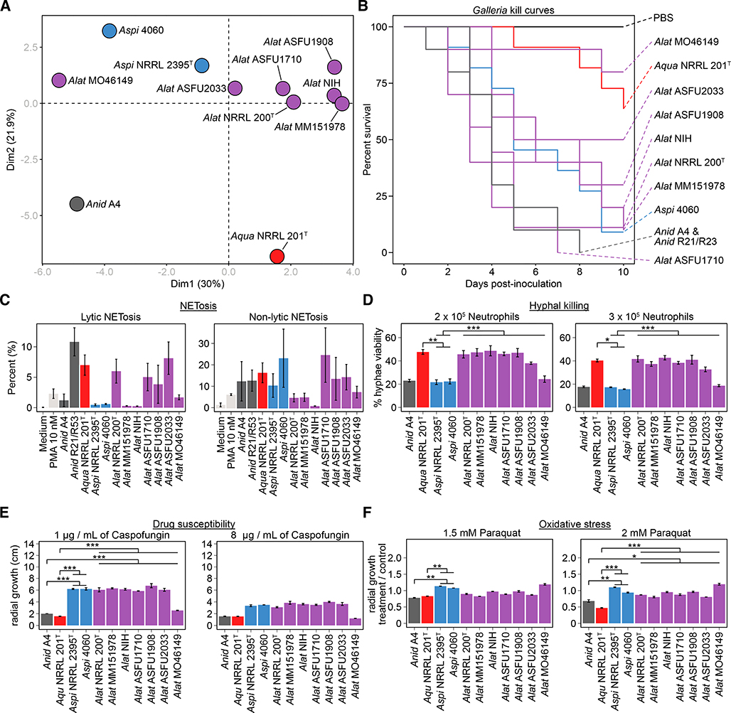
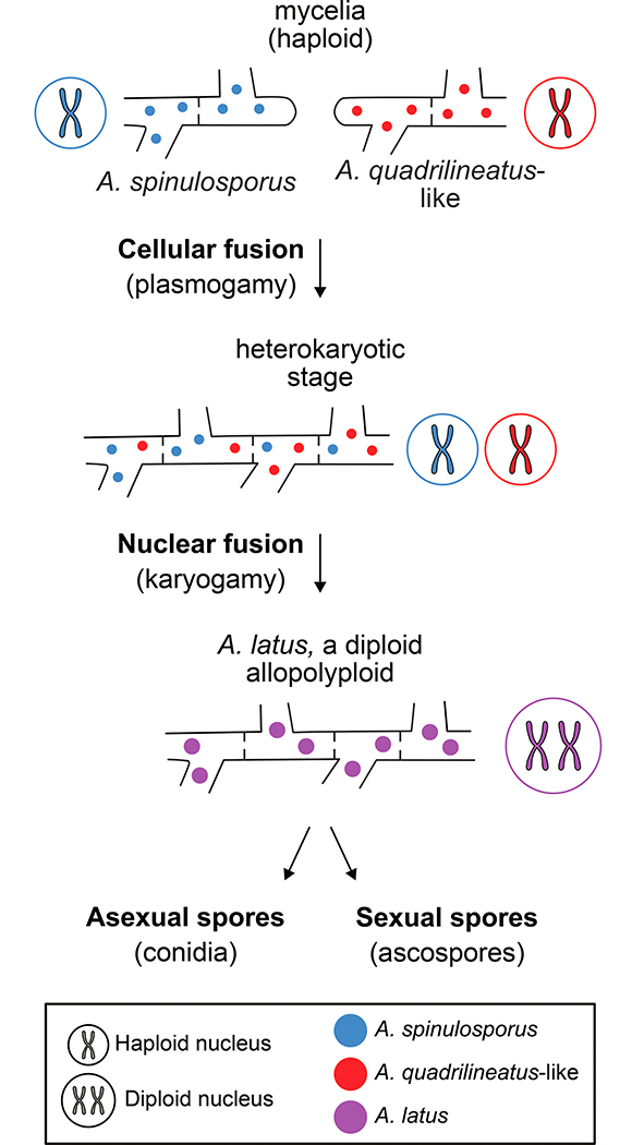
Interspecific hybridization substantially alters genotypes and phenotypes and can give rise to new lineages. Hybrid isolates that differ from their parental species in infection-relevant traits have been observed in several human-pathogenic yeasts and plant-pathogenic filamentous fungi but have yet to be found in human-pathogenic filamentous fungi. We discovered 6 clinical isolates from patients with aspergillosis originally identified as Aspergillus nidulans (section Nidulantes) that are actually allodiploid hybrids formed by the fusion of Aspergillus spinulosporus with an unknown close relative of Aspergillus quadrilineatus, both in section Nidulantes. Evolutionary genomic analyses revealed that these isolates belong to Aspergillus latus, an allodiploid hybrid species. Characterization of diverse infection-relevant traits further showed that A. latus hybrid isolates are genomically and phenotypically heterogeneous but also differ from A. nidulans, A. spinulosporus, and A. quadrilineatus. These results suggest that allodiploid hybridization contributes to the genomic and phenotypic diversity of filamentous fungal pathogens of humans.

Keywords: Eurotiomycetes; allopolyploidy; ascomycota; aspergillosis; cryptic species; fungal pathogen evolution; hybridization; nonvertical evolution; pathogenicity; virulence.


Conflict of interest statement

Declaration of Interests The authors declare no competing interests.

Figures

Figure 1.
Figure 1.. Six clinical isolates previously characterized as Aspergillus nidulans and the type strain of Aspergillus latus are diploids.
(A) Fluorescence-assisted cell sorting analysis suggests that the type strain of Aspergillus latus NRRL 200T and 6 clinical isolates (MM151978, NIH, ASFU1710, ASFU1908, ASFU2033, and MO46149) previously identified as Aspergillus nidulans have diploid genomes. In contrast, Aspergillus spinulosporus NRRL2395 and clinical isolate 4060 have haploid genomes. The haploid A. nidulans strain A4 and the laboratory-induced diploid A. nidulans strain R21/R23 were used as references of haploid and diploid genomes, respectively. (B) Asexual spore diameter is significantly different between the 6 diploid clinical isolates, the haploid A. quadrilineatus, A. spinulosporus, and A. nidulans, and the laboratory-induced diploid A. nidulans2 = 399.54, df = 2, p < 0.001; Kruskal-Wallis rank sum test). Additional pairwise comparisons are shown by brackets; all comparisons used Dunn’s test with Benjamini-Hochberg method of multi-test correction. *** p-value ≤ 0.001. (C) The 6 diploid clinical isolates and the A. latus NRRL 200T strain have substantially larger genome sizes, gene numbers, and percent duplicated BUSCO genes compared to haploid genomes of representative Aspergillus species (A. clavatus NRRL 1, A. flavus NRRL 3357, A. fumigatus Af293, A. nidulans A4, A. niger CBS 513.88, A. sydowii CBS 593.65, and A. versicolor CBS 583.65). Genus and species names are abbreviated using the following scheme: A. latus (Alat), A. spinulosporus (Aspi), A. quadrilineatus (Aqua), and A. nidulans (Anid). CI represents clinical isolates. Dark grey represents A. nidulans; red represents A. quadrilineatus; blue represents A. spinulosporus and CI 4060; purple represents A. latus and diploid isolates. See also Table 1, S1, S2, S3, and Figure S1.
Figure 2.
Figure 2.. The 6 clinical diploids belong to A. latus, an allodiploid species formed via hybridization of A. spinulosporus and a close relative of A. quadrilineatus.
(A) The type strain of A. latus NRRL 200T and the 6 diploid clinical isolates have each two copies of the taxonomic markers β-tubulin and calmodulin. Phylogenetic analysis of their β-tubulin and calmodulin sequences together with sequences from representative taxa in section Nidulantes [36] suggests that clinical isolate 4060 belongs to A. spinulosporus whereas A. latus NRRL 200T and the 6 diploid clinical isolates are derived from two parental genomes. Interestingly, neither of the parental genomes is A. nidulans; rather one is A. spinulosporus and the other is a species closely related to Aspergillus quadrilineatus. Newly sequenced isolates are shown in red and blue. (B) Examination of sequence divergence (Ks; x-axis) between each gene in an allodiploid and its best blast hit in A. spinulosporus confirms that the 6 diploid clinical isolates and the type strain of A. latus are allodiploid hybrids. In contrast, the 7th clinical isolate (4060) is a haploid A. spinulosporus. Similarly, we found no evidence of A. quadrilineatus NRRL 201T forming via allodiploid hybridization. (Bi) Examination of the haploid, non-hybrid genome of A. fumigatus strain Af293 (negative control) shows a unimodal distribution, whereas examination of the diploid, hybrid genome Zygosaccharomyces parabailii strain NBRC1047/ATCC56075 (Zpar) shows a bimodal distribution (positive control; grey represents genes from one parent; black represents genes from the other parent). Red represents genes assigned to the A. quadrilineatus-like parental genome; blue represents genes assigned to the A. spinulosporus parental genome. See also Figure S2, S3, S4, and Data S1.
Figure 3.
Figure 3.. A. latus hybrids exhibit strain heterogeneity and differ from parental species, A. quadrilineatus, and A. nidulans in infection-relevant phenotypes.
(A) Principal component analysis of diverse infection-relevant phenotypes reveals strain heterogeneity among A. latus hybrids and that they differ from the closest relative of the unknown parent, A. quadrilineatus, and the A. nidulans A4 strain. Data for each phenotype was scaled prior to principal component analysis. (B, C) Wide phenotypic variation among strains of A. latus hybrids as well as the various species tested was observed for virulence in Galleria moth model of disease and NETosis. (D) Examination of the percentage of hyphal viability between the various species revealed significant differences (F(3) = 24.514, p < 0.001; Multi-factor ANOVA). (E) Examination of the caspofungin drug susceptibility profiles among A. nidulans, A. spinulosporus, and the A. latus revealed differences between the three species (F(3) = 56.01, p < 0.001; Multi-factor ANOVA). At 1 μg / mL caspofungin treatment, A. spinulosporus and A. latus hybrids grew more than A. nidulans and A. quadrilineatus (p < 0.001 for both comparisons; Tukey honest significant differences test). We found no statistically significant differences between the various species at 8 μg / mL of caspofungin but observed a qualitative difference similar to growth in 1 μg / mL of caspofungin. (F) Examination of growth in the presence of the oxidative stress agent paraquat revealed differences among the various species (F(3) = 30.25, p < 0.001; Multi-factor ANOVA). Genus and species names are abbreviated using the following scheme: A. latus (Alat), A. spinulosporus (Aspi), A. quadrilineatus (Aqua), and A. nidulans (Anid). Dark grey represents A. nidulans; red represents A. quadrilineatus; blue represents A. spinulosporus; purple represents A. latus. All pairwise comparisons shown by brackets were examined using a Tukey honest significant differences test. * 0.01 ≤ p-value ≤ 0.05; ** 0.001 ≤ p-value ≤ 0.01; *** p-value ≤ 0.001. See also Figure S5.
Figure 4.
Figure 4.. Proposed model for the evolution of A. latus via allodiploid hybridization.
Under the model, haploid nuclei of an A. spinulosporus isolate and of an isolate from an A. spinulosporus-like species underwent cellular fusion (plasmogamy) forming a heterokaryotic mycelium (i.e., a mycelium where cells contain two distinct nuclei). Next, nuclear fusion (karyogamy) resulted in the merging of the two genetically distinct nuclei and their genomes into a single one, giving rise to the allodiploid species A. latus which, is capable of undergoing asexual and sexual reproduction to produce asexual spores (conidia) or sexual spores (ascospores).

Similar articles

Cited by

References

    1. Abbott R, Albach D, Ansell S, Arntzen JW, Baird SJE, Bierne N, Boughman J,Brelsford A, Buerkle CA, Buggs R, et al. (2013). Hybridization and speciation. J. Evol. Biol. 26, 229–246. - PubMed
    1. Baack EJ, and Rieseberg LH (2007). A genomic view of introgression and hybrid speciation. Curr. Opin. Genet. Dev. 17, 513–518. - PMC - PubMed
    1. Rieseberg LH (2003). Major Ecological Transitions in Wild Sunflowers Facilitated by Hybridization. Science (80-. ). 301, 1211–1216. - PubMed
    1. Rieseberg LH, Kim S-C, Randell RA, Whitney KD, Gross BL, Lexer C, and Clay K (2007). Hybridization and the colonization of novel habitats by annual sunflowers. Genetica 129, 149–165. - PMC - PubMed
    1. Mable BK, Alexandrou MA, and Taylor MI (2011). Genome duplication in amphibians and fish: an extended synthesis. J. Zool. 284, 151–182.

Publication types

Supplementary concepts

LinkOut - more resources