Three-Dimensional Zero Echo Time Magnetic Resonance Imaging Versus 3-Dimensional Computed Tomography for Glenoid Bone Assessment
- PMID: 32502712
- PMCID: PMC7483823
- DOI: 10.1016/j.arthro.2020.05.042
Three-Dimensional Zero Echo Time Magnetic Resonance Imaging Versus 3-Dimensional Computed Tomography for Glenoid Bone Assessment
Abstract
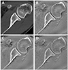
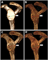
Purpose: To evaluate the 3-dimensional (3D) zero echo time (ZTE) magnetic resonance imaging (MRI) technique and compare it with 3D computed tomography (CT) for the assessment of the glenoid bone.
Methods: ZTE MRI using multiple resolutions and multislice CT were performed in 6 shoulder specimens before and after creation of glenoid defects and in 10 glenohumeral instability patients. Two musculoskeletal radiologists independently generated 3D volume-rendered images of the glenoid en face. Post-processing times and glenoid widths were measured. Inter-modality and inter-rater agreement was assessed.
Results: Intraclass correlation coefficients (ICCs) for inter-modality assessment showed almost perfect agreement for both readers, ranging from 0.949 to 0.991 for the ex vivo study and from 0.955 to 0.987 for the in vivo patients. Excellent interobserver agreement was found for both the ex vivo (ICCs ≥ 0.98) and in vivo (ICCs ≥ 0.92) studies. For the ex vivo study, Bland-Altman analyses for CT versus MRI showed a mean difference of 0.6 to 1 mm at 1.0-mm3 MRI resolution, 0.3 to 0.6 mm at 0.8-mm3 MRI resolution, and 0.3 to 0.6 mm at 0.6-mm3 MRI resolution for both readers. For the in vivo study, Bland-Altman analyses for CT versus MRI showed a mean difference of 0.6 to 0.8 mm at 1.0-mm3 MRI resolution, 0.5 to 0.6 mm at 0.8-mm3 MRI resolution, and 0.4 to 0.8 mm at 0.7-mm3 MRI resolution for both readers. Mean post-processing times to generate 3D images of the glenoid ranged from 32 to 46 seconds for CT and from 33 to 64 seconds for ZTE MRI.
Conclusions: Three-dimensional ZTE MRI can potentially be considered as a technique to determine glenoid width and can be readily incorporated into the clinical workflow.
Level of evidence: Level II, development of diagnostic criteria (consecutive patients with consistently applied reference standard and blinding).
Copyright © 2020 Arthroscopy Association of North America. All rights reserved.
Figures


Comment in
-
Editorial Commentary: Can We Evaluate Glenoid Bone With Magnetic Resonance Imaging? Yes, If You Have the Right Sequence.Arthroscopy. 2020 Sep;36(9):2401-2402. doi: 10.1016/j.arthro.2020.07.029. Arthroscopy. 2020. PMID: 32891242
References
-
- Enger M, Skjaker SA, Melhuus K, et al. Shoulder injuries from birth to old age: A 1-year prospective study of 3031 shoulder injuries in an urban population. Injury. 2018;49:1324–1329. - PubMed
-
- Zacchilli MA, Owens BD. Epidemiology of shoulder dislocations presenting to emergency departments in the United States. J Bone Joint Surg Am. 2010;92:542–549. - PubMed
-
- Burkhart SS, De Beer JF. Traumatic glenohumeral bone defects and their relationship to failure of arthroscopic Bankart repairs: significance of the inverted-pear glenoid and the humeral engaging Hill-Sachs lesion. Arthroscopy. 2000;16:677–694. - PubMed
-
- Chalmers PN, Christensen G, O’Neill D, Tashjian RZ. Does Bone Loss Imaging Modality, Measurement Methodology, and Interobserver Reliability Alter Treatment in Glenohumeral Instability? Arthroscopy. 2020;36:12–19. - PubMed
Publication types
MeSH terms
Grants and funding
LinkOut - more resources
Full Text Sources
Medical
Research Materials

