Review
. 2020 Jun 3;9(2):21.
doi: 10.3390/antib9020021.

Construction of Antibody Phage Libraries and Their Application in Veterinary Immunovirology

Affiliations
Review

Construction of Antibody Phage Libraries and Their Application in Veterinary Immunovirology

Shahbaz Bashir et al. Antibodies (Basel). .

Abstract

Antibody phage display (APD) technology has revolutionized the field of immunovirology with its application in viral disease diagnostics and antiviral therapy. This robust and versatile technology allows the expression of an antibody fused to a phage coat protein on the surface of a filamentous phage. The DNA sequence coding for the antibody is packaged within the phage, linking the phenotype to genotype. Antibody phage display inherits the ability to rapidly generate and modify or improve high-affinity monoclonal antibodies, rendering it indispensable in immunology. In the last two decades, phage-display-derived antibodies have been extensively used in human medicine as diagnostic and therapeutic modalities. Recently, they are also gaining significant ground in veterinary medicine. Even though these advancements are mainly biased towards economically important animals such as chicken, cattle, and pigs, they are laying the foundation of fulfilling the unmet needs of veterinary medicine as antibody-based biologics in viral diagnostics, therapeutics, and immunoprophylaxis. This review provides a brief overview of the construction of antibody phage libraries and their application in diagnosis, prevention, and control of infectious viral diseases in veterinary medicine in detail.

Keywords: immunovirology; monoclonal antibody; phage display; veterinary medicine.


Conflict of interest statement

The authors declare no conflict of interest.

Figures

Figure 1
Figure 1
(A) Structure of a conventional antibody and its derivatives. Abbreviations: Fv, fragment variable; Fc, fragment crystallizable; Fab, fragment antigen binding; scFv, single-chain variable fragment with linker (yellow); scFab, single-chain Fab; VH, variable domain heavy chain; F(ab)2, two Fab fragments joined together; scFv–Fc, scFv linked to Fc (inter- and intra-disulfide linkages are not shown here in the all formats). (B) Camelid heavy chain antibody (HCAb); VHH, nanobody (modified from [3]).
Figure 2
Figure 2
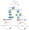
Synthesis and cloning of scFv and Fab fragments in a phagemid vector. It starts with the preparation of peripheral blood mononuclear cells (PBMCs) using density gradient from whole blood. Following RNA extraction from PBMCs, cDNA is synthesized through reverse transcription. From cDNA, the variable region of heavy (VH) and light chain (VL) is amplified through PCR using specific primers. scFv and Fab are constructed through splicing by overlap extension PCR(SOE-PCR). The amplified scFv and Fab are ligated into the phagemid vector following restriction digestion of both.
Figure 3
Figure 3
Schematic diagram of M13 phage. It carries ~6.4-kb sized circular single-stranded DNA (ssDNA), which encodes for 10 proteins, i.e., p I, p II, p III, p IV, p V, p VI, p VII, p VIII, p IX, and p X.
Figure 4
Figure 4
Panning of an antibody phage display library for the generation of high-affinity antibodies. The antigen is presented in its native form either immobilized on a plastic surface or expressed on the cell surface. For membrane protein targets, nanodisc provides a membrane-like environment. Antibody library displaying phages are incubated with antigen, and non-bound or weakly bound phages are removed by washing. The tightly bound phages are recovered by elution via trypsin or pH shift for the re-infection of E. coli cells. Following the coinfection with helper phages, new phage particles are produced for usage in subsequent rounds of panning. This cycle continues for 3–4 rounds, leading to enrichment of binders.

Similar articles

Cited by

References

    1. Hoogenboom H.R., De Brune A.P., Hufton S.E., Hoet R.M., Arends J.W., Roovers R.C. Antibody phage display technology and its applications. Immunotechnology. 1998;4:1–20. doi: 10.1016/S1380-2933(98)00007-4. - DOI - PubMed
    1. Ahmad Z.A., Yeap S.K., Ali A.M., Ho W.Y., Alitheen N.B.M., Hamid M. ScFv antibody: Principles and clinical application. Clin. Dev. Immunol. 2012;2012:1–15. - PMC - PubMed
    1. Kuhn P., Fühner V., Unkauf T., Moreira G.M.S.G., Frenzel A., Miethe S., Hust M. Recombinant antibodies for diagnostics and therapy against pathogens and toxins generated by phage display. Proteom.-Clin. Appl. 2016;10:922–948. - PMC - PubMed
    1. Hairul Bahara N.H., Tye G.J., Choong Y.S., Ong E.B.B., Ismail A., Lim T.S. Phage display antibodies for diagnostic applications. Biologicals. 2013;41:209–216. doi: 10.1016/j.biologicals.2013.04.001. - DOI - PubMed
    1. Burkovitz A., Ofran Y. Understanding differences between synthetic and natural antibodies can help improve antibody engineering. MAbs. 2016;8:278–287. doi: 10.1080/19420862.2015.1123365. - DOI - PMC - PubMed

LinkOut - more resources