

Anti-Tumor Effects of Biomimetic Sulfated Glycosaminoglycans on Lung Adenocarcinoma Cells in 2D and 3D In Vitro Models
- PMID: 32503108
- PMCID: PMC7321182
- DOI: 10.3390/molecules25112595
Anti-Tumor Effects of Biomimetic Sulfated Glycosaminoglycans on Lung Adenocarcinoma Cells in 2D and 3D In Vitro Models
Abstract
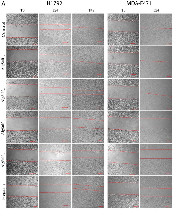
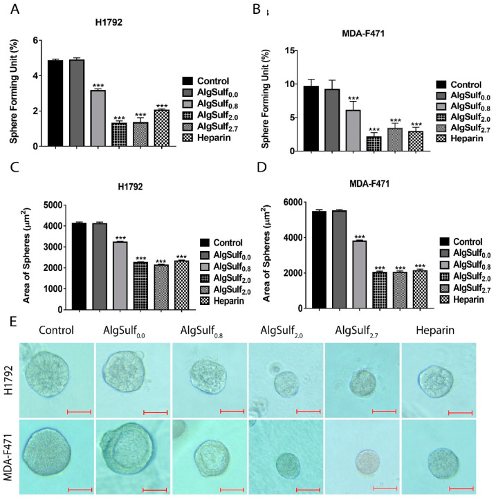
Lung cancer development relies on cell proliferation and migration, which in turn requires interaction with extracellular matrix (ECM) components such as glycosaminoglycans (GAGs). The mechanisms through which GAGs regulate cancer cell functions are not fully understood but they are, in part, mediated by controlled interactions with cytokines and growth factors (GFs). In order to mechanistically understand the effect of the degree of sulfation (DS) of GAGs on lung adenocarcinoma (LUAD) cells, we synthesized sulfated alginate (AlgSulf) as sulfated GAG mimics with DS = 0.0, 0.8, 2.0, and 2.7. Human (H1792) and mouse (MDA-F471) LUAD cell lines were treated with AlgSulf of various DSs at two concentrations 10 and 100 µg/mL and their anti-tumor properties were assessed using 3-(4,5-dimethylthiazol-2-yl)-2,5-diphenyltetrazolium bromide (MTT), trypan blue exclusion, and wound healing assays for 2D models and sphere formation assay for the 3D model. The proliferation and number of live MDA-F471 cells at the concentration of 100 µg/mL decreased significantly with the increase in the DS of biomimetic GAGs. In addition, the increase in the DS of biomimetic GAGs decreased cell migration (p < 0.001 for DS = 2.0 and 2.7 compared to control) and decreased the diameter and number of spheres formed (p < 0.001). The increased DS of biomimetic GAGs attenuated the expression of cancer stem cell (CSC)/progenitor markers in the 3D cultures. In conclusion, GAG-mimetic AlgSulf with increased DS exhibit enhanced anti-proliferative and migratory properties while also reducing growth of KRAS-mutant LUAD spheres in vitro. We suggest that these anti-tumor effects by GAG-mimetic AlgSulf are possibly due to differential binding to GFs and consequential decreased cell stemness. AlgSulf may be suitable for applications in cancer therapy after further in vivo validation.
Keywords: biomimetic; cancer stem cells; glycosaminoglycans; lung adenocarcinoma; sulfated alginates.
Conflict of interest statement
The authors declare no conflict of interest.
Figures


Similar articles
-
Biomimetic sulfated glycosaminoglycans maintain differentiation markers of breast epithelial cells and preferentially inhibit proliferation of cancer cells.Acta Biomater. 2021 Mar 1;122:186-198. doi: 10.1016/j.actbio.2020.12.049. Epub 2021 Jan 11. Acta Biomater. 2021. PMID: 33444795
-
The sulfation of biomimetic glycosaminoglycan substrates controls binding of growth factors and subsequent neural and glial cell growth.Biomater Sci. 2019 Oct 1;7(10):4283-4298. doi: 10.1039/c9bm00964g. Epub 2019 Aug 13. Biomater Sci. 2019. PMID: 31407727
-
Sulfated Alginate as a Mimic of Sulfated Glycosaminoglycans: Binding of Growth Factors and Effect on Stem Cell Behavior.Adv Biosyst. 2017 Jul;1(7):e1700043. doi: 10.1002/adbi.201700043. Epub 2017 Jun 16. Adv Biosyst. 2017. PMID: 32646173
-
Glycosaminoglycans (GAGs) and GAG mimetics regulate the behavior of stem cell differentiation.Colloids Surf B Biointerfaces. 2017 Feb 1;150:175-182. doi: 10.1016/j.colsurfb.2016.11.022. Epub 2016 Nov 20. Colloids Surf B Biointerfaces. 2017. PMID: 27914254 Review.
-
Sulfated glycosaminoglycans as promising artificial extracellular matrix components to improve the regeneration of tissues.Curr Med Chem. 2013;20(20):2501-23. doi: 10.2174/0929867311320200001. Curr Med Chem. 2013. PMID: 23521682 Review.
Cited by
-
Glycosaminoglycan from Ostrea rivularis attenuates hyperlipidemia and regulates gut microbiota in high-cholesterol diet-fed zebrafish.Food Sci Nutr. 2021 Jul 26;9(9):5198-5210. doi: 10.1002/fsn3.2492. eCollection 2021 Sep. Food Sci Nutr. 2021. PMID: 34532028 Free PMC article.
-
Extracellular Matrix Sulfation in the Tumor Microenvironment Stimulates Cancer Stemness and Invasiveness.Adv Sci (Weinh). 2024 Sep;11(36):e2309966. doi: 10.1002/advs.202309966. Epub 2024 Jul 31. Adv Sci (Weinh). 2024. PMID: 39083319 Free PMC article.
-
Sulfoconjugation of protein peptides and glycoproteins in physiology and diseases.Pharmacol Ther. 2023 Nov;251:108540. doi: 10.1016/j.pharmthera.2023.108540. Epub 2023 Sep 28. Pharmacol Ther. 2023. PMID: 37777160 Free PMC article. Review.
-
Construction of a multiple-class classifier based on mRNAs and lncRNA FAM66A and PSORS1C3 for predicting distant metastasis in lung adenocarcinoma.Ann Transl Med. 2022 Oct;10(20):1129. doi: 10.21037/atm-22-4651. Ann Transl Med. 2022. PMID: 36388820 Free PMC article.
References
MeSH terms
Substances
LinkOut - more resources
Full Text Sources
Medical
Miscellaneous
