

Pentadecanoic Acid, an Odd-Chain Fatty Acid, Suppresses the Stemness of MCF-7/SC Human Breast Cancer Stem-Like Cells through JAK2/STAT3 Signaling
- PMID: 32503225
- PMCID: PMC7352840
- DOI: 10.3390/nu12061663
Pentadecanoic Acid, an Odd-Chain Fatty Acid, Suppresses the Stemness of MCF-7/SC Human Breast Cancer Stem-Like Cells through JAK2/STAT3 Signaling
Abstract
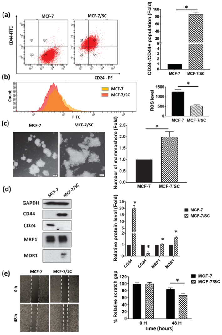
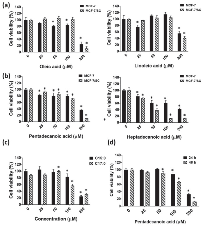
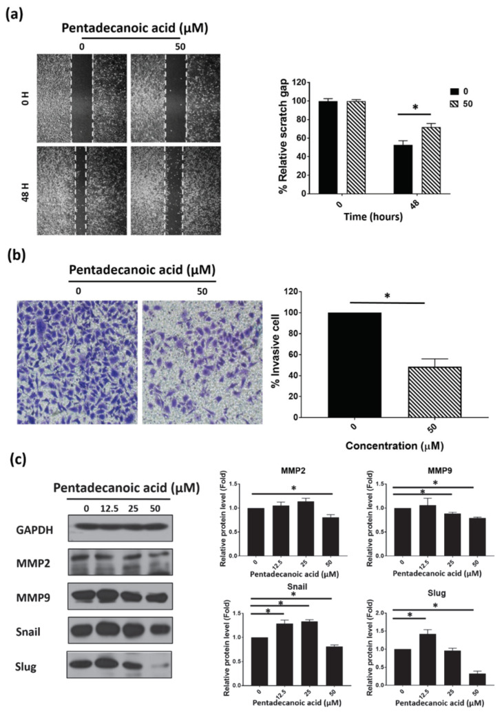
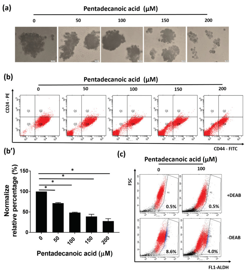
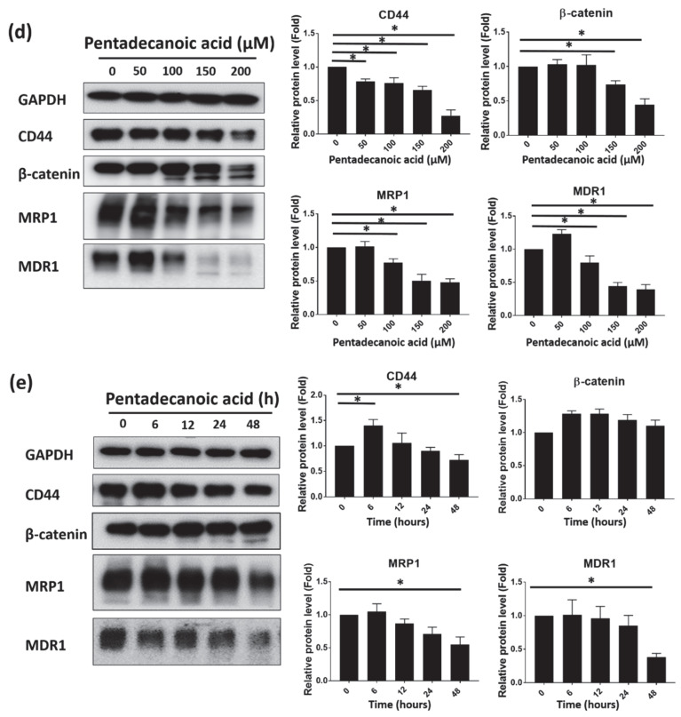
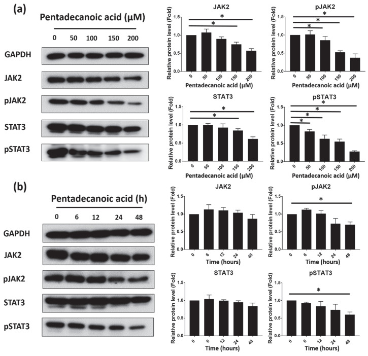
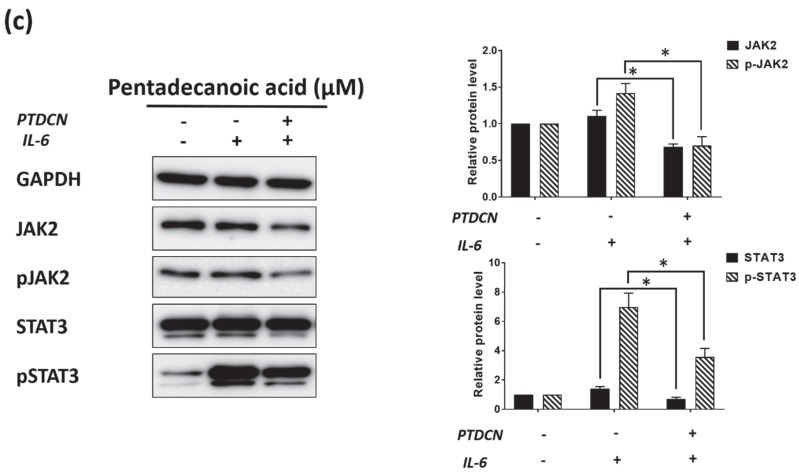
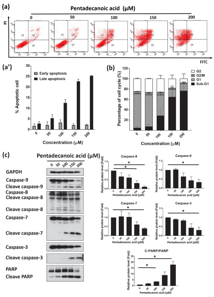
Saturated fatty acids possess few health benefits compared to unsaturated fatty acids. However, increasing experimental evidence demonstrates the nutritionally beneficial role of odd-chain saturated fatty acids in human health. In this study, the anti-cancer effects of pentadecanoic acid were evaluated in human breast carcinoma MCF-7/stem-like cells (SC), a cell line with greater mobility, invasiveness, and cancer stem cell properties compared to the parental MCF-7 cells. Pentadecanoic acid exerted selective cytotoxic effects in MCF-7/SC compared to in the parental cells. Moreover, pentadecanoic acid reduced the stemness of MCF-7/SC and suppressed the migratory and invasive ability of MCF-7/SC as evidenced by the results of flow cytometry, a mammosphere formation assay, an aldehyde dehydrogenase activity assay, and Western blot experiments conducted to analyze the expression of cancer stem cell markers-CD44, β-catenin, MDR1, and MRP1-and epithelial-mesenchymal transition (EMT) markers-snail, slug, MMP9, and MMP2. In addition, pentadecanoic acid suppressed interleukin-6 (IL-6)-induced JAK2/STAT3 signaling, induced cell cycle arrest at the sub-G1 phase, and promoted caspase-dependent apoptosis in MCF-7/SC. These findings indicate that pentadecanoic acid can serve as a novel JAK2/STAT3 signaling inhibitor in breast cancer cells and suggest the beneficial effects of pentadecanoic acid-rich food intake during breast cancer treatments.
Keywords: JAK2/STAT3 signaling; apoptosis; breast cancer; cancer stem cells; odd-chain fatty acids; pentadecanoic acid.
Conflict of interest statement
The authors declare no conflict of interest.
Figures
Similar articles
-
Loss of miR-6844 alters stemness/self-renewal and cancer hallmark(s) markers through CD44-JAK2-STAT3 signaling axis in breast cancer stem-like cells.J Cell Biochem. 2023 Aug;124(8):1186-1202. doi: 10.1002/jcb.30441. Epub 2023 Jul 12. J Cell Biochem. 2023. PMID: 37436061
-
2-Phenylnaphthyridin-4-one Derivative LYF-11 Inhibits Interleukin-6-mediated Epithelial-to-Mesenchymal Transition via the Inhibition of JAK2/STAT3 Signaling Pathway in MCF-7 Cells.Anticancer Res. 2018 May;38(5):2849-2859. doi: 10.21873/anticanres.12530. Anticancer Res. 2018. PMID: 29715108
-
The Effect of a Newly Synthesized Ferrocene Derivative against MCF-7 Breast Cancer Cells and Spheroid Stem Cells through ROS Production and Inhibition of JAK2/STAT3 Signaling Pathway.Anticancer Agents Med Chem. 2020;20(7):875-886. doi: 10.2174/1871520620666200101151743. Anticancer Agents Med Chem. 2020. PMID: 31893999
-
SLUG/SNAI2 and tumor necrosis factor generate breast cells with CD44+/CD24- phenotype.BMC Cancer. 2010 Aug 6;10:411. doi: 10.1186/1471-2407-10-411. BMC Cancer. 2010. PMID: 20691079 Free PMC article.
-
New insights on pentadecanoic acid with special focus on its controversial essentiality: A mini-review.Biochimie. 2024 Dec;227(Pt B):123-129. doi: 10.1016/j.biochi.2024.10.008. Epub 2024 Oct 10. Biochimie. 2024. PMID: 39395658 Review.
Cited by
-
Effects of Combined Pentadecanoic Acid and Tamoxifen Treatment on Tamoxifen Resistance in MCF-7/SC Breast Cancer Cells.Int J Mol Sci. 2022 Sep 26;23(19):11340. doi: 10.3390/ijms231911340. Int J Mol Sci. 2022. PMID: 36232636 Free PMC article.
-
Antioxidant activity of banana flesh and antiproliferative effect on breast and pancreatic cancer cells.Food Sci Nutr. 2022 Jan 26;10(3):740-750. doi: 10.1002/fsn3.2702. eCollection 2022 Mar. Food Sci Nutr. 2022. PMID: 35311172 Free PMC article.
-
The Effect of Genistein Supplementation on Cholesterol Oxidation Products and Fatty Acid Profiles in Serums of Rats with Breast Cancer.Foods. 2022 Feb 20;11(4):605. doi: 10.3390/foods11040605. Foods. 2022. PMID: 35206081 Free PMC article.
-
Impairment of Glucose Metabolism and Suppression of Stemness in MCF-7/SC Human Breast Cancer Stem Cells by Nootkatone.Pharmaceutics. 2022 Apr 21;14(5):906. doi: 10.3390/pharmaceutics14050906. Pharmaceutics. 2022. PMID: 35631492 Free PMC article.
-
Aging-Associated Amyloid-β Plaques and Neuroinflammation in Bottlenose Dolphins (Tursiops truncatus) and Novel Cognitive Health-Supporting Roles of Pentadecanoic Acid (C15:0).Int J Mol Sci. 2025 Apr 16;26(8):3746. doi: 10.3390/ijms26083746. Int J Mol Sci. 2025. PMID: 40332352 Free PMC article.
References
-
- Nandy S.B., Lakshmanaswamy R. Progress in Molecular Biology and Translational Science. Volume 151. Elsevier; Amsterdam, The Netherlands: 2017. Cancer stem cells and metastasis; pp. 137–176. - PubMed
MeSH terms
Substances
Grants and funding
LinkOut - more resources
Full Text Sources
Other Literature Sources
Medical
Research Materials
Miscellaneous
