Review
. 2020 Jun 3;9(6):1390.
doi: 10.3390/cells9061390.

A Proximity Mapping Journey into the Biology of the Mammalian Centrosome/Cilium Complex

Affiliations
Review

A Proximity Mapping Journey into the Biology of the Mammalian Centrosome/Cilium Complex

Melis Dilara Arslanhan et al. Cells. .

Abstract

The mammalian centrosome/cilium complex is composed of the centrosome, the primary cilium and the centriolar satellites, which together regulate cell polarity, signaling, proliferation and motility in cells and thereby development and homeostasis in organisms. Accordingly, deregulation of its structure and functions is implicated in various human diseases including cancer, developmental disorders and neurodegenerative diseases. To better understand these disease connections, the molecular underpinnings of the assembly, maintenance and dynamic adaptations of the centrosome/cilium complex need to be uncovered with exquisite detail. Application of proximity-based labeling methods to the centrosome/cilium complex generated spatial and temporal interaction maps for its components and provided key insights into these questions. In this review, we first describe the structure and cell cycle-linked regulation of the centrosome/cilium complex. Next, we explain the inherent biochemical and temporal limitations in probing the structure and function of the centrosome/cilium complex and describe how proximity-based labeling approaches have addressed them. Finally, we explore current insights into the knowledge we gained from the proximity mapping studies as it pertains to centrosome and cilium biogenesis and systematic characterization of the centrosome, cilium and centriolar satellite interactomes.

Keywords: APEX; BioID; TurboID; centriolar satellites; centrosome; cilia; ciliopathies; microtubules; proximity-labeling.


Conflict of interest statement

The authors declare no conflict of interest.

Figures
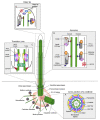
Figure 1
Figure 1
Overview of the anatomy of the centrosome/cilium complex and its sub-compartments. The centrosome/cilium complex is composed of the centrosome, the primary cilium and the centriolar satellites. At the core of the centrosome are two microtubule-based barrel-shaped centrioles, which recruit pericentriolar material (PCM). PCM contains gamma-tubulin ring complexes and functions in microtubule nucleation and organization. In interphase cells, centrioles are tethered to each other by the filamentous structure termed the “G1-G2 tether”. The two centrioles of the centrosome differ in age, structure and maturity. The older centriole is called the mother centriole and the younger centriole is called the daughter centriole. Mother centriole harbors distal and subdistal appendages at its distal end and functions as the basal body to template primary cilium assembly. The primary cilium is compartmentalized into structurally and functionally distinct regions, which include the transition zone, the ciliary axoneme and the ciliary tip. Centriolar satellites are membrane-less granules that cluster around the centrosome. They exhibit microtubule- and molecular motor-dependent active motility as well as Brownian diffusion. (A) Cross-section of the proximal end of the centrioles. Centriole barrel contains symmetrically arranged nine microtubule triplets connected by A–C linkers. The microtubule triplets are connected to the inner core by radial spokes. The inner core is a helical scaffold of a dense matrix that provides structural integrity and flexibility to the centriole barrel. PCM is organized into concentric layers of proteins that are spanned by radially extended filamentous structures formed by CEP152 and pericentrin. (B) The transition zone connects the outer microtubule doublets to the plasma membrane and functions as the diffusion barrier that regulates protein entry into and exit out of the primary cilium. The transition zone is composed of the NPHP-MKS-JBTS module. Transition fibers are the distal appendages of the basal body, which anchor the basal body to the ciliary membrane. In fact, transition fibers correspond to the distal appendages of the mother centriole. (C) The microtubule-based ciliary axoneme forms the core of the primary cilium and serves as tracks for ciliary transport complexes including the IFT-A, IFT-B and BBSome. The anterograde movement of the IFT-B complex and the retrograde movement of the IFT-A complex is powered by molecular motors kinesin-2 (blue) and cytoplasmic dynein-2 (red). BBSome complex interacts with IFT particles and mediates removal of GPCRs from the cilium. (D) The ciliary tip is the specialized region at the distal end of the cilium, which contains IFT particles, Hedgehog pathway components and microtubule-associated proteins and regulates IFT remodeling, cilium length and Hedgehog signaling.
Figure 2
Figure 2
Regulation of the centriole and cilium biogenesis during the cell cycle. Centriole and cilium biogenesis are highly regulated, multi-step processes that are tightly linked to the cell cycle. In the G1 phase, the majority of animal cells have one centrosome composed of a pair of centrioles tethered by the G1-G2 tether at their proximal ends. At the G1 and S phases the two centrioles duplicate only once such that one procentriole forms adjacent to each pre-existing parental centriole. This step is governed by the sequential centriolar recruitment and activity of a conserved set of proteins including the kinase PLK4, the scaffold STIL and the building block of the cartwheel SASS6 along with regulators of these proteins. Following initiation of centriole duplication, procentrioles elongate throughout S and G2 phases. In late G2, the two centrosomes are separated by the dissolution of the G1-G2 tether. In a process termed centriole-to-centrosome conversion, fully elongated centrioles lose their cartwheel and recruit more PCM material in preparation for bipolar spindle assembly. During mitosis, centrosomes assemble the bipolar spindle, which equally segregates both a pair of centrioles and genetic material to daughter cells. Distal appendages undergo transient disassembly during mitosis. At the end of mitosis, the centriole pairs disengage and lose their orthogonal arrangement. Centriole disengagement relicenses the centrioles for centriole duplication in the next cell cycle. As cells enter quiescence by depletion of growth factors, cilium assembles. Steady-state cilium persists into S/G2/M phases and completes disassembly close to cytokinesis after nuclear envelope breakdown.
Figure 3
Figure 3
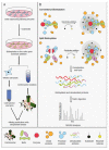
Overview of the proximity-based labeling techniques and their application to the centrosome. (A) Workflow of the application of proximity-based labeling to centrosomes. Cells that express proteins fused to proximity-based labeling enzymes are incubated with the labeling substrates (biotin or biotin-phenol). After biotinylation, cells are harvested and processed for the identification of biotinylated proteins in two different ways. First, centrosomes are enriched by sucrose gradients and the enriched fractions are solubilized under denaturing conditions. Second, cells are solubilized under denaturing conditions. Following lysis, the biotinylated proteins are captured by streptavidin beads and analyzed by mass spectrometry. (B) Schematic representation of conventional and split proximity-based labeling methods. The red circle shows the labeling enzyme that promiscuously biotinylates neighboring proteins. In the split-labeling methods, N- and C-terminal fragments of the labeling enzymes are fused to the two baits. As with protein-fragment complementation assays, the activity of the labeling enzyme is restored if the two baits associate. When substrate (biotin or biotin-phenol) is added to cells expressing enzyme fusions, the enzyme generates reactive radicals that bind to the proximal proteins (shown in blue) in the close vicinity (radius around 10 nm). The proteins that are outside of the proximity labeling radius (shown in orange) are not biotinylated. Following labeling, cells are harvested and lysed under denaturing conditions. Biotinylated proteins are captured with streptavidin beads and analyzed by mass spectrometry.

Similar articles

Cited by

References

    1. Breslow D.K., Holland A.J. Mechanism and Regulation of Centriole and Cilium Biogenesis. Annu. Rev. Biochem. 2019;88:691–724. doi: 10.1146/annurev-biochem-013118-111153. - DOI - PMC - PubMed
    1. Odabasi E., Batman U., Firat-Karalar E.N. Unraveling the mysteries of centriolar satellites: Time to rewrite the textbooks about the centrosome/cilium complex. Mol. Biol. Cell. 2020;31:866–872. doi: 10.1091/mbc.E19-07-0402. - DOI - PMC - PubMed
    1. Prosser S.L., Pelletier L. Centriolar satellite biogenesis and function in vertebrate cells. J. Cell Sci. 2020;133 doi: 10.1242/jcs.239566. - DOI - PubMed
    1. Gomes E., Shorter J. The molecular language of membraneless organelles. J. Biol. Chem. 2019;294:7115–7127. doi: 10.1074/jbc.TM118.001192. - DOI - PMC - PubMed
    1. Arquint C., Gabryjonczyk A.M., Nigg E.A. Centrosomes as signalling centres. Philos. Trans. R. Soc. Lond. B Biol. Sci. 2014;369 doi: 10.1098/rstb.2013.0464. - DOI - PMC - PubMed

Publication types

LinkOut - more resources