. 2020 Jun 3;9(6):485.
doi: 10.3390/antiox9060485.

Ethosomes for Coenzyme Q10 Cutaneous Administration: From Design to 3D Skin Tissue Evaluation

Affiliations

Ethosomes for Coenzyme Q10 Cutaneous Administration: From Design to 3D Skin Tissue Evaluation

Maddalena Sguizzato et al. Antioxidants (Basel). .

Abstract

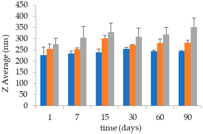
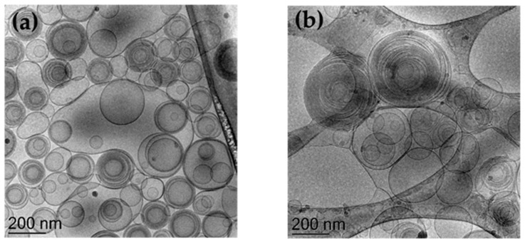
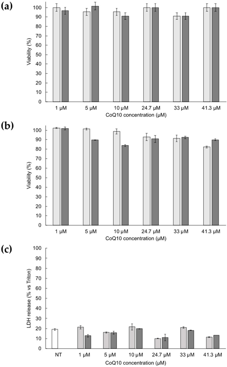
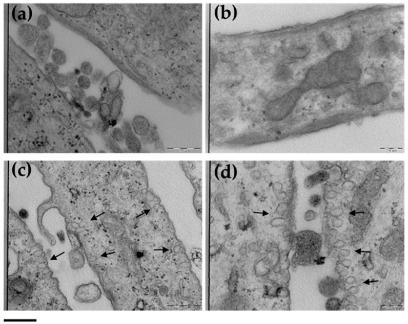
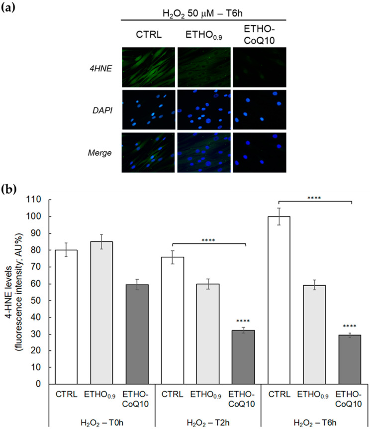
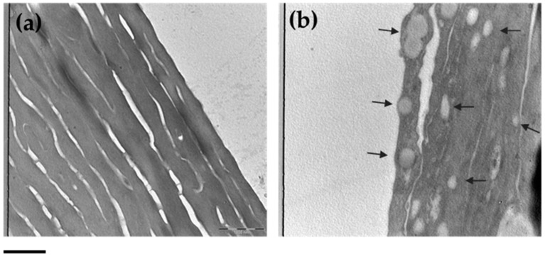
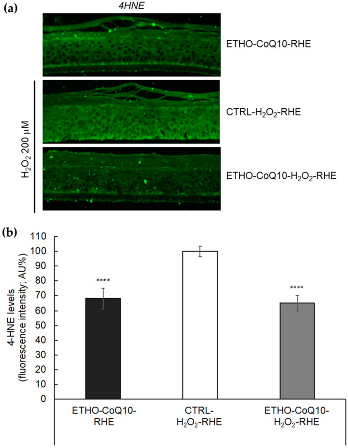
Ethosome represents a smart transdermal vehicle suitable for solubilization and cutaneous application of drugs. Coenzyme Q10 is an endogenous antioxidant whose supplementation can counteract many cutaneous disorders and pathologies. In this respect, the present study describes the production, characterization, and cutaneous protection of phosphatidylcholine based ethosomes as percutaneous delivery systems for coenzyme Q10. CoQ10 entrapment capacity in ethosomes was almost 100%, vesicles showed the typical 'fingerprint' structure, while mean diameters were around 270 nm, undergoing an 8% increase after 3 months from production. An ex-vivo study, conducted by transmission electron microscopy, could detect the uptake of ethosomes in human skin fibroblasts and the passage of the vesicles through 3D reconstituted human epidermis. Immunofluorescence analyses were carried on both on fibroblasts and 3D reconstituted human epidermis treated with ethosomes in the presence of H2O2 as oxidative stress challenger, evaluating 4-hydroxynonenal protein adducts which is as a reliable biomarker for oxidative damage. Notably, the pretreatment with CoQ10 loaded in ethosomes exerted a consistent protective effect against oxidative stress, in both models, fibroblasts and in reconstituted human epidermis respectively.

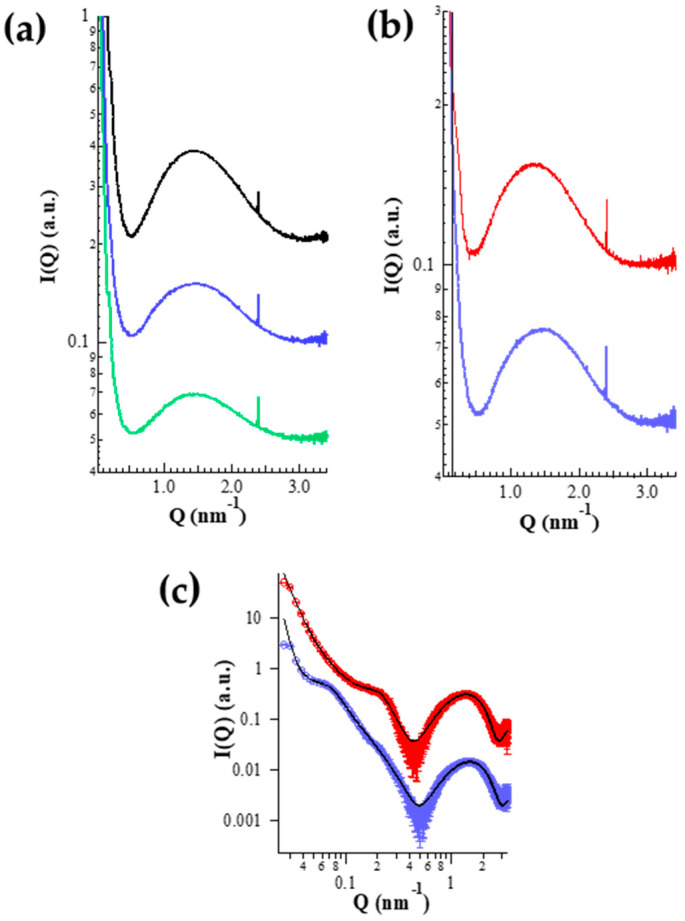
Keywords: H2O2; dermal administration; ethosome; penetration enhancers; reconstituted human epidermis; small angle X-ray scattering; ubiquinone.


Conflict of interest statement

The authors declare no conflict of interest.

Figures

Figure 1
Figure 1
Effect of PC concentration on the stability of ETHO0.6 (orange), ETHO0.9 (blue), and ETHO1.5 (grey). Diameters were measured by PCS and expressed as Z average.
Figure 2
Figure 2
Cryo-transmission electron microscopy images (Cryo-TEM) of ETHO0.9 (a) and ETHO-CoQ10 (b). Bar corresponds to 200 nm.
Figure 3
Figure 3
Small angle X-ray scattering (SAXS) profiles observed for the different ETHO samples at 25 °C. (a) ETHO0.6 (green), ETHO0.9 (blue), ETHO1.5 (black). (b) ETHO-CoQ10 (red), ETHO0.9 (blue). (c) Fitting analysis of the ETHO-CoQ10 (red) and ETHO0.9 (blue) curves by the new GENFIT model [50]. The continuous black lines are the best fit curves; the fitted Q-range corresponds to the length of the traced lines. In each frame, curves are scaled for clarity.
Figure 4
Figure 4
Cytotoxicity of ETHO0.9 (light grey) and ETHO-CoQ10 (dark grey) evaluated by trypan blue exclusion (a), MTT (b), and LDH (c) tests on primary dermal fibroblasts from healthy control subject after 24 h of treatment. Data are given as mean ± SD, representative of three independent experiments with at least three technical replicates each time.
Figure 5
Figure 5
TEM images of control cells (a,b) and cells treated with ETHO0.9 (c,d). Images of panels (a) and (c) refer to 31.5K magnification; images of panels (b) and (d) refer to 50K magnification. Arrows indicate the presence of ETHO0.9. Bar corresponds to 500 nm.
Figure 6
Figure 6
(a) Representative immunocytochemical images of 4HNE protein adducts in primary dermal fibroblasts without treatment (CTRL), treated with ETHO0.9 or ETHO-CoQ10, 6 h after exposure with 50 µM of H2O2 for 1 h. Images were taken at 40×. (b) Quantification of immunofluorescence staining for 4HNE immediately after H2O2 exposure (T0 h) or 2 (T2 h) and 6 h (T6 h) post-exposure. Data were normalized with respect to the CTRL sample treated for 6 h and expressed as arbitrary units ± SD. **** p ≤ 0.0001 vs. CTRL T0 h.
Figure 7
Figure 7
TEM images of control RHE (a) and RHE after 6 h of treatment with ETHO0.9 (b). Images refer to 20K magnification. Arrows indicate the presence of ETHO0.9. Bar corresponds to 1000 nm.
Figure 8
Figure 8
(a) Representative immunocytochemical images for 4HNE protein adducts in portions of RHE pretreated with ETHO-CoQ10 (ETHO-CoQ10-H2O2-RHE) and compared to control RHE treated with ETHO-CoQ10 not exposed to H2O2 (ETHO-CoQ10-RHE), or to RHE exposed to H2O2200 μM of H2O2 for 90 min (CTRL- H2O2-RHE). Images were taken at 40×. (b) Quantification of immunofluorescence staining for 4HNE after H2O2 exposure. Data were normalized with respect to CTRL-H2O2-RHE and expressed as arbitrary units ± SD. **** p ≤ 0.0001 vs. CTRL.

Similar articles

Cited by

References

    1. Genova M.L., Lenaz G. New developments on the functions of coenzyme Q in mitochondria. BioFactors. 2011;37:330–354. doi: 10.1002/biof.168. - DOI - PubMed
    1. Garrido-Maraver J., Cordero M.D., Oropesa-Ávila M., Fernández Vega A., De la Mata M., Delgado Pavón A., De Miguel M., Pérez Calero C., Villanueva Paz M., Cotán D., et al. Coenzyme Q10 therapy. Mol. Syndromol. 2014;5:187–197. doi: 10.1159/000360101. - DOI - PMC - PubMed
    1. Saini R. Coenzyme Q10: The essential nutrient. J. Pharm. Bioallied Sci. 2011;3:466–467. doi: 10.4103/0975-7406.84471. - DOI - PMC - PubMed
    1. Fotino A.D., Thompson-Paul A.M., Bazzano L.A. Effect of coenzyme Q10 supplementation on heart failure: A meta-analysis. Am. J. Clin. Nutr. 2013;97:268–275. doi: 10.3945/ajcn.112.040741. - DOI - PMC - PubMed
    1. Singh U., Devaraj S., Jialal I. Coenzyme Q10 supplementation and heart failure. Nutr. Rev. 2007;65:286–293. doi: 10.1111/j.1753-4887.2007.tb00306.x. - DOI - PubMed

LinkOut - more resources