Review
. 2020 Jun 3;9(6):1395.
doi: 10.3390/cells9061395.

Microtubule Organization in Striated Muscle Cells

Affiliations
Review

Microtubule Organization in Striated Muscle Cells

Robert Becker et al. Cells. .

Abstract

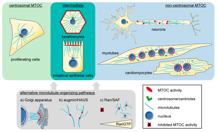
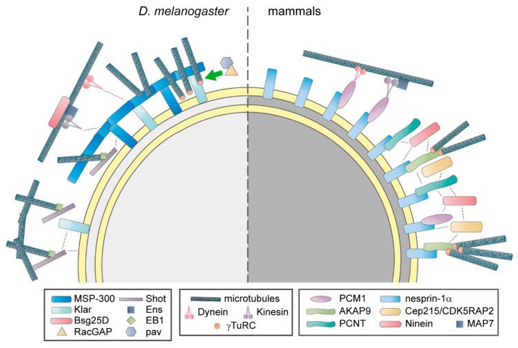
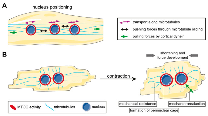
Distinctly organized microtubule networks contribute to the function of differentiated cell types such as neurons, epithelial cells, skeletal myotubes, and cardiomyocytes. In striated (i.e. skeletal and cardiac) muscle cells, the nuclear envelope acts as the dominant microtubule-organizing center (MTOC) and the function of the centrosome-the canonical MTOC of mammalian cells-is attenuated, a common feature of differentiated cell types. We summarize the mechanisms known to underlie MTOC formation at the nuclear envelope, discuss the significance of the nuclear envelope MTOC for muscle function and cell cycle progression, and outline potential mechanisms of centrosome attenuation.

Keywords: MTOC; cardiomyocytes; cell cycle; centrosome; microtubules; non-centrosomal MTOC; skeletal muscle.


Conflict of interest statement

The authors declare no conflict of interest.

Figures

Figure 1
Figure 1
Examples of microtubule organization in animal cells (depictions are simplified; see main text for details). The centrosome is the dominant microtubule-organizing center (MTOC) in proliferating cells (green panel). Differentiated cell types like neurons or striated muscle cells exhibit various non-centrosomal MTOCs and attenuate/inactivate or, as described for cardiomyocytes, disassemble their centrosomes (blue panel). Certain types of epithelial cells are “intermediates”, i.e., they retain microtubule nucleation ability at centrosomes but anchor microtubules mainly at non-centrosomal sites (cyan panel). In addition, several alternative microtubule-organizing pathways (grey panel) are active in proliferating as well as differentiated cells (neurons in particular) that act in synergy with the dominant MTOC.
Figure 2
Figure 2
The nuclear envelope microtubule-organizing center (MTOC) in Drosophila (D.) melanogaster and in mammalian muscle cells. In D. melanogaster, the outer nuclear membrane proteins MSP-300 and Klarsicht (Klar), as well as a complex of the spectraplakin Shortstop (Shot) and the microtubule plus-end tracker EB1, cooperate to maintain a circumnuclear MSP-300 ring. Potentially, this ring is constituted by isoforms of MSP-300 that lack the nuclear envelope-targeting Klarsicht, ANC-1, and Syne homology (KASH) domain. The perinuclear microtubule network is maintained by the MSP-300 ring and the Shot-EB1 complex. Additionally, RacGAP and its binding partner Pavarotti (pav) promote perinuclear γTuRC localization. Dynein and a complex of kinesin, ensconsin (Ens), and Bsg25D mediate nuclear movement along microtubules. Klar and MSP-300 have been proposed as direct interaction partners of dynein and kinesin. In mammals, nesprin-1α anchors the centrosomal proteins pericentriolar material 1 (PCM1), pericentrin (PCNT), and A-kinase anchoring protein 9 (AKAP9) to the nuclear envelope. PCM1 connects the nucleus to microtubule-associated motors. AKAP9 is specifically required for microtubule nucleation, potentially by recruiting γ-tubulin ring complexes (γTuRC). Ninein and centrosomal protein of 215 kDa (CEP215/CDK5RAP2) localize to the nuclear envelope, but the underlying mechanisms are unclear. Ortho- and paralogues have the same shape and similar color. Dashed lines represent hypothetical interactions that underlie nuclear envelope localization of MTOC components. Please note that a large part of the D. melanogaster data has been obtained in myofibers in vivo, while mammalian data derives largely from in vitro experiments in myotubes. This possibly accounts for some of the differences in ncMTOC composition.
Figure 3
Figure 3
Contribution of the non-centrosomal microtubule-organizing center (ncMTOC) to striated muscle function (see main text for details). (A) The nuclear envelope ncMTOC promotes nuclear positioning in various ways. Microtubule-associated motors are recruited to the nuclear envelope to allow nuclear movement in a cargo-like manner along microtubules. Furthermore, perinuclear microtubules enable application of pushing forces through motor-mediated microtubule sliding as well as pulling forces exerted by cortically anchored dynein on the nuclei. (B) Microtubules fulfill several functions during muscle contraction. Firstly, stable microtubules modulate contractility by providing mechanical resistance. Secondly, a perinuclear cage consisting of microtubules and MTOC proteins protects nuclear integrity. Thirdly, microtubules link the extracellular space and the contractile apparatus to the nucleus via the linker of nucleoskeleton and cytoskeleton (LINC) complex, which allows transmission of mechanical cues to regulate gene expression.

References

    1. Prosser S.L., Pelletier L. Mitotic spindle assembly in animal cells: A fine balancing act. Nat. Rev. Mol. Cell Biol. 2017;18:187–201. doi: 10.1038/nrm.2016.162. - DOI - PubMed
    1. Sanchez A.D., Feldman J.L. Microtubule-organizing centers: From the centrosome to non-centrosomal sites. Curr. Opin. Cell Biol. 2017;44:93–101. doi: 10.1016/j.ceb.2016.09.003. - DOI - PMC - PubMed
    1. Lechler T., Fuchs E. Desmoplakin: An unexpected regulator of microtubule organization in the epidermis. J. Cell Biol. 2007;176:147–154. doi: 10.1083/jcb.200609109. - DOI - PMC - PubMed
    1. Zebrowski D.C., Vergarajauregui S., Wu C.C., Piatkowski T., Becker R., Leone M., Hirth S., Ricciardi F., Falk N., Giessl A., et al. Developmental alterations in centrosome integrity contribute to the post-mitotic state of mammalian cardiomyocytes. Elife. 2015;4 doi: 10.7554/eLife.05563. - DOI - PMC - PubMed
    1. Stiess M., Maghelli N., Kapitein L.C., Gomis-Ruth S., Wilsch-Brauninger M., Hoogenraad C.C., Tolic-Norrelykke I.M., Bradke F. Axon extension occurs independently of centrosomal microtubule nucleation. Science. 2010;327:704–707. doi: 10.1126/science.1182179. - DOI - PubMed

Publication types

LinkOut - more resources