Comparative Study
. 2020 Jun 3;21(11):4010.
doi: 10.3390/ijms21114010.

Genomic Analysis of Intrinsically Disordered Proteins in the Genus Camelus

Affiliations
Comparative Study

Genomic Analysis of Intrinsically Disordered Proteins in the Genus Camelus

Manal A Alshehri et al. Int J Mol Sci. .

Abstract

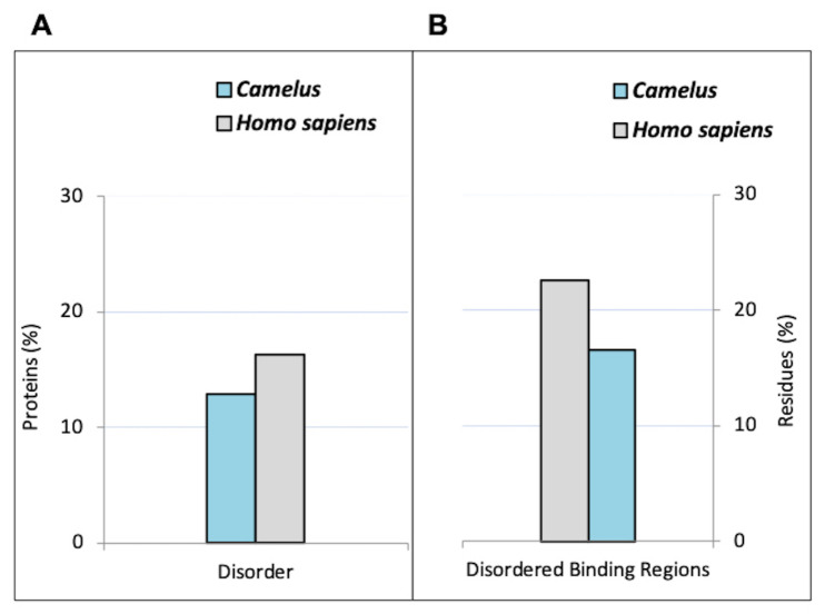
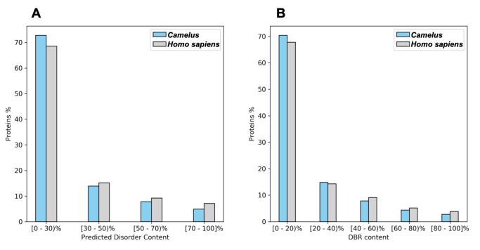
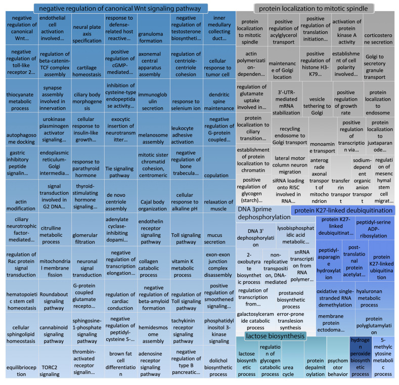
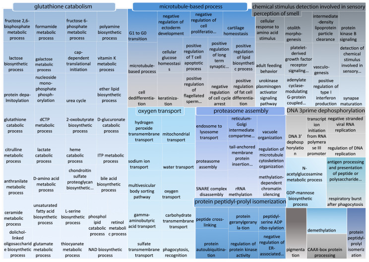
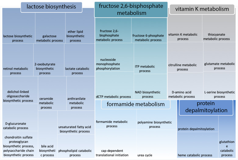
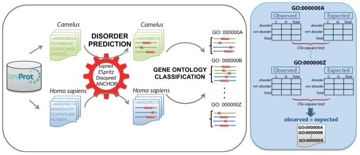
Intrinsically disordered proteins/regions (IDPs/IDRs) fail to fold completely into 3D structures, but have major roles in determining protein function. While natively disordered proteins/regions have been found to fulfill a wide variety of primary cellular roles, the functions of many disordered proteins in numerous species remain to be uncovered. Here, we perform the first large-scale study of IDPs/IDRs in the genus Camelus, one of the most important mammalians in Asia and North Africa, in order to explore the biological roles of these proteins. The study includes the prediction of disordered proteins/regions in Camelus species and in humans using multiple state-of-the-art prediction tools. Additionally, we provide a comparative analysis of Camelus and Homo sapiens IDPs/IDRs for the sake of highlighting the distinctive use of disorder in each genus. Our findings indicate that the human proteome is more disordered than the Camelus proteome. Gene Ontology analysis also revealed that Camelus IDPs are enriched in glutathione catabolism and lactose biosynthesis.

Keywords: Camelus; GO; disorder prediction; disoredered proteins.


Conflict of interest statement

The authors declare no conflict of interest.

Figures

Figure 1
Figure 1
Overall predicted disorder and disorder binding regions (DBRs) in Camelus and Homo sapiens. (A) Percentage of proteins with at least one long disordered region (LDR) with at least 50% disordered residues (according to DISOPRED predictions). (B) Percentage of disordered residues involved in binding (according to ANCHOR predictions).
Figure 2
Figure 2
Binning of Camelus and Homo sapiens proteins by degree of (A) predicted disorder (percentage of disordered residues relative to sequence length, predicted by DISOPRED) and (B) disorder binding regions (predicted by ANCHOR).
Figure 3
Figure 3
Multiple sequence alignment of four homologous proteins for Homo sapiens, Camelus dromedarius, Camelus bactrianus, and Camelus ferus.
Figure 4
Figure 4
Disorder predispositions of four homologous proteins from Homo sapiens, Camelus dromedarius, Camelus bactrianus, and Camelus ferus.
Figure 5
Figure 5
REVIGO representation of GO biological process terms (with PPV > 0.7) that are significantly enriched in disordered Camelus proteins. Disordered proteins are those containing at least one “long disordered region” based on DISOPRED predictions.
Figure 6
Figure 6
REVIGO representation of GO biological process terms (with PPV > 0.7) that are more enriched in Camelus disordered proteins than in those of Homo sapiens. Disordered proteins are those containing at least one “long disordered region” based on DISOPRED predictions.
Figure 7
Figure 7
REVIGO representation of all GO terms included within the category “glutathione catabolism”, which comprises the largest cluster of GO terms that are more enriched in Camelus disordered proteins compared to those of Homo sapiens.
Figure 8
Figure 8
Schematic representation of the methodology used for the comparative study of disordered proteins in Camelus (green) and Homo sapiens (blue). For each organism, protein sequences were retrieved from Uniprot. For each protein, disordered regions (pink) were predicted using three different methods (Iupred, ESpritz, and DISOPRED), and disordered binding regions (DBRs) were predicted using ANCHOR. Disordered proteins were then assigned to GO:BP functional classes using PANZZER, and a comparative analysis of disorder levels between the two genera was performed for each class. Contingency tables were constructed with the per-genus counts of disordered and not-disordered proteins and a Chi-squared test applied. GO classes for which the difference in disorder was positive for Camelus were considered to be more disordered in Camelus than in Homo sapiens.

References

    1. Habchi J., Tompa P., Longhi S., Uversky V.N. Introducing protein intrinsic disorder. Chem. Rev. 2014;114:6561–6588. doi: 10.1021/cr400514h. - DOI - PubMed
    1. Bernado P., Blanchard L., Timmins P., Marion D., Ruigrok R.W., Blackledge M. A structural model for unfolded proteins from residual dipolar couplings and small-angle X-ray scattering. Proc. Natl. Acad. Sci. USA. 2005;102:17002–17007. doi: 10.1073/pnas.0506202102. - DOI - PMC - PubMed
    1. Uversky V.N. Intrinsically disordered proteins and their “mysterious” (meta) physics. Front. Phys. 2019;7:10. doi: 10.3389/fphy.2019.00010. - DOI
    1. Van Der Lee R., Buljan M., Lang B., Weatheritt R.J., Daughdrill G.W., Dunker A.K., Fuxreiter M., Gough J., Gsponer J., Jones D.T., et al. Classification of intrinsically disordered regions and proteins. Chem. Rev. 2014;114:6589–6631. doi: 10.1021/cr400525m. - DOI - PMC - PubMed
    1. Romero P.R., Zaidi S., Fang Y.Y., Uversky V.N., Radivojac P., Oldfield C.J., Cortese M.S., Sickmeier M., LeGall T., Obradovic Z., et al. Alternative splicing in concert with protein intrinsic disorder enables increased functional diversity in multicellular organisms. Proc. Natl. Acad. Sci. USA. 2006;103:8390–8395. doi: 10.1073/pnas.0507916103. - DOI - PMC - PubMed

Publication types

LinkOut - more resources