The protective effects of the β3 adrenergic receptor agonist BRL37344 against liver steatosis and inflammation in a rat model of high-fat diet-induced nonalcoholic fatty liver disease (NAFLD)
- PMID: 32503411
- PMCID: PMC7275314
- DOI: 10.1186/s10020-020-00164-4
The protective effects of the β3 adrenergic receptor agonist BRL37344 against liver steatosis and inflammation in a rat model of high-fat diet-induced nonalcoholic fatty liver disease (NAFLD)
Abstract
Background: Our objective was to investigate the efficacy of the beta-3 adrenergic receptor (β3-AR) agonist BRL37344 for the prevention of liver steatosis and inflammation associated with nonalcoholic fatty liver disease (NAFLD).
Methods: Four groups were established: a control group (given a standard diet), a high-fat diet (HFD) group, an HFD + β3-AR agonist (β3-AGO) group, and an HFD + β3-AR antagonist (β3-ANT) group. All rats were fed for 12 weeks. The β3-AR agonist BRL37344 and the antagonist L748337 were administered for the last 4 weeks with Alzet micro-osmotic pumps. The rat body weights (g) were measured at the end of the 4th, 8th, and 12th weeks. At the end of the 12th week, the liver weights were measured. Serum alanine aminotransferase (ALT) and aspartate aminotransferase (AST) were analyzed with a Hitachi automatic analyzer. The lipid levels of the triglycerides (TGs), total cholesterol (TC), and low-density lipoprotein cholesterol (LDL-C) and the concentrations of free fatty acids (FFAs) were also measured. An oil red O kit was used to detect lipid droplet accumulation in hepatocytes. Steatosis, ballooning degeneration and inflammation were histopathologically determined. The protein and mRNA expression levels of β3-AR, peroxisome proliferator-activated receptor-alpha (PPAR-α), peroxisome proliferator-activated receptor-gamma (PPAR-γ), mitochondrial carnitine palmitoyltransferase-1 (mCPT-1), and fatty acid translocase (FAT)/CD36 were measured by western blot analysis and RT-qPCR, respectively.
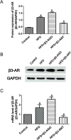
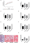
Results: After treatment with the β3-AR agonist BRL37344 for 4 weeks, the levels of ALT, AST, TGs, TC, LDL-C and FFAs were decreased in the NAFLD model group compared with the HFD group. Body and liver weights, liver index values and lipid droplet accumulation were lower in the HFD + β3-AGO group than in the HFD group. Decreased NAFLD activity scores (NASs) also showed that liver steatosis and inflammation were ameliorated after treatment with BRL37344. Moreover, the β3-AR antagonist L748337 reversed these effects. Additionally, the protein and gene expression levels of β3-AR, PPAR-α, and mCPT-1 were increased in the HFD + β3-AGO group, whereas those of PPAR-γ and FAT/CD36 were decreased.
Conclusion: The β3-AR agonist BRL37344 is beneficial for reducing liver fat accumulation and for ameliorating liver steatosis and inflammation in NAFLD. These effects may be associated with PPARs/mCPT-1 and FAT/CD36.
Keywords: Inflammation; Liver steatosis; NAFLD; β3-adrenergic receptor.
Conflict of interest statement
The authors declare that they have no competing interests.
Figures


Similar articles
-
β3-adrenoceptor mediates metabolic protein remodeling in a rabbit model of tachypacing-induced atrial fibrillation.Cell Physiol Biochem. 2013;32(6):1631-42. doi: 10.1159/000356599. Epub 2013 Dec 5. Cell Physiol Biochem. 2013. PMID: 24335437
-
Kangtaizhi Granule Alleviated Nonalcoholic Fatty Liver Disease in High-Fat Diet-Fed Rats and HepG2 Cells via AMPK/mTOR Signaling Pathway.J Immunol Res. 2020 Aug 20;2020:3413186. doi: 10.1155/2020/3413186. eCollection 2020. J Immunol Res. 2020. PMID: 32884949 Free PMC article.
-
[Effect of Nrf2 and related factors on the progression of nonalcoholic steatohepatitis].Zhongguo Ying Yong Sheng Li Xue Za Zhi. 2014 Sep;30(5):465-70. Zhongguo Ying Yong Sheng Li Xue Za Zhi. 2014. PMID: 25571645 Chinese.
-
Models of non-Alcoholic Fatty Liver Disease and Potential Translational Value: the Effects of 3,5-L-diiodothyronine.Ann Hepatol. 2017 Sep-Oct;16(5):707-719. doi: 10.5604/01.3001.0010.2713. Ann Hepatol. 2017. PMID: 28809727 Review.
-
Deciphering the molecular pathways of saroglitazar: A dual PPAR α/γ agonist for managing metabolic NAFLD.Metabolism. 2024 Jun;155:155912. doi: 10.1016/j.metabol.2024.155912. Epub 2024 Apr 11. Metabolism. 2024. PMID: 38609038 Review.
Cited by
-
Hepatic Innervations and Nonalcoholic Fatty Liver Disease.Semin Liver Dis. 2023 May;43(2):149-162. doi: 10.1055/s-0043-57237. Epub 2023 May 8. Semin Liver Dis. 2023. PMID: 37156523 Free PMC article. Review.
-
mTOR: A Potential New Target in Nonalcoholic Fatty Liver Disease.Int J Mol Sci. 2022 Aug 16;23(16):9196. doi: 10.3390/ijms23169196. Int J Mol Sci. 2022. PMID: 36012464 Free PMC article. Review.
-
Liver fibrosis and MAFLD: the exploration of multi-drug combination therapy strategies.Front Med (Lausanne). 2023 Apr 20;10:1120621. doi: 10.3389/fmed.2023.1120621. eCollection 2023. Front Med (Lausanne). 2023. PMID: 37153080 Free PMC article. Review.
-
Phytic acid-based nanomedicine against mTOR represses lipogenesis and immune response for metabolic dysfunction-associated steatohepatitis therapy.Life Metab. 2024 Jun 18;3(6):loae026. doi: 10.1093/lifemeta/loae026. eCollection 2024 Dec. Life Metab. 2024. PMID: 39873005 Free PMC article.
-
C-type natriuretic peptide is a pivotal regulator of metabolic homeostasis.Proc Natl Acad Sci U S A. 2022 Mar 29;119(13):e2116470119. doi: 10.1073/pnas.2116470119. Epub 2022 Mar 25. Proc Natl Acad Sci U S A. 2022. PMID: 35333648 Free PMC article.
References
-
- Balligand JL. Beta3-adrenoreceptors in cardiovasular diseases: new roles for an "old" receptor. Curr Drug Deliv. 2013. 10.2174/1567201811310010011. - PubMed
-
- Begriche K, Igoudjil A, Pessayre D, Fromenty B. Mitochondrial dysfunction in NASH: causes, consequences and possible means to prevent it. Mitochondrion. 2006. 10.1016/j.mito.2005.10.004. - PubMed
-
- Bhatia LS, Curzen NP, Calder PC, Byrne CD. Non-alcoholic fatty liver disease: a new and important cardiovascular risk factor? Eur Heart J. 2012. 10.1093/eurheartj/ehr453. - PubMed
-
- Bligh EG, Dyer WJ. A rapid method of total lipid extraction and purification. Can J Biochem Physiol. 1959. 10.1139/o59-099. - PubMed
Publication types
MeSH terms
Substances
LinkOut - more resources
Full Text Sources
Medical
Research Materials
Miscellaneous

