

Pulmonary cryptococcosis coexisting with central type lung cancer in an immuocompetent patient: a case report and literature review
- PMID: 32503511
- PMCID: PMC7275487
- DOI: 10.1186/s12890-020-01200-z
Pulmonary cryptococcosis coexisting with central type lung cancer in an immuocompetent patient: a case report and literature review
Abstract
Background: Pulmonary Cryptococcosis is a common fungal infection mainly caused by Cryptococcus neoformans/C.gattii species in immunocompromised patients. Cases of pulmonary cryptococcosis in patients with normal immune function are increasingly common in China. Clinical and radiographic features of pulmonary cryptococcosis are various and without obvious characteristics, so it is often misdiagnosed as pulmonary metastatic tumor or tuberculosis. When coexisting with malignant lung tumors, it was more difficult to differentiate from metastatic lung cancer, although the coexistence of pulmonary cryptococcosis and central type lung cancer is rare. Reviewing the imaging manifestations and diagnosis of the case and the relevant literature will contribute to recognition of the disease and a decrease in misdiagnoses.
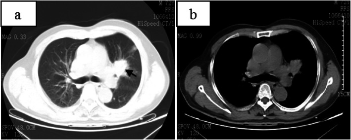
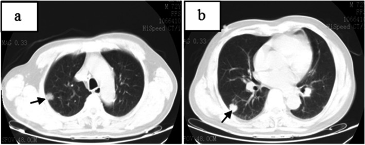
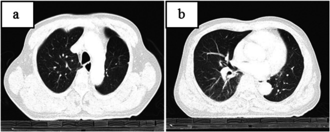
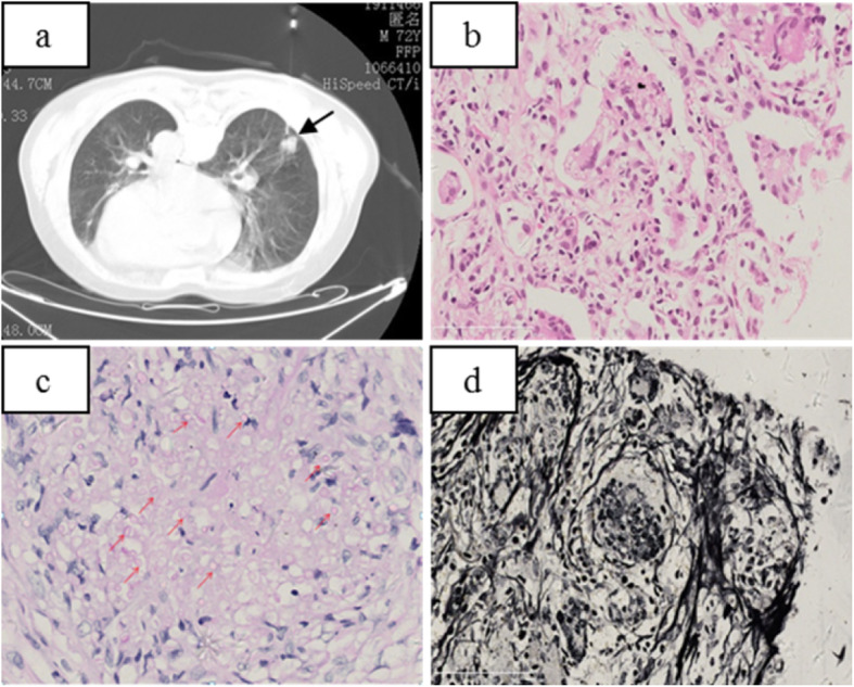
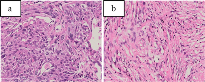
Case presentation: A 72-year-old immunocompetent Han Chinese man had repeated dry cough for more than half a year. CT examination of chest showed an irregular mass at the left hilum of the lung, and two small nodules in the right lung, which were considered as the left central lung cancer with right lung metastasis. However, the patient was diagnosed with pulmonary cryptococcosis coexisting with central type lung cancer based on the results of laboratory examination, percutaneous lung biopsy, fiberoptic bronchoscopy, and surgical pathology. The patient underwent surgical resection of the left central type lung cancer and was placed on fluconazole treatment after a positive diagnosis was made. Five years after the lung cancer surgery, the patient had a recurrence, but the pulmonary cryptococcus nodule disappeared.
Conclusion: Our case shows that CT findings of central type lung cancer with multiple pulmonary nodules are not necessarily metastases, but may be coexisting pulmonary cryptococcosis. CT images of cryptococcosis of the lung were diverse and have no obvious characteristics, so it was very difficult to distinguish from metastatic tumors. CT-guided percutaneous lung biopsy was a simple and efficient method for identification.
Keywords: CT-guided percutaneous lung biopsy; Central type lung cancer; Computed tomography; Cryptococcosis; Metastatic tumor; PAS periodic acid-Schiff.
Conflict of interest statement
The authors declare that they have no competing interests.
Figures
References
Publication types
MeSH terms
Substances
LinkOut - more resources
Full Text Sources
Medical
