. 2020 Jun 5;20(1):172.
doi: 10.1186/s12906-020-02895-7.

Iridoid glycosides from Morinda officinalis How. exert anti-inflammatory and anti-arthritic effects through inactivating MAPK and NF-κB signaling pathways

Affiliations

Iridoid glycosides from Morinda officinalis How. exert anti-inflammatory and anti-arthritic effects through inactivating MAPK and NF-κB signaling pathways

Qi Zhang et al. BMC Complement Med Ther. .

Abstract

Background: The root of Morinda officinalis How. (MO, the family of Rubiaceae) has long been used to treat inflammatory diseases in China and other eastern Asian countries, and iridoid glycosides extracted from MO (MOIG) are believed to contribute to this anti-inflammatory effect. However, the mechanism underlying the anti-inflammatory and anti-arthritic activities of MOIG has not been elucidated. The aim of the present study was to determine how MOIG exerted anti-inflammatory and anti-arthritic effects in vivo and in RAW 264.7 macrophages.

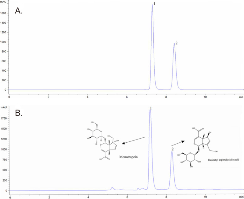
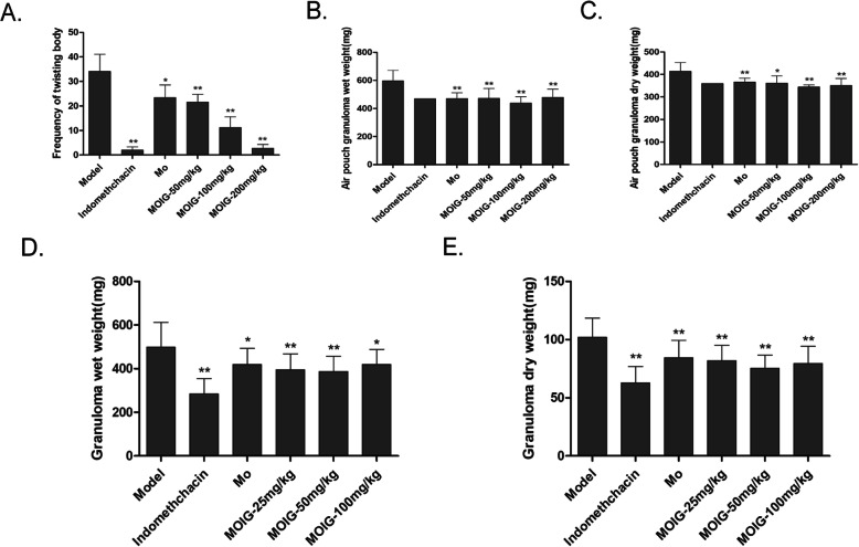
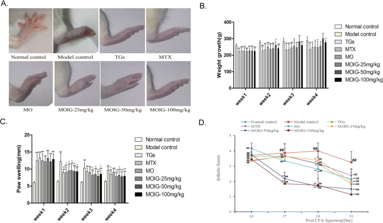
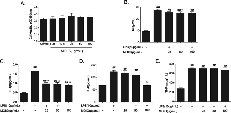
Methods: MOIG were enriched by XDA-1 macroporous resin. The maximum feasible dose method was adopted to evaluate its acute toxicity. The analgesic effect of MOIG was evaluated by acetic acid writhing test and the anti-inflammatory effect was evaluated by cotton-pellet granuloma test in rats and air pouch granuloma test in mice. The anti-arthritic effect was evaluated by establishing an adjuvant arthritis model induced by Complete Freund's Adjuvant (CFA). The viability of the cultured RAW 264.7 macrophages was assessed by 3-(4, 5-dimethylthiazol-2-yl)-2, 5-diphenyl tetrazolium bromide (MTT) assay. The anti-inflammatory activity was evaluated by measuring NO, IL-1β, IL-6 and TNF-α levels in LPS-stimulated RAW 264.7 cells. The protein level of inflammatory responsive genes was evaluated by Western blot analysis.

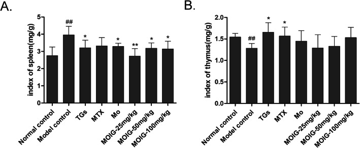
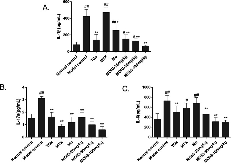
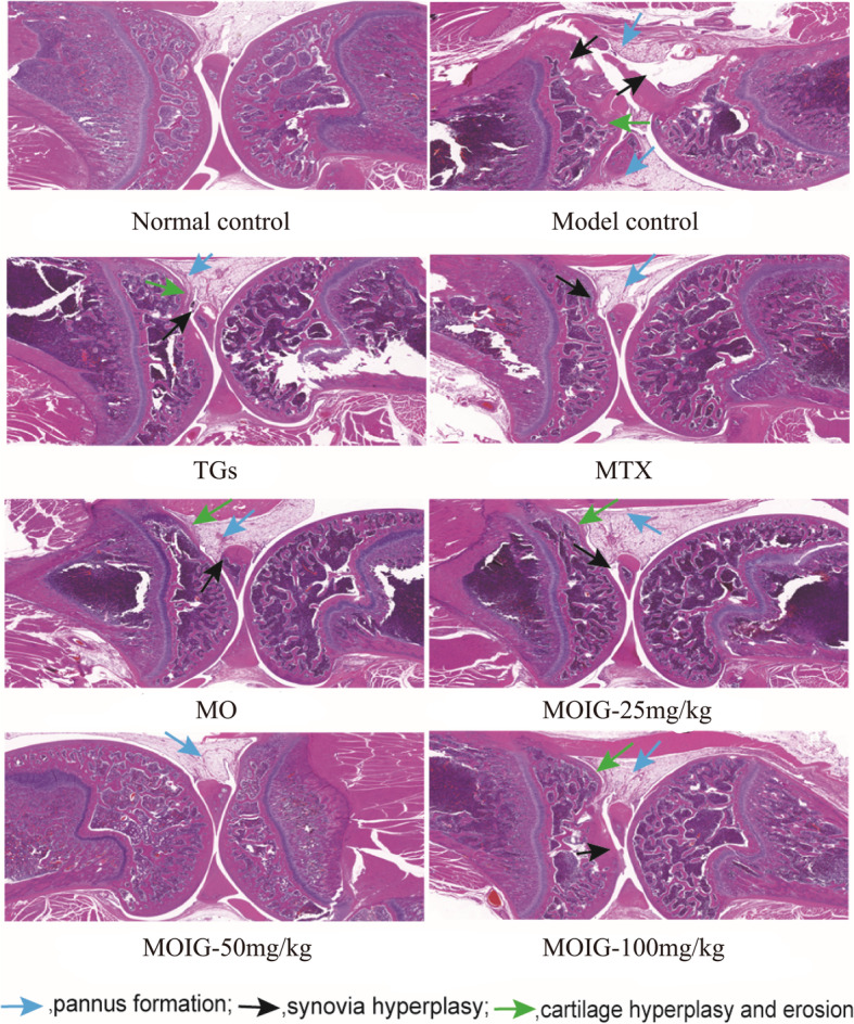
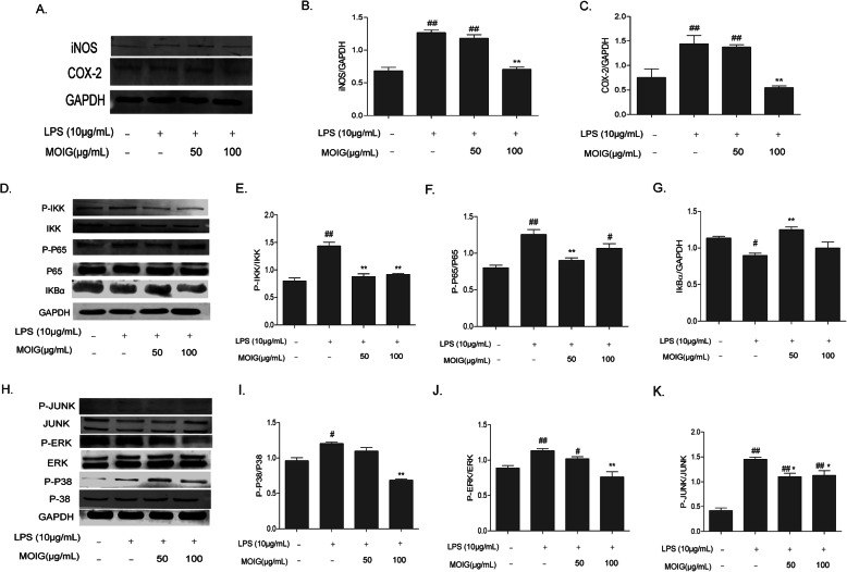
Results: MOIG had no significant toxicity at maximum feasible dose of 22.5 g/kg. MO extracts and MOIG (50,100 and 200 mg/kg) all evoked a significantly inhibitory effects on the frequency of twisting induced by acetic acid in mice compared with the model control group. Administration of MO extracts and MOIG markedly decreased the dry and wet weight of cotton pellet granuloma in rats and air pouch granuloma in mice. MOIG significantly attenuated the paw swelling and decreased the arthritic score, weight loss, spleen index, and the serum level of inflammatory factors IL-1β, IL-6 and IL-17a in CFA-induced arthritic rats. MOIG inhibited the production of inflammatory cytokines in LPS-stimulated RAW264.7 cells, and the expressions of iNOS, COX-2 and proteins related to MAPK and NF-κB signaling pathways in LPS-stimulated RAW 264.7 macrophages.

Conclusion: MOIG exerted anti-inflammatory and anti-arthritic activities through inactivating MAPK and NF-κB signaling pathways, and this finding may provide a sound experimental basis for the clinical treatment of rheumatoid arthritis with MOIG.

Keywords: Anti-arthritis; Anti-inflammation; Iridoid glycoside; MAPK pathway; Morinda officinalis how.; NF-κB; RAW 264.7 macrophages.


Conflict of interest statement

The authors declare that there are no competing interests.

Figures

Fig. 1
Fig. 1
HPLC analysis of MOIG. a HPLC chromatogram of standard reference; b HPLC chromatogram of MOIG. Peaks were detected at 240 nm. 1: monotropein; 2: Deacetyl asperulosidic acid
Fig. 2
Fig. 2
Analgesic and anti-inflammatory effects of MOIG. a MOIG inhibited the twisting induced by acetic acid in mice (n = 10); b and c MOIG attenuated inflammatory response as evidenced by wet weight and dry weight of air pouch granuloma in mice (n = 14); d and e MOIG mitigated inflammatory response as evidenced by wet weight and dry weight of cotton pellet granuloma in rats (n = 14). Data are presented as the mean ± SD. *p < 0.05, **p < 0.01, compared with the model control group
Fig. 3
Fig. 3
Effects of MOIG on CFA -induced arthritic rats. a Appearance of paw of CFA -induced arthritic rats; b Body weight; c Paw swelling (mm); d Arthritis score. Data are presented as the mean ± SD (n = 10). ##p < 0.01, compared with normal control group; *p < 0.05, **p < 0.01, compared with model control group. Note: Normal control, normal non-arthritic rats; Model control, arthritic non treated rats; TGs, Rats with arthritic and treated with TGs; MTX, Rats with arthritic and treated with MTX; MO, Rats with arthritic and treated with MO; MOIG-25 mg/kg, Rats with arthritic and treated with MOIG at dose of 25 mg/kg; MOIG-50 mg/kg, Rats with arthritic and treated with MOIG at dose of 50 mg/kg; MOIG-100 mg/kg, Rats with arthritic and treated with MOIG at dose of 100 mg/kg
Fig. 4
Fig. 4
Effects of MOIG on the thymus index and spleen index of CFA -induced arthritic rats. a spleen index, b thymus index. Data are presented as the mean ± SD (n = 10). ##p < 0. 01 compared with the normal control group. *p < 0.05, **p < 0. 01 compared with the model control group. Note: Normal control, normal non-arthritic rats; Model control, arthritic non treated rats; TGs, Rats with arthritic and treated with TGs; MTX, Rats with arthritic and treated with MTX; MO, Rats with arthritic and treated with MO; MOIG-25 mg/kg, Rats with arthritic and treated with MOIG at dose of 25 mg/kg; MOIG-50 mg/kg, Rats with arthritic and treated with MOIG at dose of 50 mg/kg; MOIG-100 mg/kg, Rats with arthritic and treated with MOIG at dose of 100 mg/kg
Fig. 5
Fig. 5
Effects of MOIG on serum levels of IL-1 β, IL-6 and IL-17a in CFA -induced arthritic rats. a IL-1β, b IL-17α, c IL-6. Data are presented as the mean ± SD (n = 10). #p < 0.05, ##p < 0. 01 compared with the normal control group; *p < 0.05, **p < 0. 01 compared with the model control group. Note: Normal control, normal non-arthritic rats; Model control, arthritic non treated rats; TGs, Rats with arthritic and treated with TGs; MTX, Rats with arthritic and treated with MTX; MO, Rats with arthritic and treated with MO; MOIG-25 mg/kg, Rats with arthritic and treated with MOIG at dose of 25 mg/kg; MOIG-50 mg/kg, Rats with arthritic and treated with MOIG at dose of 50 mg/kg; MOIG-100 mg/kg, Rats with arthritic and treated with MOIG at dose of 100 mg/kg
Fig. 6
Fig. 6
Effects of MOIG on the histopathological characteristics of joint and synovial tissue in CFA -induced arthritic rats (HE, × 200). CFA -induced arthritic rats exhibited severe synovial hyperplasia, infiltration of inflammatory cells, and pannus formation, which severely lead to cartilage hyperplasia and erosion. TGs, MTX, MO and MOIG reversed these pathological alterations in joint and synovial tissue of CFA -induced arthritic rats. Note: Normal control, normal non-arthritic rats; Model control, arthritic non treated rats; TGs, Rats with arthritic and treated with TGs; MTX, Rats with arthritic and treated with MTX; MO, Rats with arthritic and treated with MO; MOIG-25 mg/kg, Rats with arthritic and treated with MOIG at dose of 25 mg/kg; MOIG-50 mg/kg, Rats with arthritic and treated with MOIG at dose of 50 mg/kg; MOIG-100 mg/kg, Rats with arthritic and treated with MOIG at dose of 100 mg/kg
Fig. 7
Fig. 7
Effects of MOIG on the production of inflammatory cytokines in LPS -stimulated RAW264.7 cells. a cell viability; b NO level; c IL-1β; d IL-6; e TNF-α. Data are presented as the mean ± SD (n = 6) ##p < 0. 01 compared with the normal control group; *p < 0.05, **p < 0. 01 compared with LPS - stimulated control group
Fig. 8
Fig. 8
MOIG modulated the expression of COX-2, iNOS, and NF-κB and MAPK pathway in LPS -stimulated RAW264.7 cells. a, b and c Western -blot analysis for COX-2 and iNOS; d, e, f and g Western -blot analysis for related proteins of the NF-κB pathway; h, i, j and k Western -blot analysis for related proteins of the MAPK pathway. The experiments were repeated 3 times. Data are presented as the mean ± SD (n = 3) #p < 0. 05, ##p < 0. 01 compared with the normal control group; *p < 0.05, **p < 0. 01 compared with LPS - stimulated control group

Similar articles

Cited by

References

    1. Awasthi A, Kuchroo VK. Th17 cells: from precursors to players in inflammation and infection. Int Immunol. 2009;21:489–498. doi: 10.1093/intimm/dxp021. - DOI - PMC - PubMed
    1. Tateiwa D Yoshikawa H Kaito T. Cartilage and Bone Destruction in Arthritis: Pathogenesis and Treatment Strategy: A Literature Review. Cells. 2019;8(8):818. - PMC - PubMed
    1. Wang S, Wang Y, Liu X, Guan L, Yu L, Zhang X. Anti-inflammatory and anti-arthritic effects of taraxasterol on adjuvant-induced arthritis in rats. J Ethnopharmacol. 2016;187:42–48. doi: 10.1016/j.jep.2016.04.031. - DOI - PubMed
    1. Hu Y, Liu X, Xia Q, Yin T, Bai C, Wang Z, Du L, Li X, Wang W, Sun L, Liu Y, Zhang H. Deng L4, Chen Y: comparative anti-arthritic investigation of iridoid glycosides and crocetin derivatives from Gardenia jasminoides Ellis in Freund's complete adjuvant-induced arthritis in rats. Phytomedicine. 2019;53:223–233. doi: 10.1016/j.phymed.2018.07.005. - DOI - PubMed
    1. Ahmed S, Anuntiyo J, Malemud CJ, Haqqi TM. Biological basis for the use of botanicals in osteoarthritis and rheumatoid arthritis: a review. Evid Based Complement Alternat Med. 2005;2:301–308. doi: 10.1093/ecam/neh117. - DOI - PMC - PubMed