. 2020 Jun 5;13(1):68.
doi: 10.1186/s13045-020-00897-z.

Th17 cells inhibit CD8+ T cell migration by systematically downregulating CXCR3 expression via IL-17A/STAT3 in advanced-stage colorectal cancer patients

Affiliations

Th17 cells inhibit CD8+ T cell migration by systematically downregulating CXCR3 expression via IL-17A/STAT3 in advanced-stage colorectal cancer patients

Dan Wang et al. J Hematol Oncol. .

Abstract

Background: CD8+ T cell trafficking to the tumor site is essential for effective colorectal cancer (CRC) immunotherapy. However, the mechanism underlying CD8+ T cell infiltration in colorectal tumor tissues is not fully understood. In the present study, we investigated CD8+ T cell infiltration in CRC tissues and the role of chemokine-chemokine receptor signaling in regulation of T cell recruitment.

Methods: We screened chemokines and cytokines in healthy donor and CRC tissues from early- and advanced-stage patients using multiplex assays and PCR screening. We also utilized transcription factor activation profiling arrays and established a xenograft mouse model.

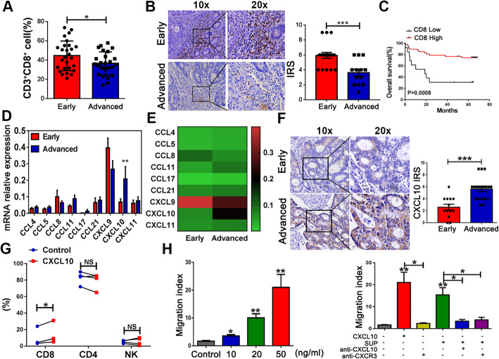
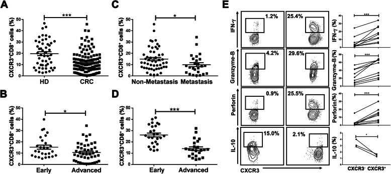
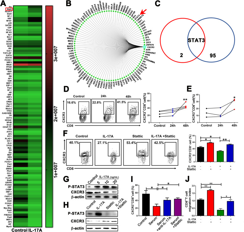
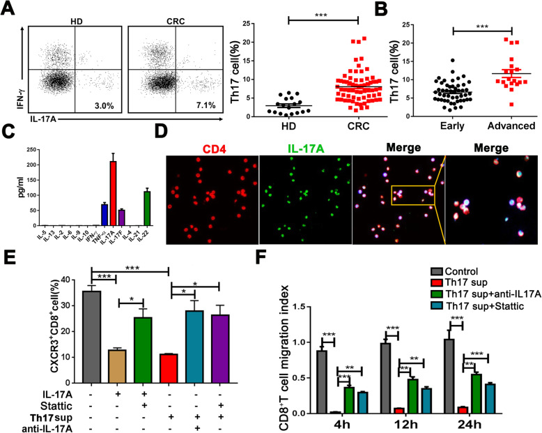
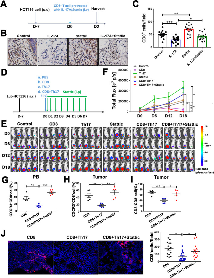
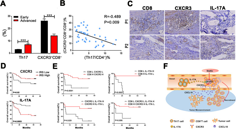
Results: Compared with tumor tissues of early-stage CRC patients, CD8+ T cell density was lower in advanced-stage tumor tissues. PCR screening showed that CXCL10 levels were significantly increased in advanced-stage tumor tissues. CXCR3 (the receptor of CXCL10) expression on CD8+ T cells was lower in the peripheral blood of advanced-stage patients. The migratory ability of CD8+ T cells to CXCL10 depended on CXCR3 expression. Multiplex arrays showed that IL-17A was increased in advanced-stage patient sera, which markedly downregulated CXCR3 expression via activating STAT3 signaling and reduced CD8+ T cell migration. Similar results were found after CD8+ T cells were treated with Th17 cell supernatant. Adding anti-IL-17A or the STAT3 inhibitor, Stattic, rescued these effects in vitro and in vivo. Moreover, survival analysis showed that patients with low CD8 and CXCR3 expression and high IL-17A levels had significantly worse prognosis.

Conclusions: CD8+ T cell infiltration in advanced-stage tumor was systematically inhibited by Th17 cells via IL-17A/STAT3/CXCR3 axis. Our findings indicate that the T cell infiltration in the tumor microenvironment may be improved by inhibiting STAT3 signaling.

Keywords: CD8; CXCR3; Colorectal cancer; IL-17A; Th17 cells.


Conflict of interest statement

The authors declare no potential conflicts of interest.

Figures

Fig 1
Fig 1
Decreased infiltration of CD8+ T cells and increased CXCL10 secretion in tumor tissues from advanced-stage CRC patients. a FACS analysis of CD8+ T cell infiltration in tumor tissues of early- and advanced-stage CRC patients. b CD8 expression was analyzed by immunohistochemistry in tumor tissues; magnification, × 200 (left). Right panel, IRS (0–9) = intensity score (0–3) × percentage score (0–3). c Kaplan-Meier OS curves of CRC patients, presented as high-CD8 or low-CD8 expression groups based on the log-rank statistic test (n = 70). High, IRS > 4; low, IRS ≤ 4. d, e Relative expression levels of effective immune cell-associated chemokines in CRC tumor tissues determined by real-time PCR. f CXCL10 expression was analyzed by immunohistochemistry. g Conditioned media were cultured in the lower chambers of a Transwell plate with or without rhCXCL10. Migrated PBMCs from HDs were collected and assessed via flow cytometry. Each line represented a different HD. h The migratory ability of purified CD8+ T lymphocytes (purity > 90%) from HDs to rhCXCL10 at different concentrations (left). Supernatants of primary tumor tissues were added alone or with CXCL10 (50 ng/mL)-specific neutralizing antibodies as indicated. After incubation, CD8+ T cells alone or cells pretreated with anti-CXCR3 that migrated into the lower chambers were collected and counted. The migration index was calculated by dividing the number of cells that migrated in the indicated groups by the number that migrated in the control groups (right). *P < 0.05, **P < 0.001, ***P < 0.0001; NS non-significant
Fig. 2
Fig. 2
Decreased CXCR3 expression on CD8+ T cells in the PB of advanced-stage CRC patients. b PBMCs were isolated from the PB of CRC patients (n = 125) and HDs (n = 50) and analyzed by flow cytometry. Representative dot plots are shown. Data are presented as the percentage of CXCR3+CD8+ T cells in total CD8+ T cells. b, c The percentage of CXCR3+CD8+ T cells in the PBMCs of early-stage, advanced-stage, non-metastasis, and metastasis CRC patients. d The percentage of CXCR3+CD8+ T cells in tumor tissues of early-stage and advanced-stage patients. e The expression of IFN-γ-, granzyme-B-, perforin-, and IL-10-producing CD8+ T cells in total CD8+ T cells from PBMCs of CRC patients. The results for gating cells on the CXCR3 negative population are shown in the left panels and for gating on the CXCR3 positive population on the right. Each line represented a different patient.*P < 0.05, ***P < 0.0001
Fig. 3
Fig. 3
IL-17A downregulates CXCR3 expression on CD8+ T cells. a The expression of 13 cytokines in the sera of CRC patients (CRC-sera) vs. HD (HD-sera), sera of advanced-stage (advanced-sera) vs. early-stage CRC patients (early-sera), and supernatants of tumor tissues (tumor-sup) vs. normal tissues (normal-sup) were analyzed by multiplex assays. Fold changes were calculated with concentrations normalized with log2. b The intersection of the screened cytokines (fold changes ≥ 2) from a (red circle), b (blue circle), and c (green circle) of Fig. 3a. c FACS analysis of CXCR3+CD8+ T cell expression in CD8+ T cells treated with recombinant human proteins (20 ng/mL IL-17A, 20 ng/mL IL-17F, or 10 ng/mL IFN-γ) for 48 h. Each line represented a different HD. d Purified CD8+ T cells treated with or without rhIL-17A were subjected to immunofluorescence for CXCR3 (red) and DAPI (blue). e, f Concentrations of IL-17A (pg/mL) in sera obtained from CRC patients and HDs were measured using ELISA. g Correlation between the concentration of IL-17A and the percentage of CXCR3+CD8+ T cells in CD8+ T cells. Data were analyzed by Spearman’s rank correlation. *P < 0.05, NS non-significant
Fig. 4
Fig. 4
IL-17A inhibits CXCR3 expression on CD8+ T cells via STAT3 phosphorylation. a HD PB-derived CD8+ T cells were treated with rhIL-17A (20 ng/mL) for 48 h. Nuclei proteins were then subjected to multiplex profiling analysis for transcriptional factor activation. Activity was normalized with transcription factor IID and expressed as the relative light unit (RLU) of the transcriptional factor. b The upstream transcriptional factor of CXCR3 was identified via radar tools of the GCBI website. c The intersection of the screened transcriptional factors. Red circle: screened transcriptional factors (fold changes ≥ 4) from Fig. 4a; blue circle: upstream transcriptional factor of CXCR3 from Fig. 4b. d, e PBMCs from HDs (d) or purified CD8+ T cells (e) were treated with or without Stattic (10 nM). After 24 h and 48 h, CXCR3+CD8+ T cell expression was measured by flow cytometric analysis. Each line represented a different HD. f CXCR3+CD8+ T cell expression was measured after CD8+ T cells were treated with rhIL-17A (20 ng/mL) or Stattic (10 nM) for 48 h. g CD8+ T cells were incubated with rhIL-17A (5、10 and 20 ng/mL) for 48 h. P-STAT3 and CXCR3 expression levels were measured by western blotting. h CD8+ T cells were incubated with rhIL-17A (20 ng/mL) or Stattic (10 nM) for 48 h. i, j CXCR3+CD8+ T cell expression (i) or migratory ability of CD8+ T cells (j) was evaluated after treating sera collected from CRC patients (i) or RPMI-1640 conditional media (j) with rhIL-17A (20 ng/mL), anti-IL-17A (10 ng/mL), or Stattic. *P < 0.05, **P < 0.001
Fig. 5
Fig. 5
IL-17A, predominately secreted from Th17 cells, inhibits recruitment of CD8+ T cells via STAT3. a, b FACS analysis of Th17 cell (IFN-γ-IL-17+CD4+ T cells) expression in PB from CRC patients and HDs (a) as well as early- and advanced-stage CRC patients (b). c The expression of 13 cytokines in supernatants of Th17 cells induced by CD4+ T cells from HD PBMCs were analyzed by multiplex assays. d Enriched Th17 cells were subjected to immunofluorescence for IL-17A (green), CD4 (red), and DAPI (blue). e, f CXCR3+CD8+ T cell expression (e) or migratory ability of CD8+ T cell (f) after treatment with rhIL-17A (20 ng/mL), Stattic (10 nM), and/or the supernatants of enriched Th17 cells (5 × 10⁶) with or without anti-IL-17A (10 ng/mL) for 48 h. *P < 0.05, **P < 0.001, ***P < 0.0001
Fig. 6
Fig. 6
Blocking the STAT3 pathway rescues CD8+ T cell recruitment inhibited by Th17 cells in vivo. a Experimental scheme for the subcutaneous carcinoma model of BALB/c nude mice. CD8+ T cell were pretreated with rhIL-17A and/or Stattic for 48 h in vitro. Then the pretreated CD8+ T cells were intravenously injected on day 0 and tumors were harvested on day 2. Mice were divided into four groups (n = 5/group) based on CD8+ T cell pretreatment. b Immunohistochemistry of CD8 in tumor tissues; magnification, × 200. c CD8+ T cells were quantified using the ImagePro Plus software (Media Cybernetics) and expressed as the positive cells in 20 high-powered fields imaged by microscopy. Three fields were evaluated in one slice. d Experimental scheme for the subcutaneous carcinoma BALB/c nude mouse model. CD8+ T cells, Th17 cells, or CD8+ T cells + Th17 cells, were intravenously injected on day 0. Stattic was administrated intraperitoneally on day 0–7. Tumors were harvested on day 18. Mice were divided into six groups (n = 5/group). e, f Quantitative photon counting analysis of tumor progression by an in vivo imaging system. gi CD8+ and CXCR3+CD8+ T cell expression in tumors and blood were examined by flow cytometry analysis. j Immunofluorescence of CD8 (red) expression in tumor tissues. Three fields were evaluated in one slice
Fig. 7
Fig. 7
Increased expression of CD8 and CXCR3 and low IL-17A levels in the tumor stroma predicts better survival for CRC patients. a The expression levels of Th17 and CXCR3+CD8+ T cells in tumor tissues from early- and advanced-stage CRC patients were analyzed by flow cytometry. b Correlation between the concentration of the percentage of Th17 cells in CD4+T cells and CXCR3+CD8+ T cells in CD8+ T cells. Data were analyzed by Spearman’s rank correlation. c Expression of CD8, CXCR3, and IL-17A was measured in consecutive tumor sections of two patients by immunohistochemistry. d OS analysis of CRC patients with high and low IL-17A and CXCR3 expression (n = 70) presented as a Kaplan-Meier curve. High, IRS > 4; low, IRS ≤ 4. e OS of CRC patients with differential expression of CXCR3, CD8, and IL-17A. H, high, IRS > 4; L, low, IRS ≤ 4. f Diagram of the proposed mechanism. In the CRC circulation, Th17 cell-derived IL-17A activates the STAT3 pathway in CD8+ T cells, resulting in decreased CXCR3 expression and inhibiting migration of CD8+ T cells toward the CXCL10 secreted by tumor cells in tumor tissues. Targeting the STAT3 pathway leads to infiltration of CD8+ T cells into tumor tissues, eventually attenuating tumor progression

Similar articles

Cited by

References

    1. Fountzilas E, Kotoula V, Tikas I, Manousou K, Papadopoulou K, Poulios C, et al. Prognostic significance of tumor genotypes and CD8+ infiltrates in stage I-III colorectal cancer. Oncotarget. 2018;9:35623–35638. - PMC - PubMed
    1. Nazemalhosseini-Mojarad E, Mohammadpour S, Torshizi Esafahani A, Gharib E, Larki P, Moradi A, et al. Intratumoral infiltrating lymphocytes correlate with improved survival in colorectal cancer patients: independent of oncogenetic features. J Cell Physiol. 2019;234:4768–4777. - PubMed
    1. Huang CY, Chiang SF, Ke TW, Chen TW, You YS, Chen WT, et al. Clinical significance of programmed death 1 ligand-1 (CD274/PD-L1) and intra-tumoral CD8+ T-cell infiltration in stage II-III colorectal cancer. Sci Rep. 2018;8:15658. - PMC - PubMed
    1. Yoon HH, Shi Q, Heying EN, Muranyi A, Bredno J, Ough F, et al. Intertumoral heterogeneity of CD3(+) and CD8(+) T-cell densities in the microenvironment of DNA mismatch-repair-deficient colon cancers: implications for prognosis. Clinical cancer research : an official journal of the American Association for Cancer Research. 2019;25:125–133. - PMC - PubMed
    1. Trajkovski G, Ognjenovic L, Jota G, Hadzi-Manchev D, Trajkovska V, Volcevski G, et al. Tumour lymphocytic infiltration, its structure and influence in colorectal cancer progression. Open access Macedonian journal of medical sciences. 2018;6:1003–1009. - PMC - PubMed

Publication types

MeSH terms