. 2020 Jun 5;11(1):218.
doi: 10.1186/s13287-020-01736-1.

Heme oxygenase-1-modified bone marrow mesenchymal stem cells combined with normothermic machine perfusion to protect donation after circulatory death liver grafts

Affiliations

Heme oxygenase-1-modified bone marrow mesenchymal stem cells combined with normothermic machine perfusion to protect donation after circulatory death liver grafts

Huan Cao et al. Stem Cell Res Ther. .

Retraction in

Abstract

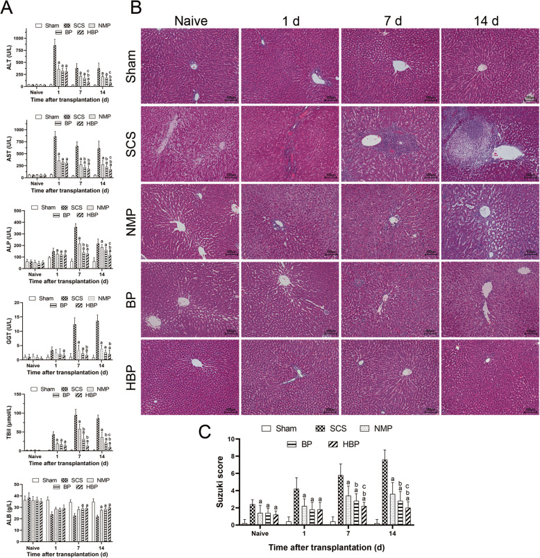
Background: Donation after circulatory death (DCD) liver grafts have a poor prognosis after transplantation. We investigated whether the outcome of DCD donor organs can be improved by heme oxygenase 1 (HO-1)-modified bone marrow-derived mesenchymal stem cells (BMMSCs) combined with normothermic machine perfusion (NMP), and explored its underlying mechanisms.

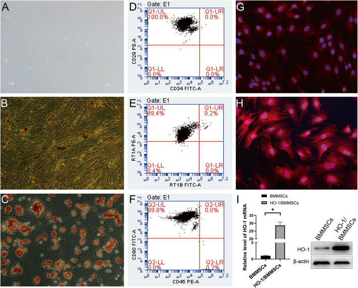
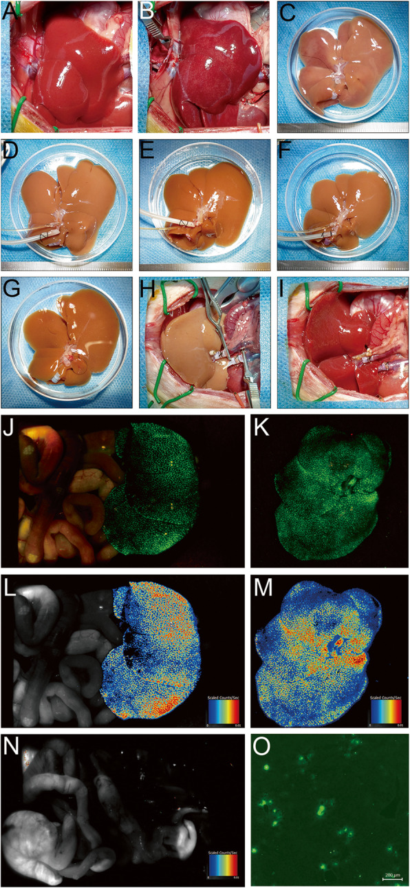
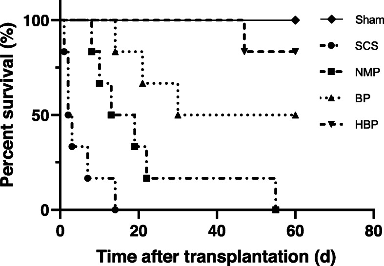
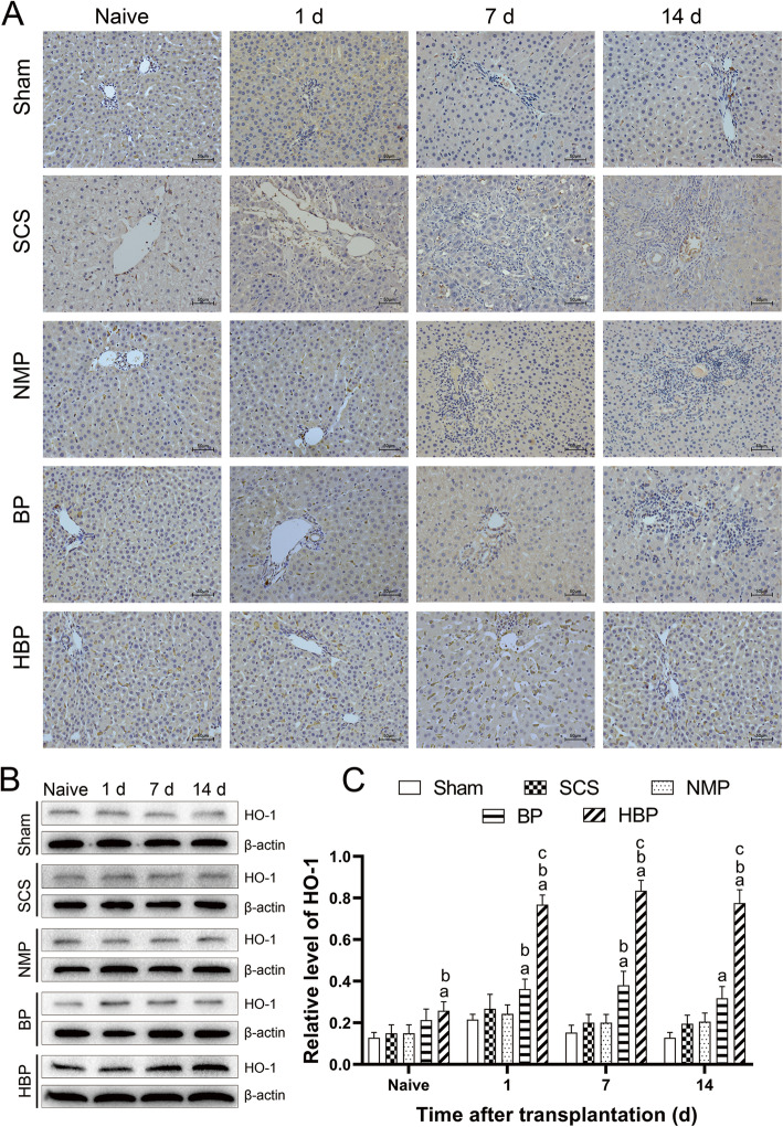
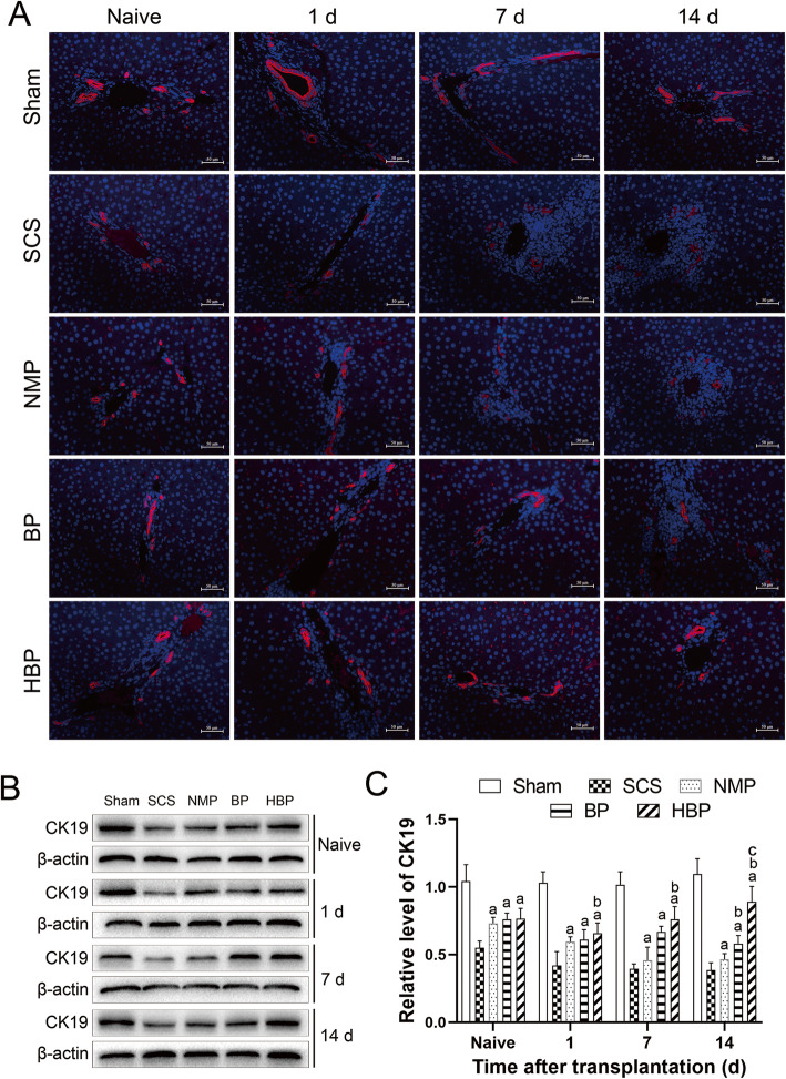
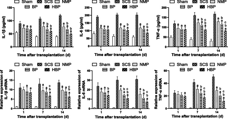
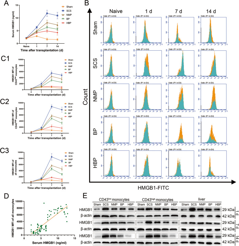
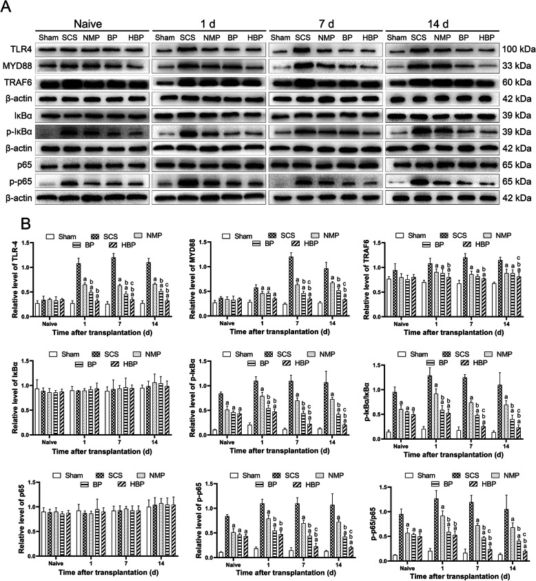
Methods: BMMSCs were isolated, cultured, and transduced with the HO-1 gene. An NMP system was established. DCD rat livers were obtained, preserved by different methods, and the recipients were divided into 5 groups: sham operation, static cold storage (SCS), NMP, BMMSCs combined with NMP, and HO-1/BMMSCs combined with NMP (HBP) groups. Rats were sacrificed at 1, 7, and 14 days after surgery; their blood and liver tissue samples were collected; and liver enzyme and cytokine levels, liver histology, high-mobility group box 1 (HMGB1) levels in monocytes and liver tissues, and expression of Toll-like receptor 4 (TLR4) pathway-related molecules were evaluated.

Results: After liver transplantation, the SCS group showed significantly increased transaminase levels, liver tissue damage, and shorter survival time. The HBP group showed lower transaminase levels, intact liver morphology, prolonged survival time, and decreased serum and liver proinflammatory cytokine levels. In the NMP and SCS groups, HMGB1 expression in the serum, monocytes, and liver tissues and TLR4 pathway-related molecule expression were significantly decreased.

Conclusions: HO-1/BMMSCs combined with NMP exerted protective effects on DCD donor liver and significantly improved recipient prognosis. The effect of HO-1/BMMSCs was greater than that of BMMSCs and was mediated via HMGB1 expression and TLR4 pathway inhibition.

Keywords: Bone marrow mesenchymal stem cells; Donation after circulatory death; Normothermic machine perfusion; Orthotopic liver transplantation.


Conflict of interest statement

The authors declare that they have no competing interests.

Figures

Fig. 1
Fig. 1
Characteristics of HO-1/BMMSCs in vitro and detection of HO-1 expression. a HO-1/BMMSCs were adherent and displayed long spindle-shaped morphology. b HO-1/BMMSCs showed osteogenic differentiation in vitro, as indicated by the calcareous deposits stained black with von Kossa staining. c Adipogenic differentiation of HO-1/BMMSCs, as indicated by oil red O-stained fat cells. df Surface biomarker identification. Results showed that 100.0, 99.8, and 99.4% of the cells were positive for CD29, CD90, and RT1A, respectively, and 100.0, 99.8, and 99.4% of the cells were negative for CD34, CD45, and RT1B, respectively. The molecular biological characteristics of HO-1/BMMSCs were not changed. g HO-1 expression in BMMSCs was identified by red fluorescence, and its intensity was weak. h HO-1 expression in HO-1/BMMSCs. The red fluorescence intensity was significantly higher in HO-1/BMMSCs than that in BMMSCs. i Western blotting and qRT-PCR results confirmed that HO-1 expression in HO-1/BMMSCs was significantly higher than that in BMMSCs
Fig. 2
Fig. 2
Liver appearance and colonization of BMMSCs in liver grafts. a The normal liver. b DCD donor liver (warm ischemia time, 30 min). c The donor liver of the SCS group was slightly edematous, with blunted edges, hepatic congestion, and uneven surface. The livers of the NMP (d), BP (e), and HBP groups (f) were pale brown in color, with sharp edges and no edema. gi OLT protocol. j Imaging of liver grafts in vivo (j) and in vitro (k). A pseudo-color image showing the fluorescence intensity of the liver graft in vivo (e) and in vitro (m). After liver resection, no GFP fluorescence signal was detected in the rat (n). In the frozen liver sections, colonization of GFP/BMMSCs in the hepatic sinusoids was observed using a fluorescence microscope (o, × 40), indicating that BMMSCs were evenly colonized in the transplanted liver
Fig. 3
Fig. 3
Kaplan-Meier survival curves of OLT recipients. Median survival time of the Sham, SCS, NMP, BP, and HBP groups was > 60, 2.5, 16, 37.5, and > 60 days, respectively. Log-rank (Mantel-Cox) test results showed that the survival rate of HBP group was significantly higher than that of other liver transplantation groups (P = 0.0005 vs. SCS group; P = 0.0013 vs. NMP group; P = 0.044 vs. BP group)
Fig. 4
Fig. 4
HO-1 expression in liver grafts. A Immunohistochemical staining of HO-1 (× 200) showed that there were more number of HO-1-positive cells in the liver interstitial cells of the HBP group compared to that of other groups. B Western blot analysis of HO-1 expression. C Relative quantification of HO-1 protein in different groups (HO-1/β-actin) showed that HO-1 level in the HBP group was significantly higher than that in other groups (aP < 0.05 vs. SCS group; bP < 0.05 vs. NMP group; cP < 0.05 vs. HBP group)
Fig. 5
Fig. 5
Liver function, histopathology of liver grafts, and the Suzuki score at different time points. A Liver function after transplantation. On POD 1, ALT levels between the SCS (882.01 ± 139.46 U/L) and NMP groups (359.42 ± 85.13 U/L) were significantly different (P < 0.05), but the difference was not significant between the NMP, BP, and HBP groups (P > 0.05). However, on POD 1 and 7, ALT levels in the HBP (96.78 ± 35.76 U/L) and BP groups (189.46 ± 47.21 U/L) were lower than those in the SCS (395.20 ± 112.57 U/L) and NMP groups (217.46 ± 36.42 U/L), and levels in the HBP group were lower than those in the BP group. The difference was significant (P < 0.05), and the AST, ALP, GGT, and TBil trends were similar to ALT. B Pathology of liver grafts at different time points (H&E staining, × 100). On POD 1, hepatic sinusoids were enlarged and vacuolar degeneration was detected in the SCS group. A small amount of vacuolar degeneration was also observed in the NMP, BP, and HBP groups. On POD 7, partial lobular destruction was observed in the SCS group, along with liver sinus congestion and infiltration of a large number of inflammatory cells; however, these effects were improved on POD 14. The liver tissue structure was more intact in the BP group than in the SCS and NMP groups. The hepatic lobular structure of the HBP group was relatively intact, with infiltration of a small number of inflammatory cells, and its tissue structure was improved compared to that of BP group. C The Suzuki score at different time points (aP < 0.05 vs. SCS group; bP < 0.05 vs. NMP group; cP < 0.05 vs. HBP group)
Fig. 6
Fig. 6
Expression of CK19 in biliary epithelial cells of the transplanted liver. A Immunofluorescence staining for CK19 (× 200, red tag) showed that the percentage of CK19-positive cells among biliary epithelial cells in the HBP group was significantly higher than that in the other groups. B Expression of CK19 in the transplanted liver analyzed by western blotting. C Relative expression of the CK19 protein (CK19/β-actin) showed that the relative expression of CK19 in the HBP group was significantly higher than that in the other groups
Fig. 7
Fig. 7
Levels of IL-1β, IL-6, and TNF-α in the serum and liver tissue. ELISA was used to determine the serum levels of IL-β, IL-6, and TNF-α, and qRT-PCR was used to determine the relative mRNA levels of IL-β, IL-6, and TNF-α in the liver tissue (aP < 0.05 vs. SCS group; bP < 0.05 vs. NMP group; cP < 0.05 vs. HBP group). On POD 1, the serum levels of IL-1β, IL-6, and TNF-α in the SCS group were significantly higher than those in other groups (P < 0.05), and no significant difference in levels between the NMP, BP, and HBP groups was observed. On POD 7, proinflammatory cytokine levels in the BP and HBP groups were lower than those in the SCS and NMP groups, and no significant difference in their levels between the BP and HBP groups was observed. On POD 14, their level in the HBP group decreased to near normal level and was lower than that in the BP group (P < 0.05). The mRNA expression pattern was similar to that of serum. The SCS group showed higher mRNA levels of IL-β, IL-6, and TNF-α than other groups on POD 1, and no difference in IL-β and TNF-α levels was observed between the NMP, BP, and HBP groups. However, on POD 7 and 14, the mRNA levels of IL-β, IL-6, and TNF-α in the BP and HBP groups were significantly lower than those in the SCS and NMP groups (P < 0.05), and on POD 14, their levels in the HBP group were significantly lower than those in the BP group
Fig. 8
Fig. 8
Serum HMGB1, MFI of HMGB1 on monocytes, and HMGB1 expression in liver grafts. A Serum HMGB1 level. The serum HMGB1 level in the HBP group was lower than that in the SCS, NMP, and BP groups (P < 0.05). B MFI of HMGB1 expression on CD43low monocytes (blue) and CD43high monocytes (yellow). C1 MFI of HMGB1 on CD43low monocytes. C2 MFI of HMGB1 on CD43high monocytes. C3 The MFI of HMGB1 on all monocytes in the HBP group decreased slowly after an increase on day 1, stabilized to near-normal levels on day 14, and was lower than that in the BP group (P < 0.05). The MFI of HMGB1 on all monocytes in the SCS group was significantly higher than those in the other groups at each time point (P < 0.05). D Correlation analysis. A strong positive correlation was observed between serum HMGB1 levels and the MFI of HMGB1 on all monocytes after OLT (r = 0.7931, P < 0.0001). E The protein expression of HMGB1 in monocytes and liver tissues. (aP < 0.05 vs. SCS group; bP < 0.05 vs. NMP group; cP < 0.05 vs. HBP group)
Fig. 9
Fig. 9
Western blot analysis of TLR4/NF-κB signaling pathway-related molecules. A Expression of key molecules involved in the TLR4/NF-κB pathway as detected by western blotting. B Relative gray value quantitative evaluation of TLR4/NF-κB pathway-related molecules. On POD 0 (naive), the expression of molecules upstream of TLR4 was not significantly changed, but that of the downstream molecules (p-IκBα and p-p65) was altered. The expression of downstream molecules in the SCS group was higher than that in NMP, BP, and HBP groups (P < 0.05), but no significant difference was observed between these three groups. On POD 1, the expression of MYD88, TRAF6, p-IκBα, and p-p65 in the BP and HBP groups was lower than that in the SCS and NMP groups (P < 0.05), but no difference in expression was observed between the BP and HBP groups. On POD 7 and 14, the expression of TLR4 pathway-related molecules (MYD88, TRAF6, p-IκBα, and p-p65) in the HBP and BP groups was significantly lower than that in the SCS and NMP groups, and its expression in HBP group was lower than that in BP group (P < 0.05). (aP < 0.05 vs. SCS group; bP < 0.05 vs. NMP group; cP < 0.05 vs. HBP group)

References

    1. Victor DR, Monsour HJ, Boktour M, Lunsford K, Balogh J, Graviss EA, et al. Outcomes of liver transplantation for hepatocellular carcinoma (HCC) beyond the University of California San Francisco (UCSF) criteria: a single center experience. Transplantation. 2019. 10.1097/TP.0000000000002835. - PubMed
    1. Chedid MF, Pinto MA, Juchem J, Grezzana-Filho T, Kruel C. Liver preservation prior to transplantation: past, present, and future. World J Gastrointest Surg. 2019;11(3):122–125. doi: 10.4240/wjgs.v11.i3.122. - DOI - PMC - PubMed
    1. Szilagyi AL, Matrai P, Hegyi P, Tuboly E, Pecz D, Garami A, et al. Compared efficacy of preservation solutions on the outcome of liver transplantation: Meta-analysis. World J Gastroenterol. 2018;24(16):1812–1824. doi: 10.3748/wjg.v24.i16.1812. - DOI - PMC - PubMed
    1. Leon DF, Fernandez AJ, Nicolas DCS, Perez RM, Sanchez PB, Montiel CC, et al. Combined flush with histidine-tryptophan-ketoglutarate and University of Wisconsin solutions in liver transplantation: preliminary results. Transplant Proc. 2018;50(2):539–542. doi: 10.1016/j.transproceed.2017.12.033. - DOI - PubMed
    1. Nair A, Hashimoto K. Extended criteria donors in liver transplantation-from marginality to mainstream. Hepatobiliary Surg Nutr. 2018;7(5):386–388. doi: 10.21037/hbsn.2018.06.08. - DOI - PMC - PubMed

Publication types