Review
. 2020 Jun 5;15(1):32.
doi: 10.1186/s13024-020-00375-7.

Harnessing regulatory T cell neuroprotective activities for treatment of neurodegenerative disorders

Affiliations
Review

Harnessing regulatory T cell neuroprotective activities for treatment of neurodegenerative disorders

Jatin Machhi et al. Mol Neurodegener. .

Abstract

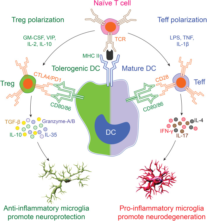
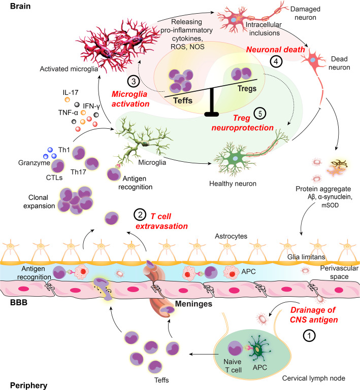
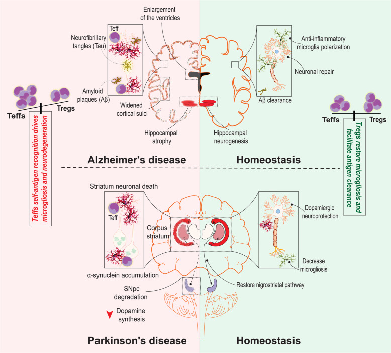
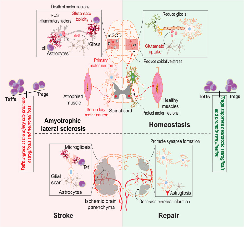
Emerging evidence demonstrates that adaptive immunity influences the pathobiology of neurodegenerative disorders. Misfolded aggregated self-proteins can break immune tolerance leading to the induction of autoreactive effector T cells (Teffs) with associated decreases in anti-inflammatory neuroprotective regulatory T cells (Tregs). An imbalance between Teffs and Tregs leads to microglial activation, inflammation and neuronal injury. The cascade of such a disordered immunity includes the drainage of the aggregated protein antigens into cervical lymph nodes serving to amplify effector immune responses. Both preclinical and clinical studies demonstrate transformation of this altered immunity for therapeutic gain. We posit that the signs and symptoms of common neurodegenerative disorders such as Alzheimer's and Parkinson's diseases, amyotrophic lateral sclerosis, and stroke can be attenuated by boosting Treg activities.

Keywords: Dendritic cells; Effector T cells (Teffs); Immune transformation; Microglia; Neurodegenerative disorders; Regulatory T cells (Tregs).


Conflict of interest statement

The authors declare no conflicts of interest.

Figures

Fig. 1
Fig. 1
DCs and T cell polarization. Immature DCs take up antigen, process and present it to immunocytes. Antigen uptake induces maturation signals in DCs in cooperation with the upregulation of co-stimulatory molecules. Mature DCs encounter naïve T cells through MHCII-T cell receptor (TCR) interactions, leading to T cell activation, Teff differentiation, and secretion of pro-inflammatory molecules (IL-10, IL-35, TGF-β, granzymes, etc.). In contrast, some DCs can maintain central and peripheral immune tolerance called tolerogenic DCs. The tolerogenic DCs exhibit low levels of costimulatory molecules and as such, provide insufficient stimulatory signals to naïve T cells to induce Treg and anti-inflammatory cytokines (IL-4, IL17, IFN-γ, etc.). Tregs suppress Teff function and proliferation to maintain immune tolerance. Teffs secreted molecules govern pro-inflammatory microglia polarization. In contrast, Tregs favor anti-inflammatory microglia polarization, supporting neuroprotection. Balance between Teffs and Tregs is essential to maintain homeostasis while their imbalance leads to neurodegeneration through microglia responses
Fig. 2
Fig. 2
Teff activities promote neuroinflammation. In neurodegenerative disorders, CNS antigens including Aβ, α-syn, mSOD and MBP provoke microglia immune responses leading to neuroinflammatory cascade in affected brain regions. (1) Self-antigen or misfolded proteins are generated from damaged neuronal cells. The neural antigens drain to the peripheral lymphoid nodes by meningeal lymphatic vessels where they are taken up by local APCs including macrophages and DCs. Natural self-antigens are presented to peripheral T cells in MHCII dependent manner. (2) Naïve T cells, upon recognition of cognate antigen, differentiate into antigen-specific Teffs. Reactive microglia secrete cytokine-chemokine milieu to upregulate cell adhesion molecules (CAM) by blood-brain barrier (BBB) endothelial cells, opening the gate for peripheral primed T cells. Teffs (Th1 and Th17) with upregulated integrins and CAM ligands readily cross the BBB. Teffs also cross the blood-CSF barrier through choroid plexus meninges. After extravasation into the brain, Teffs are reactivated upon recognition of cognate antigen on common CNS APCs. These include perivascular macrophages (PVMs), choroid plexus and meningeal macrophages and DCs and parenchymal microglia. (3, 4) Activated Teffs secrete pro-inflammatory and neurotoxic mediators to polarize microglia to a higher activation state, producing pro-inflammatory cytokines and reactive oxygen and nitrogen species which further perpetuate the inflammatory cascade to induce neurotoxicity. (5) Tregs maintain immune tolerance by suppressing effector immune responses. Naïve T cells can also differentiate into Tregs upon recognition of cognate antigen on peripheral APCs in secondary lymphoid tissues. Differentiated Tregs exert neuroprotective responses through multiple mechanisms. The inflammatory immune responses observed in neurodegenerative disorders are the outcome of Teff-Treg imbalance with upregulated Teff responses
Fig. 3
Fig. 3
Teff and Treg immunity in AD and PD. Pathogenic changes observed in AD brain include accumulation of intraneuronal neurofibrillary tangles (NFTs) of Tau protein and extracellular amyloid beta (Aβ) plaques. The accumulated peptides facilitate neuroinflammatory Teff entry into the brain where they affect resident microglia cells to induce local inflammatory responses. In PD, α-synuclein accumulation promotes immunoreactive Teff entry into the brain to activate microglia, compromising nigrostriatal axis between the substantia nigra pars compacta (SNpc) and striatum that execute motor commands. Immunoregulatory Tregs can restore homeostatic balance in the brain through clearance of neuroimmunogens, microglial polarization and restoration of normal neural functions
Fig. 4
Fig. 4
Teff and Treg immunity in ALS and stroke. In ALS, Teffs perpetrate innate microglial inflammation by misfolded SOD inciting oxidative stress and affecting astrocyte function linked to glutamate uptake, ensuring motor neuronal cell death with the primary clinical manifestations of disease. Parallel responses are operative in Stroke. Following ischemic stroke, peripheral Teffs accumulate at the brain injury site to participate in local inflammatory responses manifested by micro- and astrogliosis and secondary neuronal injuries. Following acute episode, injuries are contained, in part, through the emergence of Tregs that serve to reduce astrogliosis, promote synapse formation, and decrease the extent of injury. Tregs are neuroprotective mediators in each of these pathological processes and herald slow disease progression and control of neurodegenerative activities

References

    1. Reynolds AD, Stone DK, Hutter JA, Benner EJ, Mosley RL, Gendelman HE. Regulatory T cells attenuate Th17 cell-mediated nigrostriatal dopaminergic neurodegeneration in a model of Parkinson's disease. J Immunol. 2010;184:2261–2271. - PMC - PubMed
    1. Anderson KM, Olson KE, Estes KA, Flanagan K, Gendelman HE, Mosley RL. Dual destructive and protective roles of adaptive immunity in neurodegenerative disorders. Transl Neurodegener. 2014;3:25. - PMC - PubMed
    1. Gendelman HE, Mosley RL. A Perspective on Roles Played by Innate and Adaptive Immunity in the Pathobiology of Neurodegenerative Disorders. J Neuroimmune Pharmacol. 2015;10:645–650. - PMC - PubMed
    1. Engelhardt B, Vajkoczy P, Weller RO. The movers and shapers in immune privilege of the CNS. Nat Immunol. 2017;18:123–131. - PubMed
    1. Prinz M, Priller J. The role of peripheral immune cells in the CNS in steady state and disease. Nat Neurosci. 2017;20:136–144. - PubMed

Publication types

MeSH terms