. 2021 Mar;70(3):485-498.
doi: 10.1136/gutjnl-2020-321339. Epub 2020 Jun 5.

SETDB1 is required for intestinal epithelial differentiation and the prevention of intestinal inflammation

Affiliations

SETDB1 is required for intestinal epithelial differentiation and the prevention of intestinal inflammation

Lea Južnić et al. Gut. 2021 Mar.

Abstract

Objective: The intestinal epithelium is a rapidly renewing tissue which plays central roles in nutrient uptake, barrier function and the prevention of intestinal inflammation. Control of epithelial differentiation is essential to these processes and is dependent on cell type-specific activity of transcription factors which bind to accessible chromatin. Here, we studied the role of SET Domain Bifurcated Histone Lysine Methyltransferase 1, also known as ESET (SETDB1), a histone H3K9 methyltransferase, in intestinal epithelial homeostasis and IBD.

Design: We investigated mice with constitutive and inducible intestinal epithelial deletion of Setdb1, studied the expression of SETDB1 in patients with IBD and mouse models of IBD, and investigated the abundance of SETDB1 variants in healthy individuals and patients with IBD.

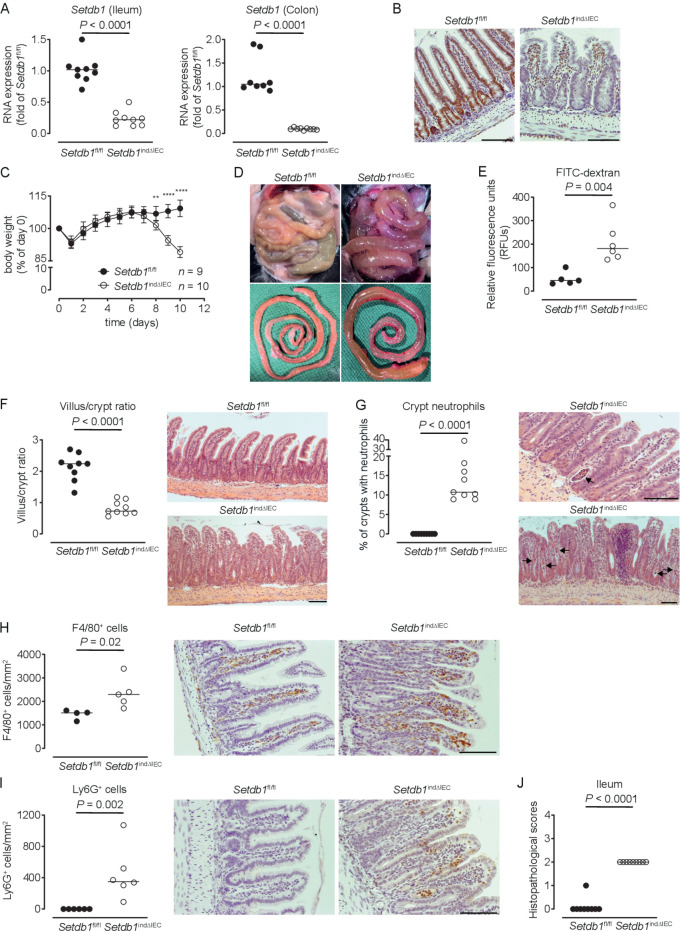
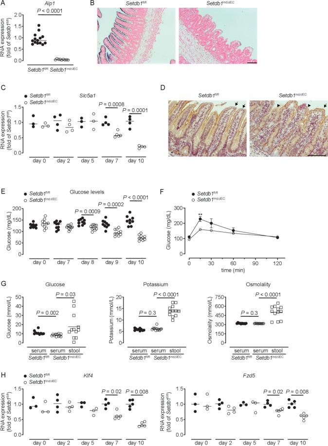
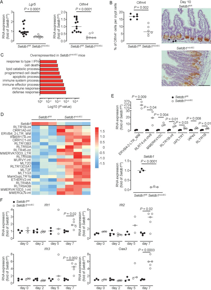
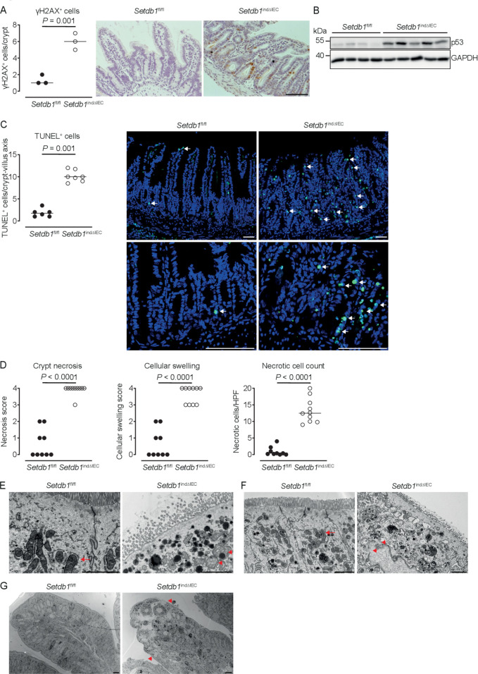
Results: Deletion of intestinal epithelial Setdb1 in mice was associated with defects in intestinal epithelial differentiation, barrier disruption, inflammation and mortality. Mechanistic studies showed that loss of SETDB1 leads to de-silencing of endogenous retroviruses, DNA damage and intestinal epithelial cell death. Predicted loss-of-function variants in human SETDB1 were considerably less frequently observed than expected, consistent with a critical role of SETDB1 in human biology. While the vast majority of patients with IBD showed unimpaired mucosal SETDB1 expression, comparison of IBD and non-IBD exomes revealed over-representation of individual rare missense variants in SETDB1 in IBD, some of which are predicted to be associated with loss of function and may contribute to the pathogenesis of intestinal inflammation.

Conclusion: SETDB1 plays an essential role in intestinal epithelial homeostasis. Future work is required to investigate whether rare variants in SETDB1 contribute to the pathogenesis of IBD.

Keywords: IBD; IBD - genetics; epithelial differentiation; gut inflammation; intestinal epithelium.


Conflict of interest statement

Competing interests: None declared.

Figures

Figure 1
Figure 1
Induced intestinal epithelial Setdb1 deletion is associated with inflammation and mortality. (A) RNA expression of Setdb1 in ileum and colon of the indicated mouse strains 10 days after the first tamoxifen injection as determined by quantitative PCR. (B) Representative immunohistochemistry staining of SETDB1 in the ileum of the indicated mouse strains on 10 days after the first tamoxifen injection. Note loss of the epithelial signal and conserved staining of SETDB1 in the lamina propria of Setdb1 indΔIEC mice. (C) Body weight of the indicated mouse strains following tamoxifen injection start at day 0. Mean±SD is shown. **P≤0.01; ****P≤0.0001. (D) Representative images at necropsy of the indicated mouse strains 10 days after the first tamoxifen injection. (E) Fluorescein isothiocyanate (FITC)-dextran measurement in serum of the indicated mouse strains 10 days after the first tamoxifen injection. (F) Villus-crypt ratio as well as representative H&E staining of ileum of the indicated mouse strains 10 days after the first tamoxifen injection. (G) Percentage of crypts containing neutrophil granulocytes (left) and representative H&E stainings (right) of ileum of the indicated mouse strains 10 days after the first tamoxifen injection. The arrows highlight crypt abscesses. (H–I) F4/80 (H) and Ly6G (I) immunohistochemistry with quantification (left) and representative images (right) in the ileum of the indicated mouse strains 10 days after the first tamoxifen injection. (J) Inflammation scores in the small intestine of the indicated mice as obtained by analysis of H&E stainings. Representative results of two-three independent experiments are shown (A–J). In (A, E–J), dots represent individual mice and the bar indicates the median. Size bars indicate 100 µm. The Mann-Whitney U test (A, E–J) or Student’s t-test (C) were applied. SETDB1, SET Domain Bifurcated Histone Lysine Methyltransferase 1, also known as ESET.
Figure 2
Figure 2
Altered intestinal epithelial differentiation in Setdb1 indΔIEC mice. (A) Representative H&E stainings of ileal villus tips of the indicated mouse strains 10 days after the first tamoxifen injection. Arrows indicate bright, round nuclei in Setdb1 indΔIEC mice. (B–C) Representative electron microscopy (EM) images of ileum of the indicated mouse strains 10 days after the first tamoxifen injection. Arrows indicate disorganised epithelium (B). In (C), a cuboid Setdb1 indΔIEC enterocyte is shown and the arrowhead highlights an irregularly shaped nucleus (N). The red line shows the basal membrane. (D, F) RNA expression of the indicated genes as determined by quantitative PCR in the ileum of the indicated mouse strains 10 days after the first tamoxifen injection. (E) Representative immunofluorescence staining of mucin-2 (Muc2) (red) in the ileum of the indicated mouse strains 10 days after the first tamoxifen injection. Arrows indicate goblet cells in wild-type (WT) mice. (G–H) Representative immunofluorescence stainings of Chga (G), green, Lyz1 (H), green and double stainings for Lyz1 (green) and Muc2 (red) (I) in the ileum of the indicated mouse strains 10 days after the first tamoxifen injection. Quantification of Lyz1-positive cells is shown on the left in (H). Arrowheads in (I) indicate Lyz1 and Muc2 double-positive cells. Representative results of three independent experiments are shown (A, D–I). EM images (B–C) are representative of three mice per group. In (D, F, H), dots represent individual mice and the bar indicates the median. Size bars indicate 50 µm (A, E, G–I), 10 µm (B, C). The Mann-Whitney U test (D, F, H) was applied. SETDB1, SET Domain Bifurcated Histone Lysine Methyltransferase 1, also known as ESET.
Figure 3
Figure 3
Loss of enterocyte differentiation in Setdb1 indΔIEC mice is associated with hypoglycaemic. (A, C, H) RNA expression of the indicated genes as determined by quantitative PCR in the ileum of the indicated mouse strains (A), 10 days after the first tamoxifen injection. (B) Representative alkaline phosphatase enzyme histochemistry in the ileum of the indicated mouse strains 10 days after the first tamoxifen injection. (D) Representative immunohistochemistry staining for SGLT1 in the ileum of the indicated mouse strains 10 days after the first tamoxifen injection. Arrows show intact SGLT1 staining and arrowheads loss of SGLT1 staining. (E–F) Blood glucose levels (E) and glucose tolerance test (F) of the indicated mouse strains at the indicated time points (E) or 10 days after the first tamoxifen injection (F). **P<0.01. (G) Glucose and potassium concentrations as well as osmolality in the serum and small intestinal luminal content (stool) of the indicated mice 10 days after tamoxifen administration. Representative results of two-three independent experiments are shown. In (A, C, E, H), dots represent individual mice and the bar indicates the median. In (F), mean±SEM is shown. In (G), the bar indicates the mean. Size bars indicate 100 µm. The Mann-Whitney U test (A, C, E, H), the Student’s t-test (F) or one-way analysis of variance (G) were applied. SETDB1, SET Domain Bifurcated Histone Lysine Methyltransferase 1, also known as ESET.
Figure 4
Figure 4
Setdb1 deletion leads to de-repression of endogenous retroviruses and induction of a type I interferon response. (A) RNA expression of Lgr5 and Olfm4 as determined by quantitative PCR (qPCR) in the ileum of the indicated mouse strains 9 days after the first tamoxifen injection. (B) Quantification and representative immunohistochemistry staining of OLFM4 in the ileum of the indicated mouse strains 10 days after the first tamoxifen injection. Arrows show intestinal stem cells (ISCs) in Villin-CreERT2-negative Setdb1 fl/fl mice. (C) Gene ontology (GO) terms of the top 100 upregulated genes in RNA sequencing of small intestinal crypts of Setdb1 indΔIEC mice 2 days after the first tamoxifen injection. (D) Heatmap of the expression of Setdb1 (top) and endogenous retroviruses (ERVs) (below) as obtained from RNA sequencing of small intestinal crypts of Setdb1 indΔIEC mice 2 days after the first tamoxifen injection. (E) Expression of the indicated ERVs (top) and Setdb1 (below) in the ileum of the indicated mouse strains 5 days after the first tamoxifen injection as determined by qPCR. (F) RNA expression of the indicated genes as determined by qPCR in the small intestine of mice at the indicated time points after the first tamoxifen injection. Representative results of two-three independent experiments are shown (A–B, E–F). RNA sequencing was performed once (C–D). In (A–B, E–F), dots represent individual mice and the bar indicates the median. Size bars indicate 100 µm. The Mann-Whitney U test (A–B, E–F) was applied. SETDB1, SET Domain Bifurcated Histone Lysine Methyltransferase 1, also known as ESET.
Figure 5
Figure 5
Loss of epithelial SETDB1 is associated with DNA damage and cell death. (A) Percentage of histone H2AX phosphorylation (γH2AX)-positive cells per crypt as determined by immunohistochemical staining (left) and representative stainings (right) in the ileum of the indicated mouse strains. (B) Western blot analysis for p53 and GAPDH in small intestinal intestinal epithelial cells (IECs) of the indicated mouse strains. (C) Percentage of terminal deoxynucleotidyl transferase dUTP nick end labeling (TUNEL)-positive cells per crypt (left) and representative immunofluorescence stainings (right, TUNEL in green) in the ileum of the indicated mouse strains. Arrows show TUNEL-positive IECs. (D) Quantification of crypt necrosis and cellular swelling as well as of the number of necrotic cells per high power field in the ileum of the indicated mouse strains. (E–G) Representative electron microscopy images in the ileum of the indicated mouse strains. Arrows indicate normal mitochondria (E–F). Arrowheads indicated swelling and loss of the structural integrity of mitochondria (E) and loss of integrity of the nuclear membrane (F) as well as swelling of the cytoplasm and rupture of the plasma membrane (G). All data were obtained at day 10 after the first tamoxifen injection. Representative results of two independent experiments are shown (A–G). In (A, C–D), dots represent individual mice and the bar indicates the median. Size bars indicate 100 µm in (A, C), 2 µm (E, F), 10 µm (G). The Student’s t-test (A) or the Mann-Whitney U test (C–D) were applied. SETDB1, SET Domain Bifurcated Histone Lysine Methyltransferase 1, also known as ESET.
Figure 6
Figure 6
Human SETDB1 expression and variants in IBD. (A) Observed-to-expected ratio of variants in SETDB1 based on the Genome Aggregation Database (gnomAD, https://gnomad.broadinstitute.org). pLoF, predicted loss-of-function variants. (B–C) Quantile normalised read count of the indicated genes as obtained by RNA sequencing of biopsies of inflamed (IF) or non-inflamed (NI) intestinal mucosa of patients with Crohn’s disease (CD), ulcerative colitis (UC), healthy controls (CTR NI) or disease controls (CTR IF). For a detailed description of the cohort, see Häsler et al. (D–E) Relative expression of SETDB1 in mucosal transcriptomes of the indicated patients. Patient cohorts were previously described by Planell et al (D) and Olsen et al (E). (F) Allele frequency of the 1:150935127 C/T variant (rs145309946, pArg1075Cys) in the Finnish (FIN) and non-Finnish European (NFE) IBD and non-IBD population as obtained through the IBD exomes browser (http://ibd.broadinstitute.org). The Kruskal-Wallis test (B–E) or the Fisher’s exact test (F) (for further information, see Rivas et al 54) was applied. NS, not significant; SETDB1, SET Domain Bifurcated Histone Lysine Methyltransferase 1, also known as ESET.

Similar articles

Cited by

References

    1. Gehart H, Clevers H. Tales from the crypt: new insights into intestinal stem cells. Nat Rev Gastroenterol Hepatol 2019;16:19–34. 10.1038/s41575-018-0081-y - DOI - PubMed
    1. de Sousa e Melo F, de Sauvage FJ. Cellular plasticity in intestinal homeostasis and disease. Cell Stem Cell 2019;24:54–64. 10.1016/j.stem.2018.11.019 - DOI - PubMed
    1. Korinek V, Barker N, Moerer P, et al. . Depletion of epithelial stem-cell compartments in the small intestine of mice lacking Tcf-4. Nat Genet 1998;19:379–83. 10.1038/1270 - DOI - PubMed
    1. van Es JH, Haegebarth A, Kujala P, et al. . A critical role for the Wnt effector Tcf4 in adult intestinal homeostatic self-renewal. Mol Cell Biol 2012;32:1918–27. 10.1128/MCB.06288-11 - DOI - PMC - PubMed
    1. Kuhnert F, Davis CR, Wang H-T, et al. . Essential requirement for Wnt signaling in proliferation of adult small intestine and colon revealed by adenoviral expression of Dickkopf-1. Proc Natl Acad Sci U S A 2004;101:266–71. 10.1073/pnas.2536800100 - DOI - PMC - PubMed

Publication types

Substances