. 2020 Jun 5;5(48):eaba0570.
doi: 10.1126/sciimmunol.aba0570.

Oral epithelial IL-22/STAT3 signaling licenses IL-17-mediated immunity to oral mucosal candidiasis

Affiliations

Oral epithelial IL-22/STAT3 signaling licenses IL-17-mediated immunity to oral mucosal candidiasis

Felix E Y Aggor et al. Sci Immunol. .

Abstract

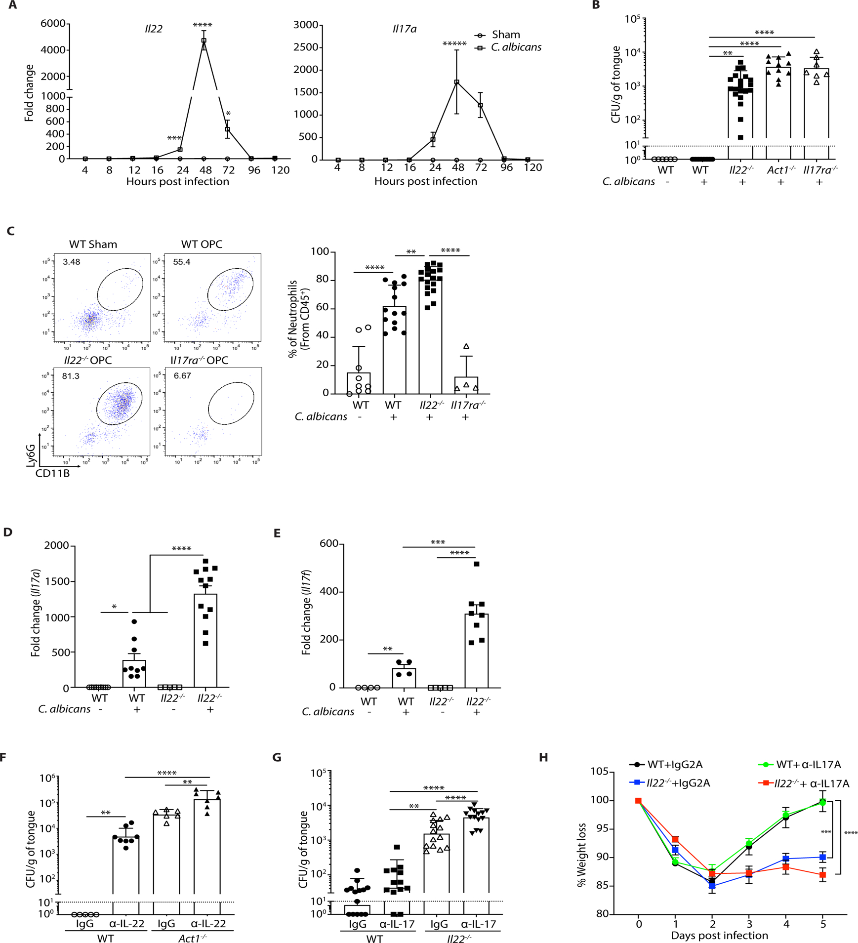
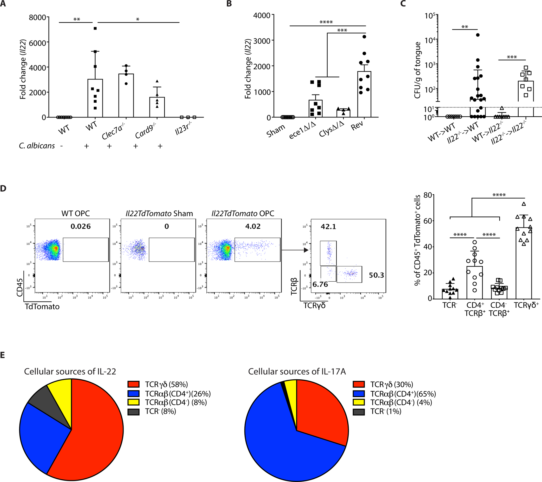
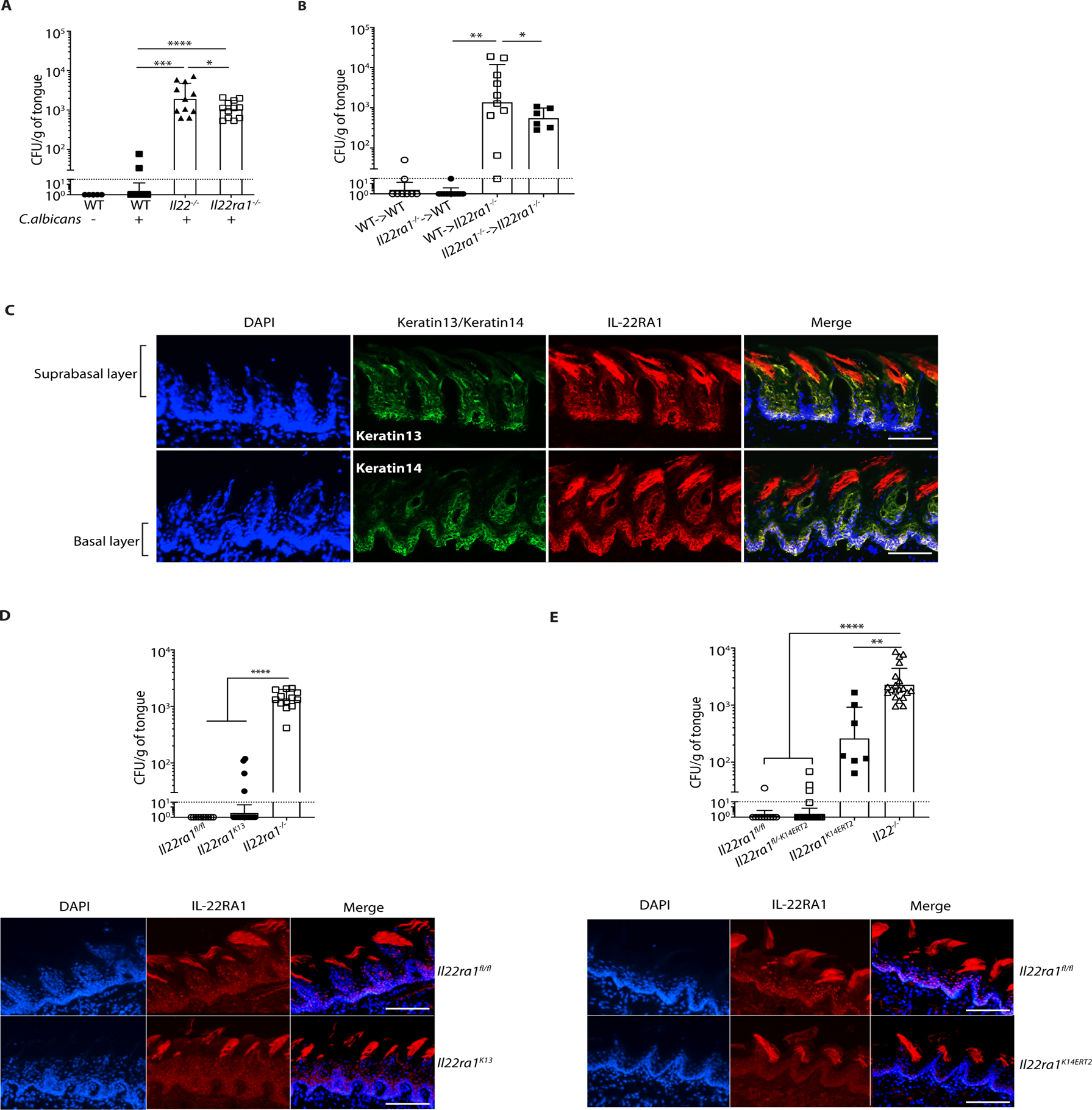
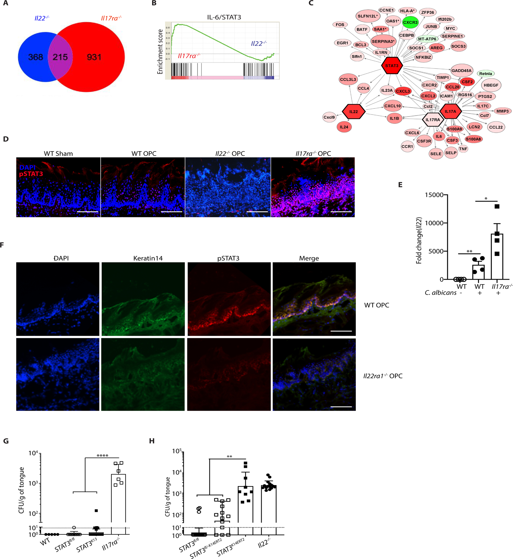
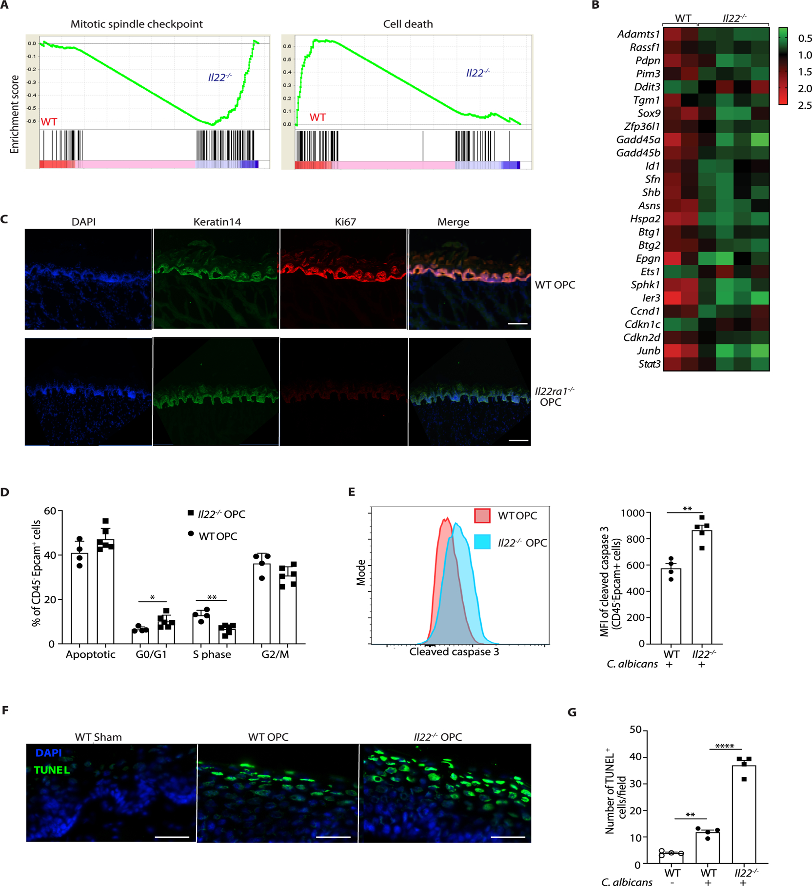
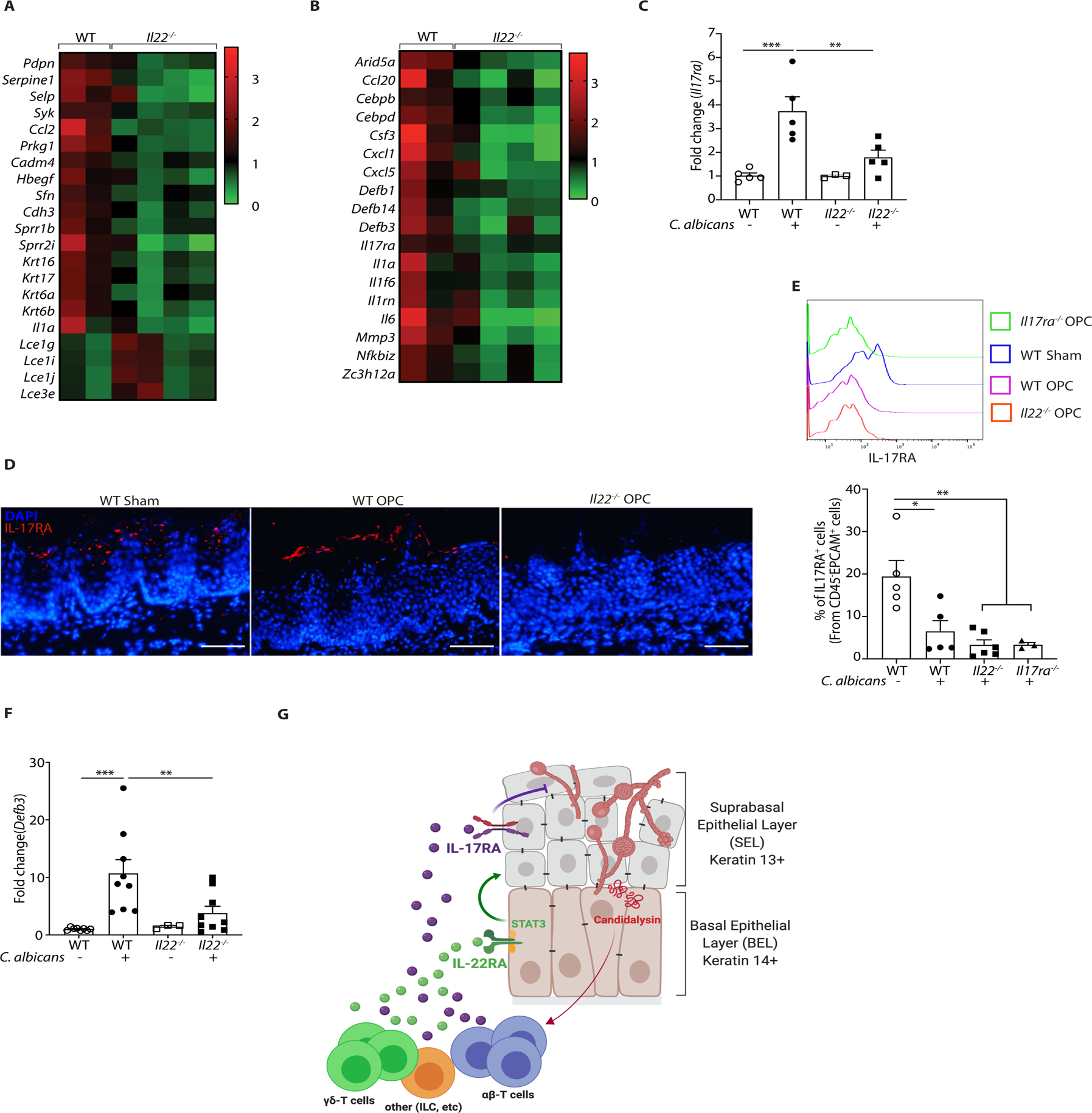
Oropharyngeal candidiasis (OPC; thrush) is an opportunistic infection caused by the commensal fungus Candida albicans Interleukin-17 (IL-17) and IL-22 are cytokines produced by type 17 lymphocytes. Both cytokines mediate antifungal immunity yet activate quite distinct downstream signaling pathways. While much is now understood about how IL-17 promotes immunity in OPC, the activities of IL-22 are far less well delineated. We show that, despite having similar requirements for induction from type 17 cells, IL-22 and IL-17 function nonredundantly during OPC. We find that the IL-22 and IL-17 receptors are required in anatomically distinct locations within the oral mucosa; loss of IL-22RA1 or signal transducer and activator of transcription 3 (STAT3) in the oral basal epithelial layer (BEL) causes susceptibility to OPC, whereas IL-17RA is needed in the suprabasal epithelial layer (SEL). Transcriptional profiling of the tongue linked IL-22/STAT3 not only to oral epithelial cell proliferation and survival but also, unexpectedly, to driving an IL-17-specific gene signature. We show that IL-22 mediates regenerative signals on the BEL that replenish the IL-17RA-expressing SEL, thereby restoring the ability of the oral epithelium to respond to IL-17 and thus to mediate antifungal events. Consequently, IL-22 signaling in BEL "licenses" IL-17 signaling in the oral mucosa, revealing spatially distinct yet cooperative activities of IL-22 and IL-17 in oral candidiasis.


Conflict of interest statement

COMPETING INTERESTS

SLG previously received a grant from Janssen to evaluate anti-IL-17A antibody function in OPC (110). Candidalysin has been patented by King’s College London, UK and Hans-Knöll Institut, Jena, DE (US Patent No.: 9,969,796; EU Patent No.: 2984103). The other authors declare no competing interests.

Figures

Figure 1.
Figure 1.. IL-22 protects against OPC non-redundantly with IL-17RA
The indicated mice were sublingually inoculated with cotton ball-saturated PBS (Sham) or C. albicans (OPC). Each symbol represents one mouse. A. Total mRNA from tongue homogenates of infected WT mice was subjected to qPCR for Il22 and Il17a and normalized to Gapdh at each time point. Graphs show mean ± SEM. Data are pooled from 4–9 mice per group. B. Fungal burdens were determined by CFU enumeration on YPD/Amp agar at day 5 p.i. Graphs show geometric mean ± SD. Data were pooled from 3 independent experiments. Dashed line indicates limit of detection (~30 CFU/g). C. Tongue homogenates were prepared on day 2 p.i. Left: A representative FACS plot showing percent of CD11b+Ly6G+ neutrophils (gated on live, CD45+ cells). Right: Data from 3 independent experiments. D-E. Il17a and Il17f in total RNA from tongue on day 2 p.i. was assessed by qPCR relative to Gapdh. Graphs show mean ± SEM relative to Sham-infected WT mice. F. Mice were injected i.p. with anti-IL-22 or isotype control IgG (150 μg) on days −1, 0, 1, 2, and 3 relative to infection. CFU was assessed on day 4, pooled from 2 independent experiments. G. Mice were treated with anti-IL-17A or isotype control (IgG2a) (200 μg) injected i.p. on days −1, 1, and 3 relative to infection. CFU was assessed on day 5, pooled from 3 independent experiments. H. Weight loss was assessed daily, shown relative to day 0 in mice from panel F. Data were analyzed by ANOVA or Student’s t-test, with Mann-Whitney U test for fungal load analysis.
Figure 2.
Figure 2.. Determinants of IL-22 induction in acute OPC
The indicated mice were subjected to OPC. Each symbol represents one mouse A. Tongues were harvested on day 2 p.i. and Il22 mRNA was assessed by qPCR, normalized to Gapdh. Graphs show mean ± SEM relative to sham-infected WT mice. B. WT mice were infected with the indicated C. albicans strains. Il22 mRNA in tongue on day 2 p.i. was assessed by qPCR, normalized to Gapdh. Data were pooled from 2 independent experiments. C. BM from indicated donors was transferred into irradiated recipients. After 6–9 weeks, mice were subjected to OPC and fungal burdens assessed on day 5 p.i. Data were pooled from 2 experiments. D. On day 2 p.i., tongue homogenates from IL22TdTomato mice were stained for the TCRβ and TCRγδ and gated on the live CD45+TdTomato+ population. Left: representative FACS plots. Right: Pooled results from 3 independent experiments. Each symbol represents data from 2 pooled tongues. Data were analyzed by ANOVA or Student’s t-test, with Mann-Whitney U test for fungal load analysis. E. Comparisons of the relative percentages of IL-22+ cells (left) or IL-17+ cells (data from (29)) isolated from tongues of mice 2 days p.i. and analyzed by flow cytometry.
Figure 3.
Figure 3.. IL-22 signaling in the oral basal epithelial layer is required for protection against OPC
The indicated mice were subjected to OPC. A. Fungal burdens were assessed on day 5 p.i. Bars show geometric mean ± SD. Data were pooled from 2 independent experiments. B. BM from indicated donors was transferred into irradiated recipients. After 6–9 weeks, mice were subjected to OPC and fungal burdens assessed on day 5 p.i. Data were pooled from 2 experiments. C. Frozen sections from WT tongues were co-stained with DAPI and Abs against K13, K14 or IL-22RA1. Suprabasal and basal epithelial layers are indicated. Images are representative of a minimum of 3 sections. Size bar = 200 μm. D. Top: Fungal burdens were assessed on day 5 p.i. Data are pooled from 3 independent experiments. Bottom: IF staining of tongues from the indicated mice were co-stained with DAPI and α-IL-22RA1 Abs. Size bar = 200 μm. E. Top: All mice except Il22−/− were administered tamoxifen for 5 days prior to OPC, and fungal burden assessed on day 5 p.i. Bars show geometric mean ± SD. Bottom: Frozen sections from tongues from the indicated mice were co-stained with DAPI and α-IL-22RA1. Size bar = 200 μm. Data were pooled from 3 independent experiments and analyzed by ANOVA with Mann-Whitney U test.
Figure 4.
Figure 4.. STAT3 in oral epithelial cells is required for protection against OPC
A. RNASeq was performed on whole tongue mRNA from WT, Il22−/− and Il17ra−/− mice subjected to OPC and harvested on day 1 p.i. Venn diagram of differentially regulated or overlapping genes in infected Il22−/− and Il17ra−/− compared to WT mice. 215 genes were regulated both by IL-22 and IL-17RA, whereas 368 genes are regulated only by IL-22, and 931 genes were regulated only by IL-17RA. B. GSEA enrichment of predicted IL-6/STAT3 gene sets in Il17ra−/− and Il22−/− mice from panel A. C. Ingenuity Pathway Analysis of RNASeq data from panel A, indicating that STAT3 is an upstream regulator integrating Il22- and Il17ra- driven transcriptional networks. D. IF staining of tongue frozen sections with DAPI and anti-pSTAT3 (Tyr705) in WT, Il22−/− and Il17ra−/− mice harvested 2 days p.i. Size bar = 200 μm. E. qPCR of Il22 in tongue mRNA from WT or Il17ra−/− mice at 2 days p.i. normalized to Gapdh F. IF staining of DAPI, pSTAT3 (Tyr705) and K14 in WT or Il22ra1−/− mice at 2 days p.i. Size bar = 200 μm. G. The indicated mice were subjected to OPC and fungal burden quantified at day 5 p.i.. Data are pooled from 3 experiments H. All mice except Il22−/− were administered tamoxifen for 5 d, subjected to OPC and fungal burden assessed on day 5 p.i. Bars show geometric mean ± SD. Data was pooled from 3 experiments. Data analyzed by ANOVA with Mann-Whitney U test.
Figure 5.
Figure 5.. IL-22 promotes cell survival and proliferation during OPC
RNASeq was performed on tongue mRNA from WT, Il22−/− or Il17ra−/− mice, isolated 24 h p.i. A. GSEA of mitotic spindle checkpoint pathway and cell death pathway genes. Normalized enrichment score is shown on Y-axis. B. Heatmap of cell cycle pathway genes in global differential gene expression analysis (Partekflow) in WT or Il22−/− mice. C. IF staining of DAPI, Ki67 and K14 in WT and Il22ra1−/− mice at 2 days p.i. Data are representative of images from 2 mice per group. Size bar = 100 μm. D. BrdU was administered 24 h p.i., and tongues harvested on day 2 p.i. Cell cycle/apoptotic status of CD45EpCAM+ cells was determined by bromodeoxyuridine (BrdU) and 7-aminoactinomycin (7AAD) staining. Data show mean ± SEM. E. Frequency of CD45EpCAM+ cells staining positive for active (cleaved) caspase-3 in tongue homogenates at day 2 pi measured by flow cytometry. Left: Representative FACS plot. Right: Pooled data from 4 independent samples showing mean fluorescence intensity (MFI) of cleaved Caspase 3 within the CD45EpCAM+ compartment. Data analyzed by Student’s t-test. F. DAPI and TUNEL staining of tongue sections from the indicated mice at day 2 p.i. Images are representative of 4 mice per group. Size bar = 200 μm. G. Quantification of TUNEL+ cells from panel. F. Data analyzed by ANOVA and post-hoc Tukey’s test.
Figure 6.
Figure 6.. IL-22 licenses IL-17 signaling during OPC
A. Heatmap of genes implicated in tissue repair, wound healing, keratinization and epithelial differentiation. B. Heatmap of IL-17 signature genes in OPC. C. qPCR of Il17ra expression normalized to Gapdh in tongue tissue from the indicated mice subjected to OPC and analyzed on day 2 p.i.. Data show mean ± SEM relative to sham-infected mice. Data analyzed by ANOVA or Student’s t-test. D. IF staining of IL-17RA and DAPI in the indicated mice on day 2 p.i. E. IL-17RA expression in CD45EpCAM+ oral epithelial cells in WT or Il22−/− mice during OPC. Top: representative FACS histogram. Bottom: Pooled data from 2 independent experiments. Size bar = 200 μm. F. Expression of BD3 (Defb3) mRNA in tongue from WT or Il22−/− mice during OPC, normalized to Gapdh. Data analyzed by ANOVA or student’s t-test. G. Diagram of stratified oral epithelium during a first encounter with C. albicans. Fungal hyphae induces cellular damage and secrete the peptide candidalysin, which facilitates tissue invasion and activates innate IL-17- and IL-22-producing lymphocytes (see Refs (29, 61)). IL-17 was shown previously to act dominantly on K13+ cells of the suprabasal epithelial layer (SEL) (31). In contrast, IL-22/STAT3 promotes proliferation of the K14+ basal epithelial layer (BEL) that serves to restore the IL-17R-expressing SEL, thus maintaining IL-17-induced antifungal signals such as β-defensins and neutrophil responses that are required to mediate clearance of C. albicans. Diagram created on Biorender.com.

Similar articles

Cited by

References

    1. Brown GD et al., Hidden killers: human fungal infections. Sci Transl Med 4, 165rv113 (2012). - PubMed
    1. Cassone A, Development of vaccines for Candida albicans: Fighting a skilled transformer. Nat Rev Microbiol 11, 884–891 (2013). - PubMed
    1. Naglik JR, Gaffen SL, Hube B, Candidalysin: discovery and function in Candida albicans infections. Curr Opin Microbiol 52, 100–109 (2019). - PMC - PubMed
    1. Fidel PL Jr., Candida-Host Interactions in HIV Disease: Implications for Oropharyngeal Candidiasis. Adv Dent Res 23, 45–49 (2011). - PMC - PubMed
    1. Conti HR, Gaffen SL, IL-17-Mediated Immunity to the Opportunistic Fungal Pathogen Candida albicans. J Immunol 195, 780–788 (2015). - PMC - PubMed

Publication types