. 2020 Jun 5;5(48):eabd0110.
doi: 10.1126/sciimmunol.abd0110. Epub 2020 Jun 5.

Inhibition of Bruton tyrosine kinase in patients with severe COVID-19

Affiliations

Inhibition of Bruton tyrosine kinase in patients with severe COVID-19

Mark Roschewski et al. Sci Immunol. .

Abstract

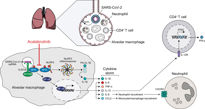
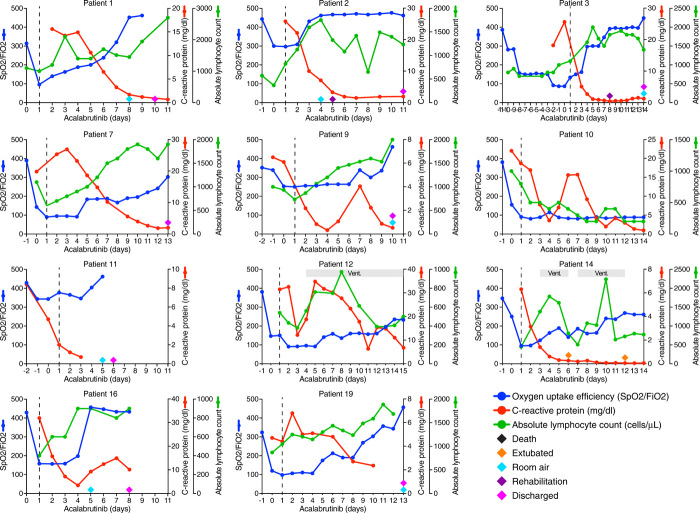
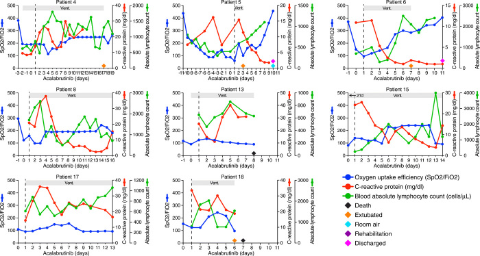
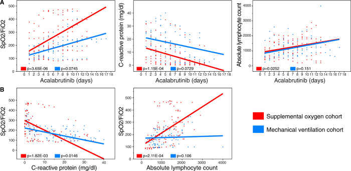
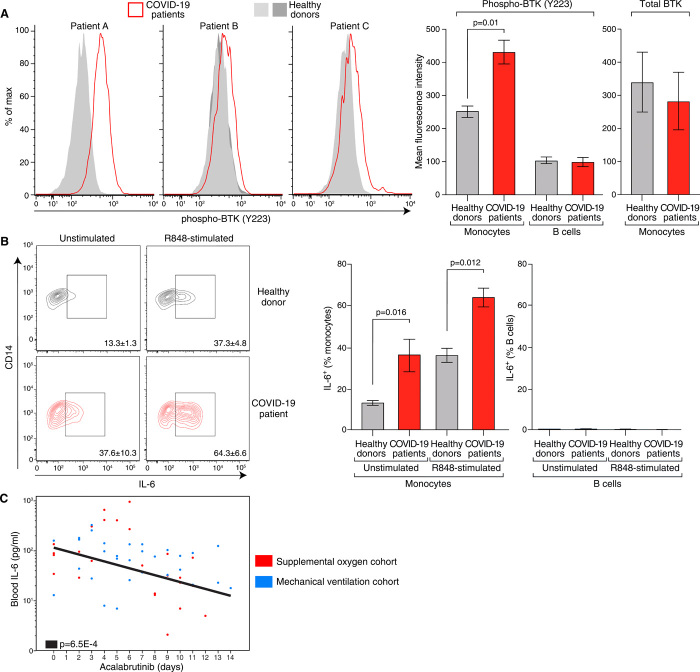
Patients with severe COVID-19 have a hyperinflammatory immune response suggestive of macrophage activation. Bruton tyrosine kinase (BTK) regulates macrophage signaling and activation. Acalabrutinib, a selective BTK inhibitor, was administered off-label to 19 patients hospitalized with severe COVID-19 (11 on supplemental oxygen; 8 on mechanical ventilation), 18 of whom had increasing oxygen requirements at baseline. Over a 10-14 day treatment course, acalabrutinib improved oxygenation in a majority of patients, often within 1-3 days, and had no discernable toxicity. Measures of inflammation - C-reactive protein and IL-6 - normalized quickly in most patients, as did lymphopenia, in correlation with improved oxygenation. At the end of acalabrutinib treatment, 8/11 (72.7%) patients in the supplemental oxygen cohort had been discharged on room air, and 4/8 (50%) patients in the mechanical ventilation cohort had been successfully extubated, with 2/8 (25%) discharged on room air. Ex vivo analysis revealed significantly elevated BTK activity, as evidenced by autophosphorylation, and increased IL-6 production in blood monocytes from patients with severe COVID-19 compared with blood monocytes from healthy volunteers. These results suggest that targeting excessive host inflammation with a BTK inhibitor is a therapeutic strategy in severe COVID-19 and has led to a confirmatory international prospective randomized controlled clinical trial.


Figures

Fig. 1
Fig. 1. Model of BTK-dependent hyper-inflammation in severe COVID-19.
Binding of SARS-CoV2 to ACE2 on respiratory epithelia initiates infection. Hypothetically, macrophages may participate in the COVID-19 inflammatory response by phagocytic uptake of viral particles or cellular debris containing viral single-stranded RNA (ssRNA). ssRNA can bind to TLR7 and TLR8, thereby recruiting and activating BTK and MYD88 (51, 52). Downstream of TLR engagement, BTK-dependent NF-κB activation results in the production of pro-inflammatory cytokines and chemokines (53), a “cytokine storm” that could increase the recruitment of monocytes/macrophages and neutrophils during the late phase of severe COVID-19 infection. BTK inhibitors such as acalabrutinib block TLR-dependent NF-κB activation in macrophages (20, 21), thereby dampening the production of pro-inflammatory mediators, as occurs in an influenza-induced lung injury model (27). During severe COVID-19, the heightened levels of IL-1β in several COVID-19 patients (11, 12) indicates the formation of an NLRP3 inflammasome that converts pro-IL-1β to mature IL-1β (54). BTK binds to and phosphorylates NLRP3, thereby promoting its oligomerization and assembly into an inflammasome (–26). BTK inhibitors such as acalabrutinib inhibit inflammasome-mediated production of IL-1β, as observed in a model of influenza-induced lung injury (27). SARS-CoV-2, severe acute respiratory syndrome coronavirus 2; COVID-19, coronavirus disease 2019; ACE2, angiotensin-converting enzyme 2; TLR, Toll-like receptor; MyD88, myeloid differentiation primary response 88; BTK, Bruton tyrosine kinase; NF-κB, nuclear factor kappa B; NLRP3, NLR family pyrin domain containing 3; ASC, Apoptosis-associated speck-like protein containing a caspase recruitment domain; ORF3a, open reading frame 3a; IFN-γ, interferon gamma; IL, interleukin; IL-12R, IL-12 receptor; CCL2, C-C motif chemokine ligand 2; CXCL1, C-X-C motif chemokine ligand 1; CXCR2, C-X-C motif chemokine receptor 2.
Fig. 2
Fig. 2. Clinical course and changes in inflammatory markers during acalabrutinib treatment in patients treated prior to intubation.
Shown are measures of oxygen uptake requirement SpO2/FiO2 (% blood oxygen saturation (SpO2)/fraction of delivered oxygen (FiO2)), a ratio that accounts for both oxygen delivery and uptake (theoretical maximum 476 for 100% oxygen saturation on room air). Also shown are measures of inflammation (C-reactive protein mg/dL) and absolute lymphocyte count (cells/μL) at all available timepoints before and after acalabrutinib treatment, which was started on day 1 (dotted line). Notable clinical parameters are shown as indicated (extubation, breathing on room air, transfer to rehabilitation, hospital discharge, death). The duration of mechanical ventilation (Vent.) is indicated.
Fig. 3
Fig. 3. Clinical course and changes in inflammatory markers during acalabrutinib treatment in patients treated while on mechanical ventilation.
See legend for Fig. 2.
Fig. 4
Fig. 4. Associations between measures of pulmonary function and inflammation following acalabrutinib treatment. A.
Plots of oxygen uptake efficiency (SpO2/FiO2), CRP and ALC levels versus days of acalabrutinib treatment for all patients at all time points. Patients in the supplemental oxygen and mechanical ventilation cohorts are indicated in red and blue, respectively. The trend lines shown represent the regression from a linear mixed-effect model blocked by patient. The reported p-values test the null hypothesis that the trend line has zero slope. B. Plots of oxygen uptake efficiency (SpO2/FiO2) versus either CRP or ALC. Trend lines and p-values as above.
Fig. 5
Fig. 5. BTK activation and IL-6 production in COVID-19. A.
Left panels: histograms of BTK phosphorylation in CD14+ blood monocytes from 3 patients with severe COVID-19 (A, B, C; Table S10) and 4 healthy volunteers, as indicated. Right panels: Summary data showing significant increase in mean fluorescence intensity of phosphorylated BTK (residue Y223) in CD14+ monocytes from 3 COVID-19 patients compared with 5 healthy volunteers, with no evident BTK phosphorylation in CD19+ B cells. Total BTK levels in blood monocytes shown in the far right panel were comparable in 3 COVID-19 patients and 5 healthy volunteers. B. Left panels: Representative contour plots of intracellular IL-6 production in CD14+ monocytes from a patient with severe COVID-19 (Patient C; Table S10) and a healthy volunteer, either as unstimulated ex vivo cells or following R848 (10 μM) stimulation, as indicated. Right panels: Summary data showing significant increase in the percent of IL-6+ CD14+ monocytes from 4 COVID-19 patients (A, B, C, D; Table S10) compared with 5 healthy volunteers, before and after R848 stimulation, with no evident IL-6 production by CD19+ B cells. C. Plot of blood IL-6 concentrations (pg/ml) on a log scale versus days of acalabrutinib treatment for patients in whom there were at least two IL-6 measurements during the plotted time course. Patients in the supplemental oxygen (n=5) and mechanical ventilation (n=3) cohorts are indicated in red and blue, respectively. The trend line shown represents the regression from a linear mixed-effect model blocked by patient for the combined cohorts due to limited data in each group. The reported p-value tests the null hypothesis that the trend line has zero slope. All quantitative data in 5A and 5B represent means ± SEM.

References

    1. Lu R., Zhao X., Li J., Niu P., Yang B., Wu H., Wang W., Song H., Huang B., Zhu N., Bi Y., Ma X., Zhan F., Wang L., Hu T., Zhou H., Hu Z., Zhou W., Zhao L., Chen J., Meng Y., Wang J., Lin Y., Yuan J., Xie Z., Ma J., Liu W. J., Wang D., Xu W., Holmes E. C., Gao G. F., Wu G., Chen W., Shi W., Tan W., Genomic characterisation and epidemiology of 2019 novel coronavirus: Implications for virus origins and receptor binding. Lancet 395, 565–574 (2020). 10.1016/S0140-6736(20)30251-8 - DOI - PMC - PubMed
    1. Guan W. J., Ni Z. Y., Hu Y., Liang W. H., Ou C. Q., He J. X., Liu L., Shan H., Lei C. L., Hui D. S. C., Du B., Li L. J., Zeng G., Yuen K. Y., Chen R. C., Tang C. L., Wang T., Chen P. Y., Xiang J., Li S. Y., Wang J. L., Liang Z. J., Peng Y. X., Wei L., Liu Y., Hu Y. H., Peng P., Wang J. M., Liu J. Y., Chen Z., Li G., Zheng Z. J., Qiu S. Q., Luo J., Ye C. J., Zhu S. Y., Zhong N. S.; China Medical Treatment Expert Group for Covid-19 , Clinical Characteristics of Coronavirus Disease 2019 in China. N. Engl. J. Med. 382, 1708–1720 (2020). 10.1056/NEJMoa2002032 - DOI - PMC - PubMed
    1. Shi H., Han X., Jiang N., Cao Y., Alwalid O., Gu J., Fan Y., Zheng C., Radiological findings from 81 patients with COVID-19 pneumonia in Wuhan, China: A descriptive study. Lancet Infect. Dis. 20, 425–434 (2020). 10.1016/S1473-3099(20)30086-4 - DOI - PMC - PubMed
    1. Shi Y., Wang Y., Shao C., Huang J., Gan J., Huang X., Bucci E., Piacentini M., Ippolito G., Melino G., COVID-19 infection: The perspectives on immune responses. Cell Death Differ. 27, 1451–1454 (2020). 10.1038/s41418-020-0530-3 - DOI - PMC - PubMed
    1. Conti P., Ronconi G., Caraffa A., Gallenga C. E., Ross R., Frydas I., Kritas S. K., Induction of pro-inflammatory cytokines (IL-1 and IL-6) and lung inflammation by Coronavirus-19 (COVI-19 or SARS-CoV-2): Anti-inflammatory strategies. J. Biol. Regul. Homeost. Agents 34, (2020). - PubMed

Publication types

MeSH terms

LinkOut - more resources