. 2020 Jun 5;11(1):2832.
doi: 10.1038/s41467-020-16721-8.

Reverse engineering synthetic antiviral amyloids

Affiliations

Reverse engineering synthetic antiviral amyloids

Emiel Michiels et al. Nat Commun. .

Erratum in

  • Author Correction: Reverse engineering synthetic antiviral amyloids.
    Michiels E, Roose K, Gallardo R, Khodaparast L, Khodaparast L, van der Kant R, Siemons M, Houben B, Ramakers M, Wilkinson H, Guerreiro P, Louros N, Kaptein SJF, Ibañez LI, Smet A, Baatsen P, Liu S, Vorberg I, Bormans G, Neyts J, Saelens X, Rousseau F, Schymkowitz J. Michiels E, et al. Nat Commun. 2023 Jun 13;14(1):3492. doi: 10.1038/s41467-023-39293-9. Nat Commun. 2023. PMID: 37311856 Free PMC article. No abstract available.

Abstract

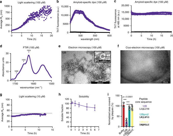
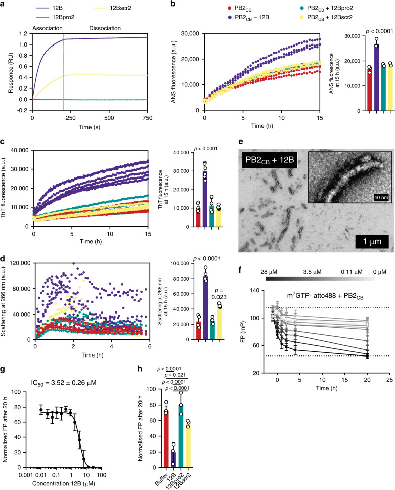
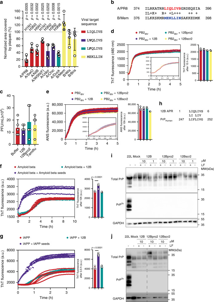
Human amyloids have been shown to interact with viruses and interfere with viral replication. Based on this observation, we employed a synthetic biology approach in which we engineered virus-specific amyloids against influenza A and Zika proteins. Each amyloid shares a homologous aggregation-prone fragment with a specific viral target protein. For influenza we demonstrate that a designer amyloid against PB2 accumulates in influenza A-infected tissue in vivo. Moreover, this amyloid acts specifically against influenza A and its common PB2 polymorphisms, but not influenza B, which lacks the homologous fragment. Our model amyloid demonstrates that the sequence specificity of amyloid interactions has the capacity to tune amyloid-virus interactions while allowing for the flexibility to maintain activity on evolutionary diverging variants.


Conflict of interest statement

The authors declare the following competing interests: J.S. and F.R. are listed as inventors of patents held by their hold institution VIB, covering the peptides described in this manuscript, and which are licensed to Aelin Therapeutics (Leuven, Belgium), of which J.S. and F.R. are the scientific founders. All other authors declare no competing interests.

Figures

Fig. 1
Fig. 1. APR-based design of a synthetic peptide with antiviral activity against influenza A.
a Graphical illustration of the peptide tandem design with the target APRs capped by charged amino acids (arginine or aspartic acid) and linked by a glycine-serine or proline-proline linker. b Cap-binding domain of the influenza A/PR8 polymerase basic protein 2 (PB2CB) (PDB = 4ENF),. The target APR, 381LIQLIVS387, is surface exposed and highlighted in red. c Plaque-size reduction assay of MDCK cells treated with cell medium, 1% DMSO, 10 µM peptide 12B, 10 µM nucleozin, 100 µM Tamiflu, or 1 µM VX-787 2 h prior to infection with influenza A/PR8. d Quantification of the area covered by plaques in c. Data are normalized to medium-treated cells and mean ± SD is shown (n = 4 independent experiments, statistics: one-way ANOVA with multiple comparison). e Dose-dependent effect of peptide 12B on area covered by plaques (red curve, left axis) and on red blood cell (RBC) lysis (purple curve, right axis). For IC50: data are normalized to buffer-treated cells and the mean ± SD is shown (n = 4 independent experiments). For toxic dose (TD50): data are normalized to buffer-treated (0% lysis) and 0.1% Triton-treated cells (100% lysis) and the mean ± SD is shown (n = 3 independent experiments). f Dose-dependent toxicity of peptide 12B, after 24-h incubation on MDCK cells. Data are normalized to buffer-treated (100% viability) and 0.1% Triton-treated cells (0% viability) and the mean ± SD is shown (n = 3 independent experiments). g Viral load in the lungs of influenza A/PR8-infected mice after daily injections of either buffer, 2.5 mg/kg peptide 12B, 2.5 mg/kg peptide 12Bpro2 (all i.v.), or 25 mg/kg Tamiflu (oral gavage) for 6 consecutive days. Data are expressed as mean ± SD (n = 13 mice over six independent experiments, statistics: one-way ANOVA with multiple comparison). h Relative concentrations (SUV) of peptide [68Ga]Ga-NODAGA-PEG2-12B in the lungs of A/PR8-infected versus healthy mice at different time points after peptide injection. The data are expressed as mean ± SD (n = 12 mice over six independent experiments, statistics: two-sided unpaired Student’s t test).
Fig. 2
Fig. 2. Peptide 12B organizes into amyloid-like structures in vitro.
a Hydrodynamic radius (RH) calculated from the regularization fit of DLS data of peptide 12B (100 µM) maturation over time. b Th-T emission spectrum after excitation at 440 nm of buffer (black line) or peptide 12B (100 µM, purple line) 5 min after solubilization (n = 3 independent experiments). c Th-T emission signal of buffer (black line) or peptide 12B (100 µM, purple line) at 485 nm after excitation at 440 nm as a function of time. Data represent three independent repeats (n = 3 independent experiments). d FTIR spectrum of 100 µM peptide 12B 5 min after solubilization. TEM (e) and cryo-TEM (f) of 100 µM freshly dissolved peptide 12B (both are one representative image of three independent experiments). g Hydrodynamic radius (RH) calculated from the regularization fit of DLS data of peptide 12B (10 µM) maturation over time (dashed line = 50 nm). h Soluble fraction of peptide 12B (10 µM) determined after ultracentrifugation (250,000 × g for 30 min), measured over time, and mean ± SD is shown (n = 3 independent experiments). i Quantification of the area covered by plaques of MDCK cells treated with 10 µM peptide in a plaque-size reduction assay with influenza A/PR8. Data are normalized to medium-treated cells and the mean ± SD is shown (n = 5 independent experiments, statistics: one-way ANOVA with multiple comparison).
Fig. 3
Fig. 3. Amyloid peptide 12B interacts with influenza via the PB2 target protein in vitro.
a Biolayer interferometry showing association and dissociation of 10 µM peptide to his-tagged, immobilized PBCB. b, c ANS (excitation 380 nm, emission 485 nm) and Th-T (excitation 440 nm, emission 485 nm) fluorescence of PB2CB (32 µM) with and without peptides (3.2 µM) over time. Quantification of the fluorescence signal after 15 h is shown on the right. Data are expressed as mean ± SD (n = 3 (b) and n = 6 (c) independent experiments, statistics: one-way ANOVA with multiple comparison to PB2CB alone). d Particle scattering (260 nm) of PB2CB (32 µM) with and without peptides (3.2 µM) over time. Quantification of scattering signal after 1 h (before precipitation) is shown on the right. Data are expressed as mean ± SD (n = 4 independent experiments, statistics: one-way ANOVA with multiple comparison to PB2CB alone). e TEM images of PB2CB (32 µM) with peptide 12B (3.2 µM) after 15 h incubation. f Binding of m7GTP-atto488 by PB2CB in absence or presence (gray scale for peptide concentration) of a twofold serial dilution of peptide 12B, determined by fluorescence polarization (FP). Mean ± SD is shown (n = 4 independent experiments). The dashed lines represent maximal binding of m7GTP-atto488 to PB2 (upper dashed line) and free m7GTP-atto488 (lower dashed line), based on control experiments (Supplementary Fig. 4h). g Concentration-dependent effect of peptide 12B on the binding of m7GTP-atto488 by PB2CB (5 µM) after 20 h incubation as shown by FP. Data represent normalized values (maximal binding = 100% and free m7GTP-atto488 = 0%) and mean ± SD is shown (n = 5 independent experiments). h Binding of m7GTP-atto488 by PB2CB (5 µM) with or without peptides (7 µM) after 20 h incubation. Data represent normalized values (similar as in g) and mean ± SD is shown (n = 3 independent experiments, statistics: one-way ANOVA with multiple comparison).
Fig. 4
Fig. 4. Amyloid peptide 12B interacts with influenza via the PB2 target protein in cellulo.
a Quantification of the area covered by plaques of A/PR8-infected MDCK cells treated with 10 µM peptide 12B at different time points relative to infection. Data are normalized to medium-treated cells and mean ± SD is shown (n = 8 from three independent experiments, statistics: one-way ANOVA with multiple comparison, p value = 0.0605). Representative images are shown in Supplementary Fig. 5a. b Quantification of the area covered by plaques in a plaque-size reduction assay of MDCK cells infected with A/PR8 virion particles that were pretreated with peptide 12B. Concentration-dependent effect of peptide 12B is shown normalized to medium-treated virion particles as mean ± SD (n = 3 independent experiments). Representative images are shown in Supplementary Fig. 5b. c MDCK cells treated with FITC-labeled peptide 12B, 12Bpro2 (10 µM), or buffer for 2 h prior to A/WSN-FLAG infection (MOI = 1, 16-h infection). Cells were fixed and stained with an anti-FLAG antibody and DAPI. One representative example of three independent experiments. d Western blot analysis of total (input) and immunoprecipitated fraction of MDCK cells treated with PEG2-biotin-labeled peptide (10 µM) or buffer for 2 h prior to A/WSN-FLAG infection (MOI = 1, 16-h infection). Quantification of immunoprecipitated fraction of PB2, relative to PB2 input levels, is shown on the right. Data represent mean ± SD (n = 5 independent experiments, statistics: one-way ANOVA with multiple comparison). Streptavidin was used as a loading control. e Western blot analysis of PB2 distribution in soluble and insoluble fraction of lysates of MDCK cells treated with peptide (10 µM) for 2 h prior to influenza A/WSN-FLAG infection (MOI = 1, 16-h infection). Vimentin and GAPDH were used as loading controls for insoluble and soluble fraction, respectively. Quantification represents soluble PB2 fraction, normalized to medium-treated cells (100% soluble PB2). Data represent mean ± SD (n = 4 independent experiments, statistics: one-way ANOVA with multiple comparison). For d and e the samples on one blot are derived from the same experiment and the gels/blots were processed in parallel. Full blots are shown in Supplementary Fig. 12.
Fig. 5
Fig. 5. Amyloid peptide 12B acts as a sequence-specific interferor of influenza A.
a Quantification of area covered by plaques of influenza-infected MDCK cells, treated with 10 µM peptide 12B. Data are normalized to buffer-treated cells and mean ± SD is shown (n = 4 independent experiments, statistics: two-sided one-sample t-test). A list of abbreviations is provided in “Methods.” b Local alignment of PB2 from influenza A/PR8 and B/Mem. Target APRs are highlighted in red (A/PR8) and blue (B/Mem). c Viral load in the lungs of influenza B/Mem-infected mice after daily injections of buffer, 2.5 mg/kg peptide 12B, 2.5 mg/kg peptide 12Bpro2 (i.v.); or 25 mg/kg Tamiflu (oral gavage) for 6 days. Data represent mean ± SD (n = 13 mice, five independent experiments, statistics: one-way ANOVA with multiple comparison, p value = 0.6027). d, e Th-T and ANS fluorescence of PB2CB(IBV) (32 µM) without and with peptides (3.2 µM, enlarged in the inserts). Fluorescence after 20 h is shown on the right as mean ± SD (n = 3 independent experiments, statistics: one-way ANOVA with multiple comparison, p value = 0.4218 for d and 0.1499 for e). f, g Th-T fluorescence of amyloid beta (10 µM) and IAPP (10 µM) without and with peptide 12B (1 µM) is shown. Self-seeded control is included by using preformed aggregates of amyloid beta and IAPP, respectively (1 µM). Th-T fluorescence at plateau is shown on the right as mean ± SD (n = 4 independent experiments, statistics: one-way ANOVA with multiple comparison). h Local alignment of the target APR in peptide 12B with human prion protein (PrPhuman). Western blot analysis of proteinase K-treated lysates of L929 15.9 cells (six passages, (i)) and CAD5 cells (eight passages, (j)) post treatment with peptides (1  or 10 µM) with or without Lipofectamine 2000 (LF). As controls, cells were exposed to brain homogenate from a terminally diseased mouse infected with scrapie strain 22L (PrPSc) or with normal (Mock) brain homogenate. GAPDH was used as loading control. The samples are derived from the same experiment and the gels/blots were processed in parallel. Full blots are shown in Supplementary Fig. 12.
Fig. 6
Fig. 6. Peptide R50 organizes into amyloid structures and interferes with ZIKV replication.
a Hydrodynamic radius (RH) calculated from the regularization fit of DLS data of peptide R50 (10 µM) maturation over time. b Th-T emission signal of 10 µM peptide R50 at 485 nm after excitation at 440 nm as a function of time (n = 3 independent experiments). c TEM image of 10 µM peptide R50 after 6 h incubation. d Soluble fraction of peptide R50 (10 µM) determined after ultracentrifugation (250,000 × g for 30 min), measured over time, and mean ± SD is shown (n = 3 independent experiments). Dose-dependent effect of 7-DMA (e), peptide R50 (f), and peptide 12B (g) on the fraction of ZIKV-infected cells following 48 h of infection with a MOI of 0.1. Data are normalized to DMSO-treated, infected cells (100% infected cells) and mean values ± SD are shown (n = 3 independent experiments). Representative images are shown in Supplementary Fig. 10. Dose-dependent effect of 7-DMA (h), peptide R50 (i), and peptide 12B (j) on cell viability of Vero E6 cells without ZIKV infection. Data are normalized to DMSO-treated, noninfected cells (100% cell viability) and the mean values ± SD are shown (n = 3 independent experiments).

References

    1. Pham CLL, Kwan AH, Sunde M. Functional amyloid: widespread in nature, diverse in purpose. Essays Biochem. 2014;56:207–219. doi: 10.1042/bse0560207. - DOI - PubMed
    1. Vogler TO, et al. TDP-43 and RNA form amyloid-like myo-granules in regenerating muscle. Nature. 2018;117:209. - PMC - PubMed
    1. Bourgade K, et al. Protective effect of amyloid-β peptides against herpes simplex virus-1 infection in a neuronal cell culture model. JAD. 2016;50:1227–1241. doi: 10.3233/JAD-150652. - DOI - PubMed
    1. Cribbs DH, Azizeh BY, Cotman CW, LaFerla FM. Fibril formation and neurotoxicity by a herpes simplex virus glycoprotein B fragment with homology to the Alzheimer’s A beta peptide. Biochemistry. 2000;39:5988–5994. doi: 10.1021/bi000029f. - DOI - PubMed
    1. Eimer WA, et al. Alzheimer’s disease-associated β-amyloid is rapidly seeded by herpesviridae to protect against brain infection. Neuron. 2018;99:56–63.e3. doi: 10.1016/j.neuron.2018.06.030. - DOI - PMC - PubMed

Publication types

MeSH terms