. 2020 Aug 3;29(13):2218-2239.
doi: 10.1093/hmg/ddaa108.

Biallelic variants in the RNA exosome gene EXOSC5 are associated with developmental delays, short stature, cerebellar hypoplasia and motor weakness

Affiliations

Biallelic variants in the RNA exosome gene EXOSC5 are associated with developmental delays, short stature, cerebellar hypoplasia and motor weakness

Anne Slavotinek et al. Hum Mol Genet. .

Abstract

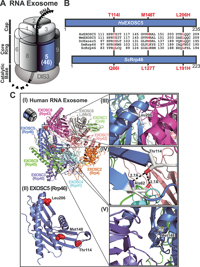
The RNA exosome is an essential ribonuclease complex required for processing and/or degradation of both coding and non-coding RNAs. We identified five patients with biallelic variants in EXOSC5, which encodes a structural subunit of the RNA exosome. The clinical features of these patients include failure to thrive, short stature, feeding difficulties, developmental delays that affect motor skills, hypotonia and esotropia. Brain MRI revealed cerebellar hypoplasia and ventriculomegaly. While we ascertained five patients, three patients with distinct variants of EXOSC5 were studied in detail. The first patient had a deletion involving exons 5-6 of EXOSC5 and a missense variant, p.Thr114Ile, that were inherited in trans, the second patient was homozygous for p.Leu206His and the third patient had paternal isodisomy for chromosome 19 and was homozygous for p.Met148Thr. The additional two patients ascertained are siblings who had an early frameshift mutation in EXOSC5 and the p.Thr114Ile missense variant that were inherited in trans. We employed three complementary approaches to explore the requirement for EXOSC5 in brain development and assess consequences of pathogenic EXOSC5 variants. Loss of function for exosc5 in zebrafish results in shortened and curved tails/bodies, reduced eye/head size and edema. We modeled pathogenic EXOSC5 variants in both budding yeast and mammalian cells. Some of these variants cause defects in RNA exosome function as well as altered interactions with other RNA exosome subunits. These findings expand the number of genes encoding RNA exosome subunits linked to human disease while also suggesting that disease mechanism varies depending on the specific pathogenic variant.


Figures

Figure 1
Figure 1
Clinical presentation and pedigrees of three patients with biallelic variants in EXOSC5. (A) Facial photographs of Patient 1, demonstrating mild ptosis, epicanthic folds, full cheeks, a high nasal bridge, broad nasal tip, prominent Cupid’s bow and mild micrognathia, and Patient 3, demonstrating a mildly broad nasal tip and prominent Cupid’s bow. Patient 3 was not considered to have significant facial anomalies by the Attending clinician. (B) Pedigrees illustrating the inheritance pattern of EXOSC5 alleles in Patients 1, 2 and 3 are shown. (C) MRI findings in EXOSC5 Patients 1 and 2 are shown. MRI scan of the brain of Patient 1 with p.Thr114Ile, showing cerebellar hypoplasia, with reduced size of the cerebellar vermis and increased cerebellar sulci on sagittal section. The brainstem was also small. MRI scan of the brain of Patient 2 with homozygosity for an EXOSC5 variant p.Leu206His at the age of 4 months. Left panel: Sagittal T1-weighted MRI revealed decreased size of pons and brainstem (asterisk) and also of the cerebellar vermis (arrow). The posterior fossa was enlarged. The corpus callosum (arrowhead) appeared thin but was within the expected physiological variation for the age. Middle panel: Axial T2-weighted MRI illustrated the coned shape of the skull and the corresponding coned shape of both frontal lobes, due to early closure of the metopic suture. The size of the ventricular system is increased (black stars). Right panel: Axial T2-weighted MRI illustrated the relative lack of reduced T2-signal intensity in the perirolandic areas (black arrows), indicating hypomyelination.
Figure 2
Figure 2
CRISPR/Cas9 targeting of exosc5 in transgenic zebrafish demonstrates larvae with morphological defects of the tail and body. (A) Typical morphology was observed in Control larvae (Uninjected Control). For CRISPR-injected larvae with targeting of exosc5, we observed larvae with a mild tail curvature (Mild phenotype), hooked tail curvature (Moderate phenotype) and larvae with severe shortening of the tail and body axis with the distortion of the body axis (Severe phenotype). Reduced eye and head size and edema were also observed in the exosc5-targeted larvae. Larvae were photographed at 54–58 h post-fertilization (hpf). (B) The relative number of morphological phenotypes (Normal, Dead, Mild, Moderate and Severe) observed in Uninjected Control (n = 230) and exosc5 crispants (n = 226) is depicted as percentage of total larvae analyzed. (C) Sections of Uninjected Control larvae and CRISPR-injected larvae with targeting of exosc5 were stained with hematoxylin and eosin. Crispant exosc5 larvae showed reduced eye and lens size, increased ventricle size and reduced brain size with aberrant formation of the mesencephalon and diencephalon as indicated. Larvae were photographed at 54–58 hpf.
Figure 3
Figure 3
CRISPR/Cas9 targeting of exosc5 in mnx1:EGFP and NBT:dsRed transgenic zebrafish demonstrates larvae with morphological defects of the tail, body and brain. (A) Transgenic zebrafish with mnx1:EGFP with sgRNA targeting of exosc5 show severe larval phenotypes, with shortened heads, bodies and tails, small or absent eyes and edema compared with Uninjected Control. Larvae with targeting of exosc5 can also have a normal appearance similar to controls as illustrated in the normal panel. Fluorescence imaging reveals decreased GFP signal and abnormal midbrain and hindbrain morphology in larvae with Severe phenotypes, marked by arrows, compared with Control and normal-appearing injected larvae. Larvae with Severe phenotypes were verified to have biallelic exosc5 targeting by Sanger sequencing. Larvae were photographed at 54–58 hpf. MB = midbrain; HB = hindbrain. (B) Transgenic zebrafish with Neurons labeled by NBT1:dsRed with sgRNA targeting of exosc5 show severe larval phenotypes, with shortened heads, bodies and tails, distorted bodies, small eyes and marked edema compared with Uninjected Control. Larvae with targeting of exosc5 can also have a Normal or Mild phenotype. Fluorescence imaging reveals extensive disruption of brain formation and morphology in one exosc5-targeted larvae, which shows a Severe phenotype. Larvae with severe phenotypes were verified to have successful exosc5 targeting by Sanger sequencing. Larvae were photographed at 54–58 hpf.
Figure 4
Figure 4
Pathogenic amino acid changes that occur in EXOSC5 are in conserved regions of the protein that could be important for interactions with other subunits of the RNA exosome complex. (A) A cartoon model of the 10-subunit RNA exosome complex is shown. The subunits of the complex are indicated by their EXOSC number (1–3 in the cap, 4–9 in the core ring), with the catalytic DIS3 subunit at the base. EXOSC5, which is a component of the core ring structure, is highlighted in marine blue. (B) Domain structure schematics are shown for both human EXOSC5 and the S. cerevisiae ortholog of EXOSC5, Rrp46. Both proteins are comprised of a single RNase PH-like domain. The amino acids changes—T114I, M148T and L206H—in EXOSC5 identified in the patients described here are indicated above EXOSC5 and the corresponding amino acid changes—Q86I, M127T and L191H—in Rrp46 are denoted below Rrp46. Between the domain structures, alignments of the amino acid sequences from human (Hs), mouse (Mm), zebrafish (Dr), fruit fly (Dm) and budding yeast (Sc), EXOSC5/Rrp46 orthologs that surround the EXOSC5 residue altered in patients (indicated in red) are shown. The alignments highlight the evolutionary conservation of the residue altered in disease and the surrounding residues. (C) Structural model of (I) the human 9-subunit RNA exosome complex and (II) the EXOSC5 subunit are shown with the position of the residues altered in patients highlighted as red side chain spheres. A miniature cartoon of the RNA exosome in the top left corner denotes the approximate orientation of the RNA exosome structure. The cap subunits EXOSC1/2/3 are depicted in lime/orange/slate blue, and the core ring subunits EXOSC4/5/6/7/8/9 are depicted in cyan/marine blue/gray/salmon red/magenta/forest green, respectively. The depicted human 9-subunit RNA exosome structure is adapted from the human 10-subunit RNA exosome-MPP6 complex (PDB: 6h25) (42) and does not show DIS3 or MPP6. A zoomed-in view of the location of the (III) EXOSC5 Leu206 residue that is changed to Histidine in patient 2 (p.Leu206His) shows that Leu206 is a buried hydrophobic residue and predicts that its replacement with a polar Histidine residue would alter EXOSC5 stability and exert a destabilizing effect on its interaction with EXOSC3. A zoomed-in view of the location of the (IV) EXOSC5 Thr114 residue that is changed to Isoleucine in patient 1 (p.Thr114Ile) shows that Thr114 makes a hydrogen bond with the backbone of the Ala62 residue in EXOSC5 and predicts that its replacement with Isoleucine would disrupt this interaction with Ala62. However, the Isoleucine residue at position 114 in EXOSC5 is not predicted to significantly affect protein stability but more likely exerts a deleterious effect due to its central location in the exosome and proximity to EXOSC3. A zoomed-in view of the location of the (V) EXOSC5 Met148 residue that is changed to Threonine in patient 3 (p.Met148Thr) predicts that Met148 substitution with a polar Threonine residue would reduce the stability of EXOSC5 and affect its interaction with EXOSC3 (Thr81).
Figure 5
Figure 5
Amino acid substitutions in the budding yeast ortholog of EXOSC5, Rrp46, that correspond to the EXOSC5 T114I, M148T and L206H substitutions identified in patients impair yeast cell growth. (A) Budding yeast cells that express the rrp46-L191H variant, corresponding to the EXOSC5 p.Leu206His variant, as the sole copy of the essential Rrp46 protein shows impaired growth at 30 and 37°C compared with cells expressing wild-type Rrp46 in a solid media growth assay. In contrast, cells expressing the rrp46-Q86I or rrp46-L127T variant, corresponding to the EXOSC5 p.T114I and p.M148T variant, show no growth defect at any temperature compared with cells expressing wild-type Rrp46. Growth of rrp46∆ yeast cells containing RRP46, rrp46-Q86I, rrp46-L127T or rrp46-L191H plasmid was analyzed by serial dilution, spotting on solid media and growth at indicated temperatures. (B) The rrp46-L191H mutant cells, but not the rrp46-Q86I or rrp46-L127T mutant cells, exhibit reduced growth at 30 and 37°C compared with wild-type RRP46 cells in a liquid culture growth assay, in which the optical density of the cultures was measured over time. (C) Immunoblotting was employed to assess the steady-state levels of the Rrp46/rrp46 proteins. Lysates of rrp46∆ yeast cells expressing only Myc-tagged wild-type Rrp46, rrp46-Q86I, rrp46-L127T or rrp46-L191H grown 30 or 37°C as indicated were analyzed by immunoblotting with anti-Myc antibody to detect Myc-tagged Rrp46 and rrp46 variants (Rrp46-Myc) and anti-Pgk1 antibody to detect 3-phosphoglycerate kinase (Pgk1) as a loading control. A representative result is shown. (D) Results from two independent immunoblotting experiments (C) were quantitated as described in Materials and Methods. The amount of wild-type Rrp46 protein detected at grown 30°C (top) or 37°C (bottom) was set to1.0, and the normalized fold-change of each rrp46 protein relative to wild-type Rrp46 is shown for each temperature. A statistically significant changes in steady-state protein level was detected for the rrp46-Q86I protein at 30°C (P = 0.0307), while the fold change in rrp46-L191H at 30°C was not statistically significant (P = 0.0710), and the remaining fold changes in rrp46 levels at 30 and 37°C were not statistically significant. * indicates P-value < 0.05.
Figure 6
Figure 6
Budding yeast cells expressing the rrp46-L191H variant, corresponding to the EXOSC5 p.Leu206His variant, show impaired processing and elevated levels of some RNA exosome target transcripts at 37°C. (A) The rrp46-L191H mutant cells show a statistically significant increase in the levels of non-coding RNA exosome target U4 snRNA precursor [P = 0.0068; very significant (**)] relative to control RRP46 cells for which the value was set to 1.0 or other rrp46 mutants analyzed. Total RNA from rrp46∆ cells containing only RRP46, rrp46-Q86I, rrp46L127T or rrp46-L191H grown at 37°C was reverse transcribed using random hexamers and measured by quantitative PCR using primers indicated in the schematic that detect the U4 snRNA precursor as described in Material and Methods. Relative RNA levels were measured in triplicate biological samples, normalized to a control ALG9 transcript by the ∆∆Ct method, averaged, and graphically shown as fold increase relative to wild-type RRP46. Error bars denote standard error of the mean. Statistically significant differences in mean RNA levels were determined using the unpaired t test. (B) The rrp46-L191H mutant cells show a statistically significant increase in the levels of non-coding RNA exosome target TLC1 telomerase precursor RNA [P = 0.0008; extremely significant (***)] relative to control RRP46 cells for which the value was set to 1.0 or other rrp46 mutants analyzed. Samples were the same as those employed for (A) and were analyzed in the same manner. The location of the primers employed to detect TLC1 precursor transcript in RT-qPCR is indicated. (C) A northern blot shows that the processing of 7S pre-rRNA to 5.8S rRNA is impaired in rrp46-L191H mutant cells, but not in rrp46-Q86I mutant cells, compared with wild-type RRP46 cells. The levels of 7S pre-rRNA and 7S rRNA processing intermediates (asterisks), potentially including 6S pre-rRNA (5.8S rRNA with an additional 5–8 nucleotides at the 3′-end), are elevated in the rrp46-L191H cells, but not in rrp46-Q86I cells. The northern blot of total RNA from biological triplicates of RRP46, rrp46-Q86I and rrp46-L191H cells grown at 37°C was probed with a 5.8S-ITS2 rRNA-specific probe. The northern blot was stained with methylene blue to detect 5.8S rRNA as a loading control.
Figure 7
Figure 7
Pathogenic amino acid substitutions in EXOSC5 can alter interactions with other RNA exosome subunits. (A) Murine EXOSC5 variants, corresponding to human EXOSC5 variants identified in patients, are expressed at a similar level to wild-type murine EXOSC5 in a mouse neuronal cell line. The steady-state levels of Myc-tagged murine EXOSC5 p.T114L (EXOSC5-T114I), EXOSC5 p.M148T (EXOSC5-M148T) and EXOSC5 p.L206H (EXOSC5-L206H) variants are similar relative to Myc-tagged wild-type EXOSC5 in mouse N2a cells. Lysates of mouse N2a cells transfected with empty vector or vectors expressing murine Myc-EXOSC5, Myc-EXOSC5-T114I, Myc-EXOSC5-M148T or Myc-EXOSC5-L206H were analyzed by immunoblotting with anti-Myc antibody to detect Myc-EXOSC5 proteins. HSP90 protein detected with anti-HSP90 antibody serves as a loading control. (B) Murine EXOSC5 variants, corresponding to human EXOSC5 variants identified in patients, show decreased interactions with exosome subunits in a mouse neuronal cell line. Myc-EXOSC5, Myc-EXOSC5-T114I, Myc-EXOSC5-M148T or Myc-EXOSC5-L206H was immunoprecipitated from N2A cells, and interactions with the RNA exosome subunits EXOSC3, EXOSC9 and EXOSC10 were analyzed by immunoblotting. As a control, we also examined the interactions of a pathogenic Myc-EXOSC2-G198D variant, which was recently found to have reduced interactions with RNA exosome subunits (46). Both the Input and Bound samples are shown for Myc-EXOSC2 and Myc-EXOSC5. The fraction of EXOSC3, EXOSC9 or EXOSC10 bound (% Bound) is indicated on the bottom right.

References

    1. Brouwer R., Allmang C., Raijmakers R., van Aarssen Y., Egberts W.V., Petfalski E., van Venrooij W.J., Tollervey D. and Pruijn G.J. (2001) Three novel components of the human exosome. J. Biol. Chem., 276, 6177–6184. - PubMed
    1. Fabre A. and Badens C. (2014) Human Mendelian diseases related to abnormalities of the RNA exosome or its cofactors. Intractable Rare Dis. Res., 3, 8–11. - PMC - PubMed
    1. Morton D.J., Kuiper E.G., Jones S.K., Leung S.W., Corbett A.H. and Fasken M.B. (2018) The RNA exosome and RNA exosome-linked disease. RNA, 24, 127–142. - PMC - PubMed
    1. Mitchell P., Petfalski E. and Tollervey D. (1996) The 3′ end of yeast 5.8S rRNA is generated by an exonuclease processing mechanism. Genes Dev., 10, 502–513. - PubMed
    1. Mitchell P., Petfalski E., Shevchenko A., Mann M. and Tollervey D. (1997) The exosome: a conserved eukaryotic RNA processing complex containing multiple 3′→5′ exoribonucleases. Cell, 91, 457–466. - PubMed

Publication types

MeSH terms

Substances

Supplementary concepts